Abstract
Mechanisms that regulate host defense after noninfectious tissue injury are incompletely understood. Our laboratory is interested in the role of the extracellular matrix glycosaminoglycan hyaluronan in the regulation of lung inflammation and fibrosis. We have identified key roles for two cell surface receptor systems that interact with hyaluronan to control lung inflammation and tissue repair. Hematopoietic CD44 is necessary to clear hyaluronan fragments that are produced after lung injury. Failure to clear hyaluronan fragments leads to unremitting inflammation. However, in the absence of CD44, alveolar macrophages continue to produce chemokines in response to hyaluronan fragments, implicating another receptor system in controlling macrophage effector function. We found that Toll-like receptors 2 and 4 (TLR2 and TLR4) are responsible for macrophage inflammatory gene expression in response to hyaluronan fragments. Although TLR2 and TLR4 initiate the innate immune response in noninfectious inflammation, they have a protective role against lung injury on alveolar epithelial cells.
Keywords: CD44, hyaluronan, innate immunity, tissue injury, Toll-like receptors
Regulation of tissue injury and repair is a carefully orchestrated host response to eradicate the offending agent and restore tissue integrity. Successful repair of tissue injury requires a coordinated host response to limit the extent of structural cell damage. The mechanisms that regulate the host response to tissue injury are incompletely understood. Abnormalities in the host repair response are associated with a variety of chronic disease states that are characterized by excessive deposition of extracellular matrix (ECM), resulting in organ fibrosis. The innate response to an insult is the component of the host response that is in place before the insult and serves as the first line of defense against injury. For example, after exposure to an infectious pathogen, the initial phase of the host response consists of macrophages recognizing the presence of the pathogen and releasing signals that trigger an inflammatory response. This innate response is mediated by Toll-like receptors (TLRs) on macrophages that orchestrate the recognition of the invading pathogen and initiation of the host inflammatory response with the goal of restoration of tissue integrity (1, 2). In concert with initiating the inflammatory response, the host must also generate signals to minimize the extent of structural cell damage. The ultimate outcome of the host depends on the balance between containment of injury, maintenance of structural cell integrity, and activating repair mechanisms. The mechanisms that regulate the host response to noninfectious tissue injury are poorly understood. This review focuses on recent work from our laboratory investigating the interactions between ECM and innate immune receptors in regulating noninfectious lung injury and repair.
A hallmark of tissue injury is increased turnover of ECM. A variety of lung diseases are associated with abnormal ECM turnover. Asthma, emphysema, and pulmonary fibrosis are three chronic lung diseases that have in common an imbalance between the synthesis and degradation of ECM. In chronic lung diseases, the ECM is modified by the inflammatory milieu, and degradation products are generated by oxidants and other mechanisms that take on unique properties not attributable to the precursor molecules. Work from our laboratory has shown that failure to remove matrix degradation products from the lung after injury results in the host succumbing to unremitting inflammation (3). We have focused our studies on hyaluronan (HA).
HA is an ECM component that undergoes dynamic regulation during tissue injury and inflammation. HA is a nonsulfated glycosaminoglycan composed of repeating polymeric disaccharides D-glucuronic acid and N-acetyl glucosamine. HA can exist as both a soluble polymer and as noncovalently linked to proteoglycan core proteins. Under physiologic conditions, HA exists as a high-molecular-weight polymer (> 106 D) and undergoes dynamic regulation resulting in accumulation of lower molecular weight species after tissue injury (3, 4). During normal development, the absence of HA by genetically targeting deletion of HA synthase 2 (HAS2) results in embryonic lethality due to severe cardiac and vascular abnormalities (5). HA degradation products generated in vitro induce the expression of a variety of genes, including chemokines, cytokines, growth factors, signal transduction molecules, and adhesion molecules, in a variety of cell types, suggesting endogenously generated HA fragments may regulate inflammatory processes (6–10). CD44 is a type 1 transmembrane glycoprotein that is expressed on most cells and is a major cell surface receptor for HA (11). The interaction of HA and CD44 has been implicated in the regulation of a variety of biological processes, including tumor growth and metastasis, wound healing, T-cell recruitment to sites of inflammation, macrophage activation, neutrophil migration, and endothelial cell activation (12). However, unlike the targeted deletion of HAS2, mice with a targeted deletion in CD44 were found to develop normally (13). To investigate the role of HA in lung injury and repair, we examined the inflammatory response in CD44-deficient mice after noninfectious lung injury (3). Instillation of the DNA-damaging chemotherapeutic agent bleomycin sulfate into the tracheas of mice causes epithelial cell injury and elicits an acute inflammatory response that usually peaks by Day 7. Over the ensuing 14 d, the inflammatory response remits and there is a robust fibroproliferative response that results in the significant deposition of ECM components such as collagen as well as the transient presence of myofibroblasts.
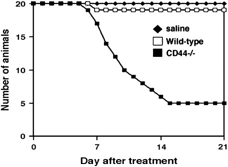
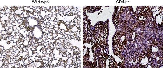
In addition to the deposition of collagen, an important aspect of the injury and repair response is the accumulation and subsequent clearance of HA. HA production increases several-fold after bleomycin treatment and peaks between Days 7 and 10. HA is subsequently cleared from the lung interstitium coincident with the accumulation of collagen. HA degradation products accumulate in the lung in the size range (10–500 kD) that can stimulate inflammatory gene expression by macrophages and other inflammatory cells in vitro. To examine the role of HA homeostasis in lung injury and repair, we examined the inflammatory response to intratracheal bleomycin sulfate in CD44-deficient mice (3). We made the unexpected observation that CD44 deficiency led to an increased susceptibility to lung injury with a marked increase in mortality after bleomycin treatment (Figure 1). Examination of the lung tissue at a time point when the wild-type mouse lung had resolved the inflammatory response demonstrated an overwhelming accumulation of inflammatory cells in the CD44-deficient mice (Figure 2).
Figure 1.
CD44 deficiency leads to increased mortality after bleomycin-induced lung injury. Reproduced by permission from Reference 3. Copyright © 2002 The American Association for the Advancement of Science.
Figure 2.
CD44 deficiency leads to unremitting lung inflammation after bleomycin-induced lung injury.
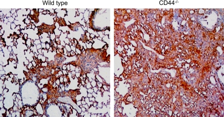
In addition to the inability to resolve the lung inflammation, we also identified a striking abnormality in the deposition of HA. As shown in Figure 3, there is abundant HA accumulation in the injured lungs in the CD44-deficient mice relative to the wild-type control mice. CD44-deficient mice were unable to clear HA degradation products from the lung interstitium. The CD44-deficient state was associated with an inability to resolve lung inflammation and properly remove HA from the lung tissue. Evaluation of the molecular mass of the lung tissue HA revealed major differences between the wild-type and CD44-deficient states. CD44 deficiency resulted in the accumulation of both higher and lower molecular mass species than observed in the wild-type mice. CD44 thus has a profound role in regulating HA homeostasis in the lung after acute injury. Because CD44 is present on both structural cells (epithelium, fibroblasts, and endothelium) and hematopoietic cells, we sought to determine which cellular compartment was responsible for HA clearance and resolution of inflammation. We generated chimeric mice by transferring bone marrow from syngeneic wild-type mice into sublethally irradiated CD44-deficient mice. We thus reconstituted CD44-positive hematopoietic cells in CD44-deficient mice. When these chimeric mice were then treated with bleomycin we found that the profound increase in mortality was prevented. This reversal in phenotype was accompanied by a restoration of the ability to resolve the inflammatory response and remove HA from the lung interstitium (3).
Figure 3.
CD44 deficiency leads to impaired clearance of hyaluronan after bleomycin-induced lung injury. Reproduced by permission from Reference 3. Copyright © 2002 The American Association for the Advancement of Science.
Collectively, these data suggest that the production of HA after acute tissue injury serves the important function of initiating the host innate immune response by providing an essential signal to macrophages to produce chemokines that recruit other leukocyte subsets required to debride the tissue injury and begin restoring tissue integrity. In addition, although it appears essential to the host to initiate inflammatory responses when HA degradation products are recognized by macrophages, it is also important that the inflammatory response be successfully resolved. An important component of the resolution of the inflammatory response is the successful removal of HA degradation products. Failure to properly clear these ECM breakdown products results in unremitting inflammation. These data suggest that a previously unrecognized component of the innate immune response is the generation and clearance of ECM components that are produced during the inflammatory response.
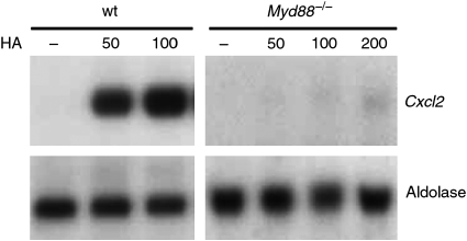
Although these studies suggested a previously unrecognized role for CD44 in resolving lung inflammation, it also became clear that CD44 was not required by macrophages to produce chemokines in response to HA fragments. This suggested that another recognition system was involved. The repeating polymeric structure of HA coupled with the knowledge that HA is a component of the cell coat of Streptococcus and is structurally similar to the polysaccharide side chain of endotoxin suggested that, under certain circumstances, perhaps HA fragments may be recognized by TLRs (1, 2). To investigate this possibility that TLRs may be involved in HA recognition, we isolated elicited peritoneal macrophages from mice that are deficient in the critical adaptor protein MyD88. MyD88 is involved in signaling through TLR2 and TLR4 (14). We found that MyD88-deficient macrophages failed to respond to HA fragments (Figure 4). To determine which TLRs were involved in HA recognition, we determined if macrophages from TLR1-, TLR2-, TLR3-, TLR4-, TLR6-, and TLR9-deficient mice responded to HA fragments. We found that chemokine gene expression remained intact in all these strains but was reduced in TLR2- and TLR4-deficient mice. To determine if both TLR2 and TLR4 were required, we generated double knockout mice and found that chemokine expression in response to HA fragments was absent (Figure 5). These data suggest that HA fragments are able to trigger innate immune responses in a manner that overlaps with both gram-positive and gram-negative organism recognition pathways (15).
Figure 4.
Hyaluronan (HA) fragments stimulate chemokine expression through MyD88. Cxcl2 mRNA expression by peritoneal macrophages from wild-type (wt) or Myd88−/− mice treated with HA fragments detected by Northern analysis. Reproduced by permission from Reference 15.
Figure 5.
HA fragments stimulate chemokine expression through both Toll-like receptor 4 (TLR4) and TLR2. Macrophage inflammatory protein (MIP)-1β mRNA expression by peritoneal macrophages from wt, TLR2−/−, TLR4−/−, or TLR2−/−/TLR4−/− mice treated with HA fragments or LPS in the presence or absence (underlined) of polymyxin B, detected by Northern analysis. Reproduced by permission from Reference 15.
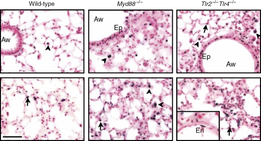
To determine the in vivo role of TLR2, TLR4, and MyD88 in regulating noninfectious lung injury, inflammation, and repair, we challenged TLR-deficient mice with intratracheal bleomycin. As expected, there was less evidence of inflammation in the alveolar spaces (Figure 4). However, we made the surprising observation that there was increased mortality in the absence of TLR2 and TLR4 as well as MyD88 after lung injury compared with control mice. The findings of impaired neutrophil migration but increased mortality and evidence of lung injury made us suspect a previously unrecognized role for TLR2 and TLR2 in the parenchymal cell response to injury. We were able to demonstrate increased epithelial cell apoptosis in the TLR2/TLR4 double knockout mice as well as in MyD88 knockout mice relative to control animals after lung injury (Figure 6). To determine if there was a physiologic role for an interaction between HA and TLR2/TLR4 in epithelial cell physiology, we treated mice with an inhibitor of HA–cell interactions and found a similar phenotype to the TLR deficiency (15). We provided further evidence for this by generating transgenic mice that overexpress high-molecular-mass HA using the Clara cell (CC10) promoter. The overexpression of high-molecular-mass HA improved mortality after lung injury (15). To directly interrogate the role of HA and TLR2/TLR4 in alveolar epithelial cell responses to injury, we isolated primary alveolar epithelial cells from wild-type and TLR2/TLR4-deficient mice. Interestingly, wild-type alveolar epithelial cells expressed HA on the cell surface and this was markedly abrogated in the absence of TLR2 and TLR4 (15).
Figure 6.
TLRs and HA regulate lung cell apoptosis. TUNEL staining of apoptosis from lung tissue sections of wild-type, Myd88−/−, and Tlr2−/−/Tlr4−/− mice 5 d after bleomycin injury. Aw = airway; Ep = airway epithelial cells; En = endothelial cells. Reproduced by permission from Reference 15.
Collectively, these data suggest that HA has several functions in lung injury and repair. After lung injury, HA fragments accumulate and stimulate alveolar macrophages to produce chemokines that recruit subsets of inflammatory cells. Chemokine production is dependent on TLR2 and TLR4 and requires MyD88. These HA fragments are then cleared from the inflamed lung by alveolar macrophages in a CD44-dependent manner. However, in addition, high-molecular-mass HA is expressed on the cell surface of alveolar epithelial cells. Cell surface HA interacts with TLR2 and TLR4 to provide a homeostatic signal to protect the distal lung from injury and promote repair. These data raise the interesting possibility that restoring high-molecular-mass HA to the lung in the context of lung injury may promote repair and restoration of lung function.
Supported by research grants from the National Institutes of Health (HL-57486 and AI-52478).
Conflict of Interest Statement: Neither author has a financial relationship with a commercial entity that has an interest in the subject of this manuscript.
References
- 1.Barton GM, Medzhitov R. Toll-like receptor signaling pathways. Science 2003;300:1524–1525. [DOI] [PubMed] [Google Scholar]
- 2.Takeda K, Kaisho T, Akira S. Toll-like receptors. Annu Rev Immunol 2003;21:335–376. [DOI] [PubMed] [Google Scholar]
- 3.Teder P, Vandivier RW, Jiang D, Liang J, Cohn L, Pure E, Henson PM, Noble PW. Resolution of lung inflammation by CD44. Science 2002;296:155–158. [DOI] [PubMed] [Google Scholar]
- 4.Fraser JR, Laurent TC, Laurent UB. Hyaluronan: its nature, distribution, functions and turnover. J Intern Med 1997;242:27–33. [DOI] [PubMed] [Google Scholar]
- 5.Camenisch TD, Spicer AP, Brehm-Gibson T, Biesterfeldt J, Augustine ML, Calabro A Jr, Kubalak S, Klewer SE, McDonald JA. Disruption of hyaluronan synthase-2 abrogates normal cardiac morphogenesis and hyaluronan-mediated transformation of epithelium to mesenchyme. J Clin Invest 2000;106:349–360. [DOI] [PMC free article] [PubMed] [Google Scholar]
- 6.Horton MR, Burdick MD, Strieter RM, Bao C, Noble PW. Regulation of hyaluronan-induced chemokine gene expression by IL-10 and IFN-gamma in mouse macrophages. J Immunol 1998;160:3023–3030. [PubMed] [Google Scholar]
- 7.McKee CM, Penno MB, Cowman M, Burdick MD, Strieter RM, Bao C, Noble PW. Hyaluronan (HA) fragments induce chemokine gene expression in alveolar macrophages: the role of HA size and CD44. J Clin Invest 1996;98:2403–2413. [DOI] [PMC free article] [PubMed] [Google Scholar]
- 8.Noble PW, McKee CM, Cowman M, Shin HS. Hyaluronan fragments activate an NF-kappa B/I-kappa B alpha autoregulatory loop in murine macrophages. J Exp Med 1996;183:2373–2378. [DOI] [PMC free article] [PubMed] [Google Scholar]
- 9.Termeer C, Benedix F, Sleeman J, Fieber C, Voith U, Ahrens T, Miyake K, Freudenberg M, Galanos C, Simon JC. Oligosaccharides of hyaluronan activate dendritic cells via Toll-like receptor 4. J Exp Med 2002;195:99–111. [DOI] [PMC free article] [PubMed] [Google Scholar]
- 10.Ohkawara Y, Tamura G, Iwasaki T, Tanaka A, Kikuchi T, Shirato K. Activation and transforming growth factor-β production in eosinophils by hyaluronan. Am J Respir Cell Mol Biol 2000;23:444–451. [DOI] [PubMed] [Google Scholar]
- 11.Aruffo A, Stamenkovic I, Melnick M, Underhill CB, Seed B. CD44 is the principal cell surface receptor for hyaluronate. Cell 1990;61:1303–1313. [DOI] [PubMed] [Google Scholar]
- 12.Ponta H, Sherman L, Herrlich PA. CD44: from adhesion molecules to signalling regulators. Nat Rev Mol Cell Biol 2003;4:33–45. [DOI] [PubMed] [Google Scholar]
- 13.Schmits R, Filmus J, Gerwin N, Senaldi G, Kiefer F, Kundig T, Wakeham A, Shahinian A, Catzavelos C, Rak J, et al. CD44 regulates hematopoietic progenitor distribution, granuloma formation, and tumorigenicity. Blood 1997;90:2217–2233. [PubMed] [Google Scholar]
- 14.Medzhitov R, Preston-Hurlburt P, Kopp E, Stadlen A, Chen C, Ghosh S, Janeway CA Jr. MyD88 is an adaptor protein in the hToll/IL-1 receptor family signaling pathways. Mol Cell 1998;2:253–258. [DOI] [PubMed] [Google Scholar]
- 15.Jiang D, Liang J, Fan J, Yu S, Chen S, Luo Y, Prestwich GD, Mascarenhas MM, Garg HG, Quinn DA, et al. Regulation of lung injury and repair by Toll-like receptors and hyaluronan. Nat Med 2005;11:1173–1179. [DOI] [PubMed] [Google Scholar]


