Abstract
Iris recognition systems are among the most accurate of all biometric technologies with immense potential for use in worldwide security applications. This study examined the effect of eye pathology on iris recognition and in particular whether eye disease could cause iris recognition systems to fail. The experiment involved a prospective cohort of 54 patients with anterior segment eye disease who were seen at the acute referral unit of the Princess Alexandra Eye Pavilion in Edinburgh. Iris camera images were obtained from patients before treatment was commenced and again at follow-up appointments after treatment had been given. The principal outcome measure was that of mathematical difference in the iris recognition templates obtained from patients' eyes before and after treatment of the eye disease. Results showed that the performance of iris recognition was remarkably resilient to most ophthalmic disease states, including corneal oedema, iridotomies (laser puncture of iris) and conjunctivitis. Problems were, however, encountered in some patients with acute inflammation of the iris (iritis/anterior uveitis). The effects of a subject developing anterior uveitis may cause current recognition systems to fail. Those developing and deploying iris recognition should be aware of the potential problems that this could cause to this key biometric technology.
Keywords: iris recognition, Hamming distance, ophthalmology, iritis
1. Introduction
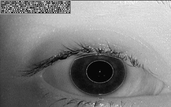
Biometric identification, or biometrics, refers to the automated recognition of an individual's identity based on his or her distinguishing characteristics (Bolle et al. 2004). In the case of iris recognition, it is the distinctive structures of a human iris that are exploited to achieve this (Nanavati et al. 2002). The concept of using iris patterns for personal identification was initially proposed in a clinical textbook (Doggart 1949) and later patented by two ophthalmologists (Flom & Safir 1987). However, the first algorithms for automated iris recognition were developed by Daugman (1994) at the University of Cambridge and patented in 1994. His pioneering automatic iris recognition system (Daugman 2004) used a special mathematical transformation employing ‘two-dimensional Gabor wavelets’ to transform iris images into ‘iris codes’ (figure 1). It has since been reported that iris recognition is one of the most reliable and accurate of all biometric identification systems (Nanavati et al. 2002). Daugman's algorithms have become the basis of all known publicly deployed iris recognition systems (Du 2006), although research into alternative methods continues.
Figure 1.
A standard iris camera image with automated iris segmentation demonstrated and the computed iris code displayed at top left.
A typical iris recognition system involves four main modules (Du 2006).
Image acquisition. Iris cameras are monochromatic, using the near-infrared band of illumination (700–900 nm).
Pre-processing. This involves techniques that detect and exclude occlusions such as eyelids, eyelashes and reflections. This step allows the iris patterns to be isolated and extracted from the image.
Template generation. In this step, an iris code template is created from the extracted iris patterns. Typically, for each iris 2 K bits of information are computed.
Pattern recognition. In this step, the iris template is finally compared with one or many other templates to detect whether a match is found. The iris matcher computes a ‘Hamming distance’ measure, which is a count of bit differences between the two templates (Bolle et al. 2004).
Iris scan technologies have an immense potential in worldwide security applications including immigration, banking and personal security. Even highly accurate automated technologies, such as fingerprint recognition for which the likelihood of a false match may be 1 out of 100 000, do not approach the false match performance of iris scan technology (Nanavati et al. 2002) unless multiple fingerprints are used simultaneously. Data formats for iris recognition have been standardized by the International Organization for Standardization, and their specification has been adopted by the International Civil Aviation Organization for the potential to store iris data on passports. Iris recognition is already deployed in United Arab Emirates in Homeland Security Border Control, Schiphol Airport, The Netherlands, and a number of US and Canadian airports. In the UK, it is operating at all five Heathrow terminals as well as Manchester, Birmingham and Gatwick airports.
Considering the vast potential and increasing market for iris recognition, the potential for problems caused by patients developing ocular pathology is considerable. These might include difficulty in obtaining access to personal funds or problems with security measures caused by the development of ocular disease any time after initial enrolment and iris capture. Roizenblatt (Roizenblatt et al. 2004) has investigated the effect of cataract surgery on iris recognition and found that the surgical intervention for cataract can lead to some eyes being no longer recognizable by the iris recognition system unless they are re-enrolled post-operatively. However, acute ocular disease might potentially cause far greater practical problems due to its unpredictable onset and course. There are no studies on the effect that the vast array of acute ophthalmic pathologies might have on iris recognition and thus the impact of ophthalmic disease on this ever-expanding security technology is currently unknown. The aim of this study was to examine the effect of eye pathology on iris recognition and in particular to determine whether eye disease could cause iris recognition systems to fail in the validation of subject identity.
2. Methods
The study was granted ethical approval by the local ethics committee and informed consent was taken from all patients. Patients who attended the Acute Referral Centre at the Edinburgh Eye Pavilion with acute eye disease were invited to take part in the study. Inclusion criteria included adult patients in whom any iris was apparent and who had some anterior segment pathology that might affect imaging or the appearance of the iris. Both eyes were enrolled at a patient's first attendance and relevant images obtained. The images were captured with an H100 IrisGuard portable tripod-mounted iris camera. It incorporates a 680 000 pixel sensor to achieve good image resolution with controlled illumination, and it controls focus distance by a voice interface to communicate with the patient.
The patients' affected eyes were treated and reviewed as necessary by the attending ophthalmologist. Eventually, when patients had recovered from their acute pathology, further iris images of both eyes were captured. Thus, images were obtained of each patient's eyes both with disease and after being fully treated. Unaffected eyes were also imaged and used as control eyes that had remained healthy throughout the course of the study.
Once acquired, the images were made anonymous and analysed in an automated process using the Daugman algorithms. The Hamming distance (mathematical difference) comparing patients before and after treatment for eye disease was calculated for each eye. Different images of the same eye taken close together in time would still never yield a Hamming distance of 0 due to variations in the subject's angle of gaze, degree of eyelid occlusion, random silhouettes of eye lashes, hippus and specular reflection from the cornea (Daugman 1993). In accordance with the deployed iris recognition systems, a Hamming distance of over 0.33 should be regarded as a false non-match result. That is, if the Hamming distance between different images of the same eye is greater than 0.33, the effect of eye disease has caused the iris recognition system to fail in its task of recognizing the images as that from the same eye. This correlates with an acceptable practical level of security and gives a false match rate of approximately one in a million, depending on the database tested (Daugman 2004).
As an additional measure, we also assessed any significant statistical difference in the Hamming distance in patients' eyes that had been diseased compared with control eyes that had stayed disease free. Power calculations were based upon preliminary studies with Hamming distances demonstrating a mean of 0.153 and standard deviation of 0.069 in diseased eyes. The smallest difference of importance required to be detected was set to 0.1, which would still leave the Hamming distance well within the limits of 0.33. According to sample size algorithms (Machin et al. 2007), at least 10 patient eyes would be required to show a 90 per cent chance of detecting a difference at the 5 per cent significance level with the unpaired t-test.
3. Results
3.1 Patient epidemiology
Fifty-six patients with a wide mix of anterior segment disease were recruited into the trial. Two patients dropped out from the trial as they did not want the inconvenience of waiting for repeat photography. Out of the 54 remaining patients, 15 had bilateral disease (30 eyes) and 39 had unilateral disease. Thus, a total of 69 eyes with pathology were investigated in the study and 39 normal eyes investigated during the same period in the control group.
Eye pathology was as follows:
three eyes with glaucoma requiring YAG laser iridotomy (small laser puncture to iris);
twenty-four eyes had anterior uveitis (inflammation of the iris); and
thirty-three eyes had corneal pathologies (12 infective and 21 non-infective disease).
The remainder of eyes with pathology had conditions affecting outer ocular layers:
two eyes with episcleritis (inflammation of episclera of eye);
one eye with scleritis (inflammation of sclera of eye); and
six eyes with conjunctivitis.
3.2 Overall change in the Hamming distance
The primary outcome measure was to determine whether any of the pathologies could cause enough of a change in the Hamming distance to render a patient's second iris scan not verifiable as being of the same patient. The Hamming distance for this to occur is set at 0.33 in deployed systems. There were five patients' eyes in whom the iris recognition failed by these criteria. All five involved cases of anterior uveitis in which the eye had been prescribed pharmacological dilation as part of the treatment. The actual Hamming distance values were 0.406, 0.387, 0.402, 0.345 and 0.349.
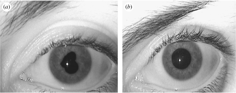
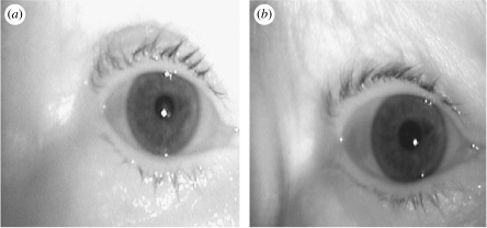
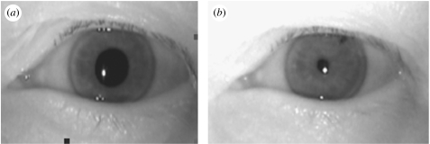
The first patient had an anterior uveitis with severe lacrimation and moderate number of cells in the anterior chamber of the eye (the anterior chamber lies between the cornea at the front of the eye and the iris). There were a few inflammatory precipitates on the inner surface of the cornea (endothelial precipitates). The eye was significantly pharmacologically dilated at the last visit compared with enrolment as well as being free of anterior chamber activity. The second eye had herpetic anterior uveitis with dendritic corneal ulcer (a branching ulcer that in this case was not large or dense enough to obscure iris view). There were endothelial precipitates and moderate anterior chamber cells. It was only moderately dilated compared with its state at enrolment. The last three eyes had anterior uveitis, which presented with posterior synechiae (stuck-down iris causing pupil distortion) and moderate anterior chamber activity. The synechiae had been broken by the time of the final visit leaving the pupil much less distorted (figure 2). Other examples of eye pathology tested are demonstrated in figures 3 and 4.
Figure 2.
Iris camera images of an eye with anterior uveitis showing synechiae (a) at first attendance and (b) resolved after treatment. The eye was not successfully recognized by iris recognition (Hamming distance 0.402).
Figure 3.
A patient with infective corneal disease with corneal haze on enrolment treated with acyclovir. The infrared imaging appears to penetrate through the corneal haze sufficiently adequately to keep the Hamming distance low (0.165). (a) Diseased eye, (b) recovered eye.
Figure 4.
Patient (a) before and (b) after laser iridotomy. Despite a pupil constriction and a small aperture in the iris at the 2 o'clock position after laser iridotomy, the Hamming distance remains low (0.236).
Overall, it was found that the large majority of eyes were resilient to change in the Hamming distance despite having developed significant pathology. Even eyes that had laser iridotomies performed and that had been pharmacologically constricted were successfully identified. Interestingly, an eye with large inferior coloboma (developmental iris defect) was successfully enrolled and identified, demonstrating that significant abnormalities of iris structure may still be conducive to successful iris recognition. Significant corneal opacities that limited full iris feature view did not seem to be a barrier to iris recognition technology nor did the existence of significant corneal oedema associated with Fuch's endothelial dystrophy. The mean Hamming distance for patients' eyes with different pathologies are shown below compared with control eyes (table 1). Only the group with anterior uveitis showed a significant increase in the Hamming distance in eyes that had recovered from disease compared with eyes that had remained healthy.
Table 1.
The mean Hamming distance of eye pathologies against control. (The control group consists of fellow eye of all patients, where there was no evidence of acute anterior segment disease. Note that while the Hamming distance difference is statistically significant for anterior uveitis, this leads to failure of recognition of only five patients' eyes.)
| no. of eyes | mean Hamming distance | variance | level of significance from control | |
|---|---|---|---|---|
| anterior uveitis | 24 | 0.252 | 0.0088 | p<0.001 |
| corneal disease | 33 | 0.136 | 0.0030 | p=0.301 |
| other anterior segment | 12 | 0.155 | 0.0030 | p=0.867 |
| control | 39 | 0.152 | 0.0057 | — |
4. Discussion
A match is never declared if the Hamming distance between two iris codes is larger than 0.33, which means that more than 33 per cent of the bits disagreed. If images from the same eye generate Hamming distances higher than 0.33, then they are erroneously declared non-matches. The frequency of such events is the false non-match rate. In our study, this false rejection was found to occur only in the presence of anterior uveitis, in 5 cases out of 24. Conversely, the false match rate is the rate at which different eyes are falsely matched with each other. We observed no such cases.
Ideally, we would have liked to have enrolled patients while they were free of disease and then examined their Hamming distance after acute eye disease onset. This would have recreated the scenario that might cause problems in the general use of iris recognition. However, due to practical difficulties of such a study, our experiment examined the reverse scenario, with patients first attending with eye disease and then being reassessed once the eye disease had resolved. This allowed us a useful means of determining the same mathematical differences in iris code via a much more practical experimental protocol.
We found that iris enrolment and subsequent recognition was remarkably resilient to compounding by acute eye disease. Iris recognition failed only in 5 out of the 24 eyes with anterior uveitis. Other forms of pathology failed to significantly affect the process. The particular iris imaging technology may have been resilient to corneal disease as the infrared light used by the camera is less attenuated by opacities than visible light, and may have thus been less affected by the corneal disruption. Interestingly, complete full thickness punctures in the iris caused by iridotomies were also insignificant in the iris recognition process, even when accompanied by pharmacological pupil constriction.
The cause of failure of recognition with the uveitis subjects is not known. Factors that may be involved are pharmacological pupil dilation and pathological pupil distortion. The iris recognition algorithms are designed to account for normal variations of pupil size, which are modelled mathematically by models of elastic change. Physiological dilation and pharmacological constriction may conform to this model while extensive pharmacological dilation may not. Similarly, morphology such as uveitis may be complicated by synechiae distorting the iris in a chaotic manner that cannot be easily modelled (J. Daugman 2007, personal communication).
Another cause for iris recognition to fail in uveitis patients may be changes in iris architecture or atrophy. These may not necessarily need to be evident at slit-lamp examination to be significant in the iris camera images. Although the striated meshwork of elastic ligament creates the predominant texture under visible light, with near-infrared wavelengths of the iris camera, deeper stromal features dominate the iris pattern (Daugman 2004). Future studies will further investigate the aetiology of failure in uveitis patients and the effect of pharmacological dilation relative to the effect of uveitic changes.
The use of biometric technology is bound to increase as security requirements continue to be important for individuals and society. Iris recognition is likely to be at the forefront of this expansion and has already been incorporated into several international airports including all five Heathrow terminals. The continuing development of image capture devices and algorithms for analysis has many continuing challenges such as image capture from a distance and detection of subterfuge attempts. This study shows that the compounding factor of potential eye pathologies in the population as a whole should also be borne in mind.
Our preliminary investigations show that the assessed iris capture camera combined with the Daugman algorithms is remarkably resistant to patients having a wide range of eye disease. However, patients with uveitis could pose a problem to iris recognition technologies particularly if they develop synechiae or require pharmacological dilation. Uveitis afflicts patients in a non-predictable manner and can be unilateral or bilateral. Current prevalence rates have been estimated at 115 out of 100 000 in the Western world (Gritz & Wong 2004). Engineers should bear this experiment's findings in mind when designing future systems, and subjects of iris recognition schemes may need to be advised about the use of those systems if they experience acute ocular disease. Further research is needed into the potential problems acute eye pathology might cause for this key biometric technology.
Acknowledgments
We thank Imad Malhas of IrisGuard for providing the iris camera and advice on camera set-up and John Daugman of the University of Cambridge for software to compute the Hamming distances between iris patterns.
References
- Bolle R.M., Connell J.H., Pankanti S., Ratha N.K., Senior A.W. Springer; New York, NY: 2004. Guide to biometrics. [Google Scholar]
- Daugman J. High confidence visual recognition of persons by a test of statistical independence. IEEE Trans. Pattern Anal. Mach. Intell. 1993;15:1148–1161. doi: 10.1109/34.244676. [DOI] [Google Scholar]
- Daugman, J. 1994 Biometric personal identification system based on iris analysis. US Patent no. 5291560.
- Daugman J. How iris recognition works. IEEE Trans. Circ. Syst. Video Technol. 2004;14:21–30. doi: 10.1109/TCSVT.2003.818350. [DOI] [Google Scholar]
- Doggart J. Kimpton; London, UK: 1949. Ocular signs in slit-lamp microscopy. [Google Scholar]
- Du Y.E. Review of iris recognition: cameras, systems, and their applications. Sensor Rev. 2006;26:66–69. doi: 10.1108/02602280610640706. [DOI] [Google Scholar]
- Flom, L. & Safir, A. 1987 Iris recognition system. US Patent no. 4641349.
- Gritz D.C., Wong I.G. Incidence and prevalence of uveitis in Northern California: the Northern California Epidemiology of Uveitis Study. Ophthalmology. 2004;111:491–500. doi: 10.1016/j.ophtha.2003.06.014. discussion 500. [DOI] [PubMed] [Google Scholar]
- Machin D., Campbell M., Walters S.J. Wiley; Chichester, UK: 2007. Medical statistics: a textbook for the health sciences; pp. 273–274. [Google Scholar]
- Nanavati S., Thieme M., Nanavati R. Wiley; New York, NY: 2002. Biometrics: identity verification in a networked world. [Google Scholar]
- Roizenblatt R., Schor P., Dante F., Roizenblatt J., Belfort R., Jr Iris recognition as a biometric method after cataract surgery. Biomed. Eng. Online. 2004;3:2. doi: 10.1186/1475-925X-3-2. [DOI] [PMC free article] [PubMed] [Google Scholar]


