

Abstract
Investigation of patients, healthy persons, and ticks in Jinghe County, Xinjiang Uygur Autonomous Region, People's Republic of China, for evidence of spotted fever group (SFG) rickettsiosis demonstrated strong evidence for a high prevalence of pathogenic SFG rickettsiae. Antibodies to SFG rickettsiae were detected in 62.5% of healthy subjects tested by enzyme-linked immunosorbent assay and 20% tested by complement fixation test. Two febrile patients were documented as having acute spotted fever rickettsiosis by complement fixation seroconversion. One, and 11-year-old Kazakh boy with eschar and regional lymphadenopathy, had an SFG rickettsia (An strain) isolated from his blood. A hemolymph test revealed that 20% of ticks contained rickettsiae. Two strains of SFG rickettsiae were isolated from male and female Dermacentor nuttalli ticks. The human SFG rickettsial isolate is the first to be obtained in the People's Republic of China.
Full text
PDF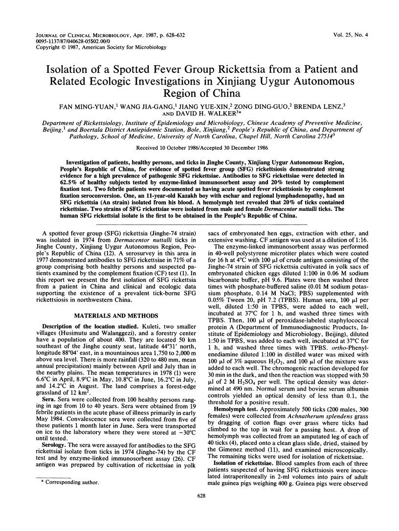
Images in this article
Selected References
These references are in PubMed. This may not be the complete list of references from this article.
- BELL E. J., KOHLS G. M., STOENNER H. G., LACKMAN D. B. NONPATHOGENIC RICKETTSIAS RELATED TO THE SPOTTED FEVER GROUP ISOLATED FROM TICKS, DERMACENTOR VARIABILIS AND DERMACENTOR ANDERSONI FROM EASTERN MONTANA. J Immunol. 1963 May;90:770–781. [PubMed] [Google Scholar]
- BELL E. J., STOENNER H. G. Immunologic relationships among the spotted fever group of rickettsias determined by toxin neutralization tests in mice with convalescent animal serums. J Immunol. 1960 Feb;84:171–182. [PubMed] [Google Scholar]
- Burgdorfer W. Hemolymph test. A technique for detection of rickettsiae in ticks. Am J Trop Med Hyg. 1970 Nov;19(6):1010–1014. [PubMed] [Google Scholar]
- Burgdorfer W., Sexton D. J., Gerloff R. K., Anacker R. L., Philip R. N., Thomas L. A. Rhipicephalus sanguineus: vector of a new spotted fever group rickettsia in the United States. Infect Immun. 1975 Jul;12(1):205–210. doi: 10.1128/iai.12.1.205-210.1975. [DOI] [PMC free article] [PubMed] [Google Scholar]
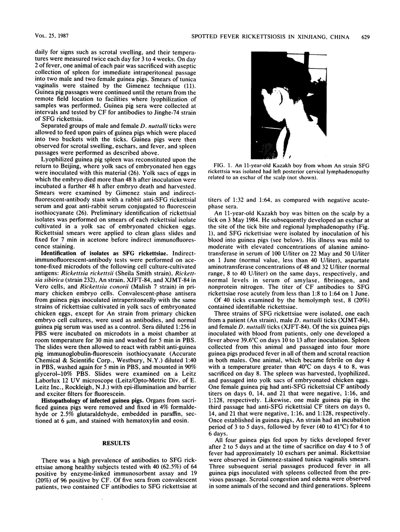
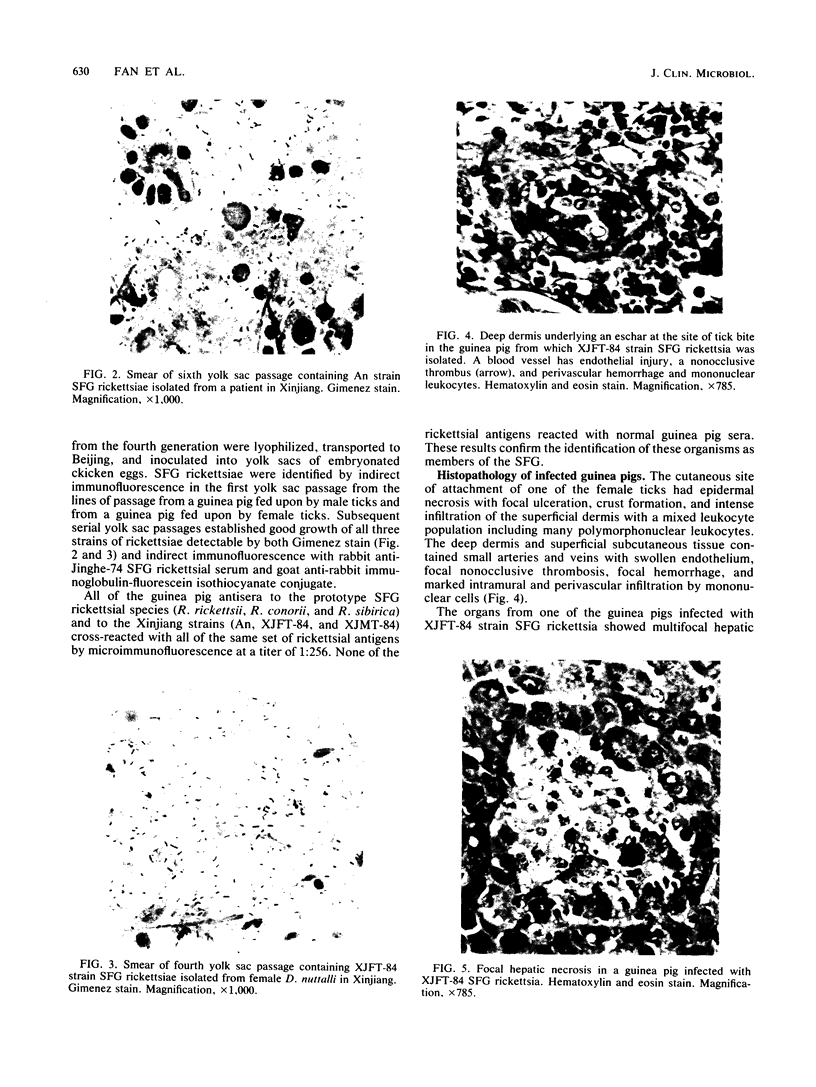
- GIMENEZ D. F. STAINING RICKETTSIAE IN YOLK-SAC CULTURES. Stain Technol. 1964 May;39:135–140. doi: 10.3109/10520296409061219. [DOI] [PubMed] [Google Scholar]
- Liu G. D. [Isolation and serological identification of the spotted fever rickettsia in Hu Meng, the Inner Mongolia Autonomous Region]. Zhonghua Liu Xing Bing Xue Za Zhi. 1985 Oct;6(5):265–268. [PubMed] [Google Scholar]
- Philip R. N., Casper E. A., Burgdorfer W., Gerloff R. K., Hughes L. E., Bell E. J. Serologic typing of rickettsiae of the spotted fever group by microimmunofluorescence. J Immunol. 1978 Nov;121(5):1961–1968. [PubMed] [Google Scholar]
- Robertson R. G., Wisseman C. L., Jr Tick-borne rickettsiae of the spotted fever group in West Pakistan. II. Serological classification of isolates from West Pakistan and Thailand: evidence for two new species. Am J Epidemiol. 1973 Jan;97(1):55–64. doi: 10.1093/oxfordjournals.aje.a121485. [DOI] [PubMed] [Google Scholar]
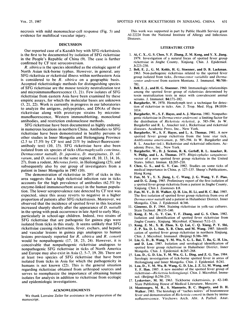
- Walker D. H., Gear J. H. Correlation of the distribution of Rickettsia conorii, microscopic lesions, and clinical features in South African tick bite fever. Am J Trop Med Hyg. 1985 Mar;34(2):361–371. doi: 10.4269/ajtmh.1985.34.361. [DOI] [PubMed] [Google Scholar]
- Walker D. H., Staiti A., Mansueto S., Tringali G. Frequent occurrence of hepatic lesions in boutonneuse fever. Acta Trop. 1986 Jun;43(2):175–181. [PubMed] [Google Scholar]