Abstract
Dermoid cysts are developmental abnormal arrangement of tissues and are often evident soon after birth. Its occurrence in the orbit is relatively rare. We report a case of orbital floor dermoid in an 18-year-old female patient who presented with progressive, painless swelling in the lower eyelid associated with mild proptosis of three months duration. The lesion was excised completely, and histopathology confirmed the diagnosis of dermoid cyst.
Keywords: Dermoid choristoma, implantation dermoid, ocular dermoid
Dermoid cysts are developmental abnormal arrangement of tissues and are often evident soon after birth. It may occur anywhere in the body, but its occurrence in the orbit is relatively rare. Dermoid cysts, account for about 3–9% of all orbital masses and 0.04–0.6% of primary orbital tumors.[1–9] The frequent site of origin is the superotemporal quadrant of orbit. Depending on the location, size, and associated abnormalities of the cyst, the patient may have proptosis, diplopia, and restriction of eye movements. However, we could find only one case presenting with progressive painless swelling in the lower eyelid, which was associated with mild proptosis without any pressure symptoms. We report here one such rare case of orbital floor dermoid in an adult female, which was investigated and managed with good results.
Case Report
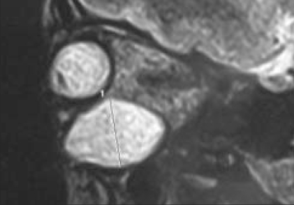
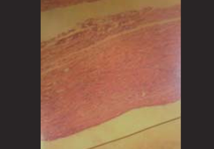
An 18-year-old female patient presented with a three-month history of progressive, painless swelling associated with mild proptosis in the lower eye lid. There was no history of diplopia. On examination, a firm, non-tender 1.5 × 2.0 cm swelling in the right lower lid was found [Fig. 1]. It was freely mobile over the bone, and skin over it was also free. The eyeball was pushed upward and laterally. Extraocular movements were normal. The vision and fundus were within normal limits. The results of blood investigations were as follows: hemoglobin 10.8 g%, total leucocyte count 8,400/mm3, blood glucose 104 mg%, and blood group O positive. X-ray of the orbit did not show any bony erosion. B-Scan ultrasonography results of the right eye were normal, except for edema in the lower eyelid. Ultrasound-guided fine-needle aspiration cytology was suggestive of dermoid cyst. Magnetic resonance imaging (MRI) of the orbit showed a well-defined intraorbital lesion with an intact eyeball [Fig. 2]. Computed tomographic scan was not performed. The patient underwent total excision of the lesion through an incision along the inferior orbital margin. There was a well-defined lesion, and after opening the lesion, a thick cheesy material was noted with few hairs suggestive of dermoid cyst. Diagnosis was confirmed on histopathology, as it showed nonkeratinizing squamous epithelium with variable numbers of admixed goblet cells [Figure 3]. Patient's proptosis was relieved immediately, and she was doing well at six months follow-up.
Figure 1.
Clinical photograph showing swelling in the lower eye lid
Figure 2.
MRI T2-weighted image showing well-defined hyperintense lesion in the lower eye lid pushing the eye ball upward
Figure 3.
Histopathology showing nonkeratinising squamous epithelium with variable numbers of admixed goblet cells suggestive of dermoid cyst (H&E, ×40)
Discussion
Dermoid cyst is an ectodermal inclusion cyst which may occur anywhere in the body, but its occurrence in orbit and intracranial sites is relatively rare. Approximately 50% of tumors that involve the head are found in or adjacent to the orbit. Forty percent of orbital dermoids are diagnosed between 15 and 40 years of age. The most frequent site of origin is the superetemporal quadrant of orbit, and the parasellar and frontobasal region are the most common intracranial sites.[1–3,9] Dermoid cyst gradually increases in size through epithelial desquamation and glandular secretions. The slow enlargement of dermoid cysts, due to the accumulation of this debris within the lumen, probably leads to attenuation of the epithelial lining and leakage of lipids and keratin into the tissues of the cyst wall. Malignant transformation is rarely seen.[1] Clinical presentation of orbital or intracranial dermoid cysts depends on the location, size, and associated abnormalities of cyst. Orbital dermoid cysts located superficially in and around the orbit present as subcutaneous or subconjunctival discrete well-circumscribed swellings in childhood. In our case, the presentation was at 18 years, and the site was in the orbital floor. Larger cysts that abut the globe or cysts that are located deep in the orbit may displace the globe and can compress the optic nerve and extraocular muscles leading to proptosis and restriction of eye movements.[4,10,11] In our case, there was mild proptosis, but there was no pressure effect or restricted eye movements. Pathologically dermoid cysts are well described.[4,12] The differential diagnosis of orbital cysts include choristoma (epidermoid, dermoid, dermolipoma), teratoma, the congenital cystic eye, and colobomatous cyst. Imaging modalities such as ultrasonography, computed tomography scan, and MRI of the dermoid cyst are valuable in the diagnosis and characterization of benign lesion and also to demonstrate their intraorbital and intracranial extension.[4,6,13,14] Leakage of contents can cause an acute or chronic inflammatory response with, in some cases, phagocytosis of lipid by macrophages, giant cell and granuloma formation, and secondary fibrosis around the cyst. Neighboring normal tissues are affected by inflammatory cells recruited to the area around the cyst and by the fibrosis occurring during the resolution of inflammation. Treatment of dermoid cyst is surgical en bloc excision, which is indicated for cosmetic purposes, confirmation of diagnosis, to relieve the symptoms created by mass in periorbital region and to prevent complications in cases of intracranial extension. Complete surgical excision with intact capsule is done to prevent dissemination of the contents which otherwise can incite an acute inflammatory response. This is also to avoid the deposition of cells that could form a new cyst at the operative site.[10,11] Orbital dermoid cysts are rare, but if untreated they can still lead to devastating complications. Periorbital dermoid cysts should be removed intact because the leakage of their contents can cause irritation and inflammation in the surrounding tissues.[8] The present case is unusual in few aspects, as the site of dermoid cyst in the orbital floor, which is uncommon, short duration of presentation with mild proptosis, and no pressure symptoms. Preoperative diagnosis became easier with the availability of MRI and excellent results could be achieved by early surgical intervention.
References
- 1.Pfeiffer RL, Nicholl RJ. Dermoid-epidermoid tumours of orbit. Arch Ophthalmol. 1948;46:39. [PMC free article] [PubMed] [Google Scholar]
- 2.Blei L, Chambers JT, Liotta LA, Di Chiro G. Orbital dermoid diagnosed by computed tomographic scanning. Am J Ophthalmol. 1978;85:58–61. doi: 10.1016/s0002-9394(14)76665-6. [DOI] [PubMed] [Google Scholar]
- 3.Jakobiec FA, Bonanno PA, Sigelman J. Conjunctival adnexal cysts and dermoids. Arch Ophthalmol. 1978;96:1404–9. doi: 10.1001/archopht.1978.03910060158012. [DOI] [PubMed] [Google Scholar]
- 4.Osborn AG. Diagnostic Neuroradiology. Mosby; 1997. pp. 66–7.pp. 631–48. [Google Scholar]
- 5.Whitney CE, Leone CR, Jr, Kincaid MC. Proptosis with mastication: An unusual presentation of an orbital dermoid cyst. Ophthalmic Surg. 1986;17:295–8. [PubMed] [Google Scholar]
- 6.Nugent RA, Lapointe JS, Rootman J, Robertson WD, Graeb DA. Orbital dermoids: Features on CT. Radiology. 1987;165:475–8. doi: 10.1148/radiology.165.2.3659368. [DOI] [PubMed] [Google Scholar]
- 7.Sathananthan N, Moseley IF, Rose GE, Wright JE. The frequency and clinical significance of bone involvement in outer canthus dermoid cysts. Br J Ophthalmol. 1993;77:789–94. doi: 10.1136/bjo.77.12.789. [DOI] [PMC free article] [PubMed] [Google Scholar]
- 8.Karatza EC, Shields CL, Shields JA, Eagle RC., Jr Calcified orbital cyst simulating a malignant lacrimal gland tumor in an adult. Ophthal Plast Reconstr Surg. 2004;20:397–9. doi: 10.1097/01.iop.0000139527.83345.64. [DOI] [PubMed] [Google Scholar]
- 9.James A. In Henderson's orbital tumors. 4th ed. Philadelphia: Lippincott Williams and Wilkins; 2007. [Google Scholar]
- 10.Sherman RP, Rootman J, Lapoint JS. Dermoids - clinical presentation and management. Br J Ophthalmol. 1984;68:642–52. doi: 10.1136/bjo.68.9.642. [DOI] [PMC free article] [PubMed] [Google Scholar]
- 11.Albert DM, Jakobief FA. Principles and practice of ophthalmology. Philadelphia: W B Saunder Company; 2000. pp. 3072–81. [Google Scholar]
- 12.Yanoff M, Fine BS. Ocular pathology. 3rd ed. Philadelphia: Harper and Row; 1988. p. 520. [Google Scholar]
- 13.Kaufman LM, Villablanca JP, Mafee MF. The Radiological Clinics of North America. Philadelphia: W B Saunders Company; 1998. pp. 1149–63. [DOI] [PubMed] [Google Scholar]
- 14.Stark DD, Bradley WG. Magnetic resonance imaging. 2nd ed. St. Louis: Mosby; pp. 801–2.pp. 1016 [Google Scholar]


