Abstract
Calotropis procera produces copious amounts of latex, which has been shown to possess several pharmacological properities. Its local application produces intense inflammatory response. In the 10 cases of Calotropis procera-induced keratitis reported here, the clinical picture showed corneal edema with striate keratopathy without any evidence of intraocular inflammation. The inflammation was reversed by the local application of steroid drops.
Keywords: Calotropis procera, keratitis, latex, striate keratopathy
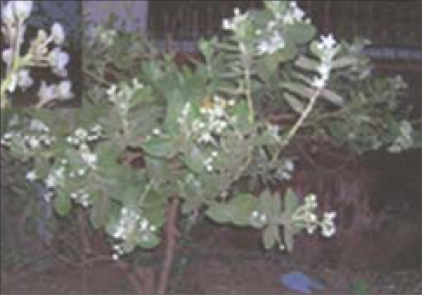
Calotropis procera, a xerophytic shrub of family Asclepiadaceae [Fig. 1] is widely distributed in the tropics of Asia, Africa, and Northeast of South America. It is commonly known as Akra and Madaar in India and is widely used in the indigenous systems of medicine.[1,2] The milky white endogenous latex, produced by the plant in appreciable amount, exhibits a variety of effects in various animal models. On oral administration, the latex produces potent anti-inflammatory, analgesic, and weak antipyretic effects, while on local administration it induces an intense inflammatory response in animal models.[3–6] These antagonistic biological activities (inflammatory and anti-inflammatory) are reported in the whole latex of Calotropis procera and depend on the extraction medium and route of administration in experimental animals. Pro-inflammatory activity seems to predominate over anti-inflammatory activity, suggesting its prevalence in the whole latex. These activities are displayed by compounds suitable to be fractionated on the basis of their molecular size.[4]
Figure 1.
Photograph of Calotropis procera tree with the inset showing latex oozing out from its stem
Accidental exposure to the latex has been reported to cause inflammation of the skin and eyes.[2,6,7] Ocular injury from this plant can be mechanical, or more commonly, toxic, due to the exposure to its latex.
Case Report
We report here 10 cases of Calotropis procera-induced keratitis who presented to us between May 2005 and May 2007. All patients except one, were females, who got the latex splashed in their eyes accidentally while plucking flowers from the plant for religious purpose. The sole male patient had inadvertent instillation of the latex while he was applying it on his knee for arthritic pain.
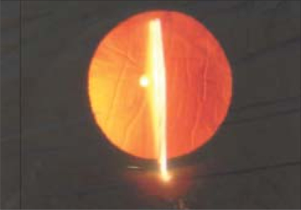
All patients reported a burning sensation and watering immediately after the accidental splashing of Calotropis latex. This was followed by grittiness and redness, and within a few hours, they noticed blurring of vision. There was mild discomfort although none of the patients reported any significant pain. There was no history of ocular trauma, surgery, or any other ophthalmic problem in any of the patients, and vision had been normal in the eyes previously, except in two patients (Cases 8 and 9) who had bilateral immature senile cataracts. The visual acuity was variably reduced in all eyes [Table 1], while in the uninvolved eyes, the best corrected visual acuity (BCVA) was 20/20, except in the two patients who had immature senile cataract in both eyes. All eyes showed mild conjunctival and circumcorneal injection. The three patients who presented within few hours of injury showed few epithelial lesions, while in all others, the corneal epithelium was intact. Corneal stroma was edematous showing striate keratopathy [Fig. 2]. The endothelium appeared normal on specular reflection. The anterior chamber was quiet with no cells or flare. Iris, pupil, and lens were also normal. Intraocular pressure was normal in all eyes (12–16 mmHg). The patient who presented to us after an interval of 10 days had consulted a local doctor a few hours after injury who had prescribed antibiotics and lubricating eye drops for local application. As the complaints persisted, she consulted us on the tenth day after injury. On examination, her BCVA was 20/80. She had conjunctival and mild circumcorneal congestion. Cornea was edematous and showed stromal keratitis similar to that seen in all the other cases. Based on the above clinical features and history of exposure to the Calotropis latex, we diagnosed these cases as toxic keratoconjunctivitis. The three patients with epithelial lesions were prescribed antibiotic (tobramycin 0.3%) and nonsteroidal anti-inflammatory (ketorolac 0.5%) drops on the first day. On follow-up the next day, the epithelial lesions had healed completely, although stromal edema and striate keratopathy remained unchanged. Corticosteroid drops (prednisolone acetate 1%) were given for application in the affected eyes six times a day. The other patients were prescribed corticosteroid drops on the day of presentation.
Table 1.
Demographics and visual acuity of all patients
| Case number | Age (years) | Sex | Eye involved | Time of presentation after injury | BCVA on presentation* | BCVA 2 days after starting steroids | BCVA 10 days after starting steroids |
|---|---|---|---|---|---|---|---|
| 1 | 51 | F | RE | 28 hours | 20/40 | 20/30 | 20/20 |
| 2 | 49 | F | RE | 40 hours | 20/40 | 20/30 | 20/20 |
| 3 | 45 | F | LE | 36 hours | 20/40 | 20/30 | 20/20 |
| 4 | 47 | F | RE | 10 days | 20/80 | 20/40 | 20/20 |
| 5 | 53 | F | RE | 4 hours | 20/30 | 20/20 | 20/20 |
| 6 | 42 | F | RE | 6 hours | 20/30 | 20/20 | 20/20 |
| 7 | 56 | F | RE | 7 hours | 20/40 | 20/20 | 20/20 |
| 8 | 56 | F | LE | 24 hours | 20/120 | 20/80 | 20/80 |
| 9 | 84 | F | LE | 28 hours | 20/200 | 20/120 | 20/120 |
| 10 | 60 | M | RE | 24 hours | 20/40 | 20/20 | 20/20 |
BCVA - Best corrected visual acuity, F- female, M- male, RE- right eye, LE- left eye
Figure 2.
Slit lamp photograph showing corneal edema and striate keratopathy on the day of presentation, 24 hours after exposure to Calotropis latex
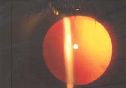
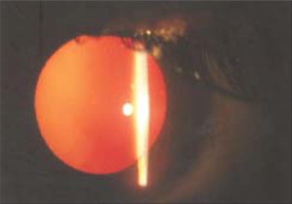
On follow-up after 2 days of starting the corticosteroid drops, the stromal edema and striate keratopathy had reduced significantly in all patients [Fig. 3], including the one who presented late. By day 10, the inflammation had resolved completely in all the eyes [Fig. 4] with BCVA recovered to 20/20 in all except two eyes with immature senile cataract (cases 8 and 9).
Figure 3.
Slit lamp photograph after the instillation of local steroid drops for two days showing significantly reduced corneal edema and striate keratopathy
Figure 4.
Slit lamp photograph of the same patient taken after 10 days of steroid instillation showing normal cornea
Discussion
Al-Mezaine et al.[7] have reported one case of Calotropis procera-induced keratitis. The clinical course of their patient was very similar to our series of patients. They attributed it to toxicity of its latex to corneal endothelial cells due to an unknown molecular mechanism. Tomar et al.[2] reported one case of toxic iridocyclitis induced by the latex of Calotropis procera. However, none of our patients showed any evidence of intraocular inflammation. Both the above-reported patients recovered after topical steroid use.
The irritant and pro-inflammatory property of latex of Calotropis procera has been well established.[4] The milky white latex of this plant irritates the mucous membrane and produces inflammatory reaction on local application or accidental exposure. It is known to produce contact dermatitis, and the latex of this plant produces intense inflammation when injected locally in animal models.[5,6] Shivkar et al.,[6] in their study on rat paw edema model, found that the injection of dried latex produces an intense inflammatory response involving edema formation and cellular infiltration. They showed that this was due to the presence of histamine in the latex itself and also due to the release of mast cell histamine by the latex. Besides, the latex has also been shown to induce prostaglandin synthesis through the induction of cyclooxygenase -2 (COX-2).[5] Both histamine and prostaglandins are the key mediators in an inflammatory response. Accordingly, we suggest the mechanism of stromal keratitis to be due to inflammation induced by exposure to latex due to its strong pro-inflammatory property. The resolution of keratitis with local corticosteroid use corroborates this notion. A reduction in endothelial count due to the direct toxic effect of the latex could be another possible mechanism, as suggested by Al-Mezaine et al.[7]
The epithelial lesions seen in three patients could be due to mechanical injury while rubbing. The painless clinical course of our patients could be attributed to the analgesic property present in the latex of Calotropis procera.[8]
Acknowledgments
We deeply acknowledge the support and guidance given to us in every step of our study, by Prof. P. K. Mukherjee (M.S.), former Head of the Department, Department of Ophthalmology, Pt. J. N. M. Medical College, Raipur. We are also thankful to Dr. Pradeep Jain, (DOMS), Nayan Jyoti Netra Chikitsalaya, Raipur, for sharing his patients with us for the study.
References
- 1.Mascolo N, Sharma R, Jain SC, Capasso F. Ethnopharmacology of Calotropis procera flowers. J Ethnopharmacol. 1988;22:211–21. doi: 10.1016/0378-8741(88)90129-8. [DOI] [PubMed] [Google Scholar]
- 2.Tomar VP, Agarwal PK, Agarwal BL. Toxic iridocyclitis caused by Calotropis. J All India Ophthalmol Soc. 1970;18:15–6. [PubMed] [Google Scholar]
- 3.Arya S, Kumar VL. Antiinflammatory efficacy of extracts of latex of Calotropis procera against different mediators of inflammation. Mediators Inflamm. 2005;4:228–32. doi: 10.1155/MI.2005.228. [DOI] [PMC free article] [PubMed] [Google Scholar]
- 4.Alencar NM, Oliveira JS, Mesquita RO, Lima MW, Vale MR, Etchells JP, et al. Pro- and anti-inflammatory activities of the latex from Calotropis procera (Ait) R.Br. are triggered by compounds fractionated by dialysis. Inflamm res. 2006;55:559–64. doi: 10.1007/s00011-006-6025-y. [DOI] [PubMed] [Google Scholar]
- 5.Kumar VL, Shivkar MY. Involvement of prostaglandins in inflammation induced by latex of Calotropis procera. Mediators Inflamm. 2004;13:151–5. doi: 10.1080/09511920410001713583. [DOI] [PMC free article] [PubMed] [Google Scholar]
- 6.Shivkar YM, Kumar VL. Histamine mediates the pro-inflammatory effect of latex of Calotropis procera in rats. Mediators Inflamm. 2003;12:299–302. doi: 10.1080/096293503310001619708. [DOI] [PMC free article] [PubMed] [Google Scholar]
- 7.Al-Mezaine HS, Al-Rajhi AA, Al-Assiri A, Wagoner MD. Calotropis procera (ushaar) keratitis. Am J Ophthalmol. 2005;391:199–202. doi: 10.1016/j.ajo.2004.07.062. [DOI] [PubMed] [Google Scholar]
- 8.Dewan S, Sangraula H, Kumar VL. Preliminary studies on the analgesic activity of Calotropis procera. J Ethnopharmacol. 2000;73:307–11. doi: 10.1016/s0378-8741(00)00272-5. [DOI] [PubMed] [Google Scholar]


