Abstract
We describe the occurrence of bilateral iridocorneal endothelial (ICE) syndrome with glaucoma in a young girl with Down's syndrome. A 16-year-old girl with Down's syndrome was found to have secondary glaucoma in the right eye with features of progressive iris atrophy in both eyes. She was uncontrolled on maximum tolerable medical therapy for glaucoma. She underwent an uneventful trabeculectomy with mitomycin-C in her right eye. Scanning electron microscopy of the trabecular meshwork obtained in this case is described.
Keywords: Down's syndrome, glaucoma, iridocorneal endothelial syndrome, scanning electron- microscopy
Iridocorneal endothelial (ICE) syndrome is typically a unilateral condition that occurs mostly in female patients in early and middle adulthood. The condition is reported to be acquired, and there are no consistently reported systemic associations. Glaucoma has been described to occur in almost 50% of these cases.We report a rare case of a young girl with Down's syndrome and bilateral ICE syndrome with unilateral glaucoma.
Case Report
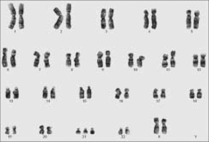
A 16-year-old girl with Down's syndrome presented with complaints of pain, redness, and watering of 5 months duration in her right eye. She had typical features of short stature, mental retardation, brachycephaly, mongoloid slant of eyelid fissures, short and broad hands with simian crease. There was a history of surgery for imperforate anus soon after birth. A karyotyping revealed an additional chromosome 21 complement [Fig. 1].
Figure 1.
A karyotypic analysis showing the trisomic 21 state
On ocular examination, her best corrected visual acuity was 20/60 in the right eye (RE) and 20/20 in left eye (LE). Her refractive error was +3.5 diopter sphere (Dsph) in the RE and +0.5 Dsph in the LE. Her near vision was N/12 RE and N/6 LE.
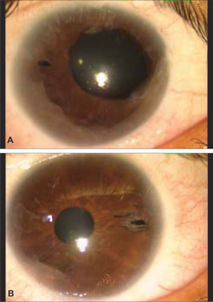
Corneas in both eyes had normal diameters of 12 mm horizontally and 11.5 mm vertically. There was no posterior embryotoxon. In the right eye, the iris pattern was lost with areas of patchy iris atrophy, 360° of ectropion uveae and multiple anterior synechiae along the entire circumference extending to the corneal periphery. There were extensive anterior synechiae even in the left eye [Fig. 2]. There were no keratic precipitates or any cells or flare in the anterior chamber. No Brushfield spots on the iris were seen in either eye. An iridotomy, which the girl had undergone 2 years previously, was also seen in both eyes. The iridotomies had been done probably because of a suspicion of secondary angle closure. No iris nodules were seen. The pupil in the right eye was 6 mm with a sluggish direct and consensual response, whereas in her left eye, the pupil was 3 mm and reactive. Ocular findings were normal in the parents and two siblings.
Figure 2.
Anterior segment photograph of RE (A) showing areas of patchy iris atrophy with ectropion uvea and LE (B) showing presence of anterior synechiae
Her highest recorded intraocular pressure (IOP) was 38 mmHg in the RE and 18 mmHg in the LE. On gonioscopy, the anterior chamber angle was totally closed, and none of the angle structures could be visualized in the RE, whereas in the LE, the angles were closed in the superior 180° because of the extensive synechiae. The right optic disc showed a cup disc ratio of 0.7:1 with an inferior thinning of the neuroretinal rim, and the left optic disc had 0.5:1 cup disc ratio with a healthy neuroretinal rim.
Pachymetry revealed central corneal thickness in the RE of 637 and 630 µm in LE. On specular microscopy, there was varying degree of pleomorphism and a lack of clear hexagonal pattern with some cells showing dark centers (disseminated ICE cells). The specular count in the RE was 1700/mm2 when compared with 3000/mm2 in the LE.
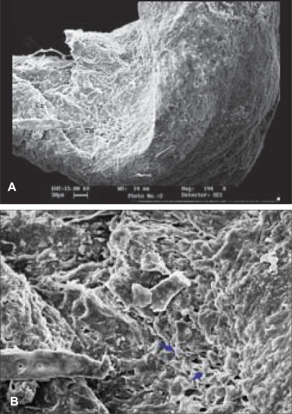
The patient was administered with antiglaucoma medication in the RE that included timolol 0.5% twice/day and latanoprost once daily, after which her IOP was 30 mmHg in the RE. Systemic acetazolamide was added, after which her IOP in the RE still remained 24 mmHg and was advised RE trabeculectomy with mitomycin-C. The patient underwent an uneventful surgery under general anesthesia. Her IOP was maintained at 12 mmHg without antiglaucoma medication till the 3-month follow-up after surgery. A scanning electron microscopy evaluation of the trabeculectomy specimen revealed the iris tissue extending onto the trabecular meshwork with collapse of trabecular beams, thickened trabecular beams, with decreased intertrabecular spaces [Fig. 3].
Figure 3.
(A) Scanning electron photomicrograph (×194) shows the concavity as the trabecular meshwork (TM) and anterior inserted iris tissue (Ir). (B) The trabecular meshwork (×447) shows thickened trabecular beams (black arrows) with decreased intertrabecular spaces
Discussion
The present case had typical features of ICE syndrome namely iris atrophy with ectropion uveae and endothelial changes. Bilaterality of the disorder has also been described earlier. ICE syndrome comprises a spectrum of one disease, encompassing three clinical variants—progressive iris atrophy, Chandler syndrome, and Cogan-Reese syndrome. Our patient had the features of progressive iris atrophy and had unilateral glaucoma, which is known to occur more frequently in eyes with progressive iris atrophy than in those with Chandler syndrome or Cogan-Reese variants of the disease.[2,3]
The mechanism of increased IOP postulated in ICE syndrome involves a dystrophic endothelium producing a membrane that extends across the anterior chamber angle onto the iris, causing synechiae formation. Scanning electron microscopy of the trabecular meshwork sample in our patient revealed the presence of collapsed trabecular beams and decreased intertrabecular spaces, which could have also been the cause of increased IOP in her RE.
Infantile glaucoma is known to occur in Down's syndrome,[4,5] and the prevalence is reported to be between 6 and 12%;[6,7] however, glaucoma in Down's syndrome is usually bilateral and is postulated to be due to the high iris insertion. To our knowledge, this is the first reported case of bilateral ICE syndrome and glaucoma in a patient with Down's syndrome. Down's syndrome is a manifestation of a chromosomal aberration, whereas ICE syndrome is known to be acquired with a viral etiology having been postulated.[8]
Axenfeld Rieger syndrome is also described to occur in patients with Down's syndrome.[9,10] However, the lack of a posterior embryotoxon and the characteristic endothelial changes were against Axenfeld Rieger syndrome in our patient.
A genetic association of ICE syndrome has not been extensively evaluated because of its rarity. Whether this was a chance association or a duplication of certain genes in the trisomic state that led to the abnormalities in the eye of our patient can only be resolved on a detailed genetic analysis. The possibility of a genetic association between the two syndromes is supported by the relatively younger age of our patient. There is only one case, to our knowledge, reported to have manifested ICE syndrome with glaucoma in an 11-year old-girl,[11] although no genetic analysis of the child was reported by these authors.
The presence of ICE syndrome as a cause of early secondary glaucoma should thus be kept in mind in young patients with Down's syndrome.
References
- 1.Laganowski HC, Kerr Muir MG, Hitchings RA. Glaucoma and Iridocorneal endothelial syndrome. Arch Ophthalmol. 1992;110:346–50. doi: 10.1001/archopht.1992.01080150044025. [DOI] [PubMed] [Google Scholar]
- 2.Teekhasaenee C, Ritch R. Iridocorneal endothelial syndrome in Thai patients. Arch Ophthalmol. 2000;118:187–92. doi: 10.1001/archopht.118.2.187. [DOI] [PubMed] [Google Scholar]
- 3.Wilson MC, Shields MB. A comparison of the clinical variations of the Iridocorneal endothelial syndrome. Arch Ophthalmol. 1989;107:1465–8. doi: 10.1001/archopht.1989.01070020539035. [DOI] [PubMed] [Google Scholar]
- 4.Traboulsi EI, Levine E, Mets MB, Parelhoff ES, O'Neill JF, Gaasterland DE. Infantile glaucoma in Down's syndrome (trisomy 21) Am J Ophthalmol. 1988;105:389–94. doi: 10.1016/0002-9394(88)90303-0. [DOI] [PubMed] [Google Scholar]
- 5.Jerndal T. Infantile glaucoma in Down's syndrome (trisomy 21) Am J Ophthalmol. 1988;106:638–9. doi: 10.1016/0002-9394(88)90611-3. [DOI] [PubMed] [Google Scholar]
- 6.Yokoyama T, Tamura H, Tsukamoto H, Yamane K, Mishima HK. Prevalence of glaucoma in adults with Down's syndrome. Jpn J Ophthalmol. 2006;50:274–6. doi: 10.1007/s10384-005-0305-x. [DOI] [PubMed] [Google Scholar]
- 7.Liza-Sharmini AT, Azlan ZN, Zilfalil BA. Ocular findings in Malaysian children with Down syndrome. Singapore Med J. 2006;47:14–9. [PubMed] [Google Scholar]
- 8.Alvarado JA, Underwood JL, Green WR, Wu S, Murphy CG, Hwang DG, et al. Detection of herpes simplex viral DNA in the iridocorneal endothelial syndrome. Arch Ophthalmol. 1994;112:1601–9. doi: 10.1001/archopht.1994.01090240107034. [DOI] [PubMed] [Google Scholar]
- 9.Dark AJ, Kirkham TH. Congenital corneal opacities in a patient with Rieger's anomaly and Down's syndrome. Br J Ophthalmol. 1968;52:631–5. doi: 10.1136/bjo.52.8.631. [DOI] [PMC free article] [PubMed] [Google Scholar]
- 10.Stokes DW, Parrish CM. Axenfeld's anomaly associated with Down's syndrome. Cornea. 1992;11:163–4. doi: 10.1097/00003226-199203000-00012. [DOI] [PubMed] [Google Scholar]
- 11.Salim S, Shields MB, Walton D. Iridocorneal endothelial syndrome in a child. J Pediatr Ophthalmol Strabismus. 2006;43:308–10. doi: 10.3928/01913913-20060901-06. [DOI] [PubMed] [Google Scholar]


