Abstract
Strain superinfection occurs when a second pathogen strain infects a host already carrying a primary strain. Anaplasma marginale superinfection occurs when the second strain carries a variant repertoire different from that of the primary strain, and the epidemiologic consequences depend on the relative efficiencies of tick-borne transmission of the two strains. Following strain superinfection in the reservoir host, we tested whether the presence of two A. marginale (sensu lato) strains that differed in transmission efficiency altered the transmission phenotypes in comparison to those for single-strain infections. Dermacentor andersoni ticks were fed on animals superinfected with the Anaplasma marginale subsp. centrale vaccine strain (low transmission efficiency) and the A. marginale St. Maries strain (high transmission efficiency). Within ticks that acquired both strains, the St. Maries strain had a competitive advantage and replicated to significantly higher levels than the vaccine strain. The St. Maries strain was subsequently transmitted to naïve hosts by ticks previously fed either on superinfected animals or on animals singly infected with the St. Maries strain, consistent with the predicted transmission phenotype of this strain and the lack of interference due to the presence of a competing low-efficiency strain. The vaccine strain was not transmitted by either singly infected or coinfected ticks, consistent with the predicted transmission phenotype and the lack of enhancement due to the presence of a high-efficiency strain. These results support the idea that the strain predominance in regions of endemicity is mediated by the intrinsic transmission efficiency of specific strains regardless of occurrence of superinfection.
Strain superinfection occurs when a second pathogen strain infects a host already persistently infected with a primary strain. Superinfection has epidemiologic and pathogenic relevance for pathogens ranging from small-genome RNA viruses, such as human immunodeficiency virus and hepatitis C virus, to complex parasites, such as Trypanosoma brucei (1, 10, 12, 20, 24, 37). The epidemiologic consequences of superinfection depend on the subsequent fitness of the two strains for onward transmission to a new host. We address this question by studying transmission of Anaplasma marginale, a tick-borne bacterial pathogen which establishes persistent infection in mammalian reservoir hosts (domestic and wild ruminants) (21) and for which the basis for strain superinfection has recently been reported (8).
A. marginale strain superinfection occurs when the second strain carries a repertoire of the antigenically variable outer membrane protein (designated major surface protein-2 [Msp2]) different from that of the primary strain, allowing the second strain to escape the immune response generated against the primary strain (8, 25). Once superinfection is established, both strains are maintained in the host, which serves as the reservoir for subsequent tick acquisition and transmission (18). Whether and how pathogen strains compete to establish infection within the tick vector are unknown. If there is no interaction among strains, the most fit strain, defined as that acquired by the highest percentage of ticks (tick infection rate) and replicated to the highest levels in the salivary glands (tick infection level), is predicted to predominate in the vector and subsequently be preferentially transmitted. Alternatively, pathogen strains may interact and either competitively inhibit or enhance the transmissibility of a second strain.
To address these hypotheses, we selected two pathogen strains with different intrinsic transmission efficiencies. The St. Maries strain is a prototypical sensu stricto A. marginale strain that is highly transmissible by Dermacentor andersoni adult ticks. This efficiency is manifested in a high infection rate, replication to high levels in the salivary gland, and consistent transmission to naïve animals by use of ≤10 ticks (7, 26, 28, 35). In contrast, the Israel vaccine strain is a sensu lato Anaplasma marginale subsp. centrale strain that is significantly less efficiently transmitted using the same D. andersoni colony, requiring >100 ticks for transmission (35, 36). We predicted that these two strains would establish superinfection in the mammalian host, given the presence of at least one distinct msp2 allele difference between the two strains (2, 30). Superinfection of the reservoir host with these two strains would then permit testing of the interaction between the strains within the vector following tick acquisition feeding and determination of whether this interaction inhibited development and subsequent transmission of the highly transmissible St. Maries strain or enhanced transmissibility of the vaccine strain. Herewith, we report testing of these hypotheses and discuss the results in the context of the epidemiological consequences of pathogen strain superinfection.
MATERIALS AND METHODS
Pathogen strains, tick vectors, and animal hosts.
The efficiencies of transmission of both the St. Maries strain and the Israel vaccine strain by Dermacentor andersoni have previously been reported (26, 28, 35, 36). The Reynolds Creek colony of D. andersoni was used in all transmission experiments (28). Age-matched male Holstein calves were used in all experiments; the calves were determined to be negative for A. marginale by an Msp5 competitive enzyme-linked immunosorbent assay (C-ELISA) prior to initiation of the experiments (34).
A. marginale superinfection of vaccine strain-infected animals.
Four calves (no. 6171, 6175, 6187, and 6188) were infected by intravenous inoculation with 108 organisms of the vaccine strain. As a control, calf no. 6170 was inoculated at the same time, but using 108 organisms of the St. Maries strain of A. marginale. All calves became infected, as determined by microscopic examination of Giemsa-stained blood smears and by Msp5 C-ELISA seroconversion. Following progression to persistent infection (bacteremia, ≤107 organisms/ml), adult male D. andersoni ticks infected with the St. Maries strain were allowed to transmission feed (n = 50 ticks/calf) for 7 days on three of the vaccine strain-infected calves (no. 6175, 6187, and 6188). Superinfection (the presence of both strains) was detected by strain-specific PCR of blood and quantified using strain-specific quantitative PCR. Both assays are described in detail below. The remaining vaccine strain-infected calf (no. 6171) and the St. Maries strain-infected calf (no. 6170) were maintained as controls for transmission of single strains.
Transmission following tick feeding on superinfected animals.
Uninfected D. andersoni adult males were allowed to attach and acquisition feed on one of the three groups of animals: (i) those superinfected with the vaccine strain and the St. Maries strain (no. 6175, 6187, and 6188), (ii) that infected with only the vaccine strain (no. 6171), and (iii) that infected with only the St. Maries strain (no. 6170). Ticks were acquisition fed for 7 days and then incubated for an additional 7 days at 26°C and 93% relative humidity with a 12-h photoperiod. Cohorts of the ticks from each animal were then dissected, and DNA was extracted from individual salivary glands and midguts to determine the presence and quantity of each strain, as described below. The remaining ticks were used for transmission feeding on naïve, C-ELISA-seronegative calves (no. 32004, 32023, 32001, 31991, and 32008) to determine if the presence of the two strains altered the transmission phenotypes. The number of ticks (n = 70 per animal) was selected to exceed the number necessary to transmit the high-efficiency St. Maries strain (≤10 ticks) and be less than the number needed to transmit the low-efficiency vaccine strain (>100 ticks), thus allowing detection of significant shifts in transmission phenotype due to inhibition and enhancement, respectively, in coinfected ticks. Following 7 days of transmission feeding, ticks were removed, salivary glands were obtained by dissection, and DNA was extracted from individual salivary glands to determine the presence and quantity of each strain (35). Transmission to the naïve animals was monitored by microscopic examination of Giemsa-stained blood smears, strain-specific PCR amplification (see below) of DNA isolated from blood, and C-ELISA seroconversion (34).
Strain-specific and quantitative PCR.
Dissected tick salivary glands were collected in cell lysis buffer and digested with proteinase K overnight at 37°C, followed by incubation at 65°C for 2 h. DNA was isolated from blood samples and tick tissues by using a Puregene DNA isolation kit (Qiagen). For strain-specific PCR, the locus containing the msp1α gene was targeted; the St. Maries strain has a prototypical msp1α gene, while the vaccine strain is highly divergent in this locus (2, 6, 9, 19). The St. Maries strain was amplified using forward primer 5′ TGCTTATGGCAGACATTTCCAT 3′ and reverse primer 5′ GGGAAAGAGGACAACCACACA 3′, generating a 155-bp amplicon. The vaccine strain was amplified using forward primer 5′ TGCAGTTGAGAAGTTCCGTATCA 3′ and reverse primer 5′ TGTTGCCTTAGCTGGGTCAAT 3′, generating a 122-bp amplicon. The identities of the strain-specific amplicons were confirmed by sequencing in both directions. To enhance sensitivity of detection of infected ticks, strain-specific amplicons were probed by Southern analysis using a digoxigenin (DIG)-labeled probe (9). The probes were generated using the strain-specific primers listed above and a PCR DIG probe synthesis kit (Roche). Following hybridization overnight at 42°C, the membranes were washed three times for 15 min in 2× SSC (1× SSC is 0.15 M NaCl plus 0.015 M sodium citrate)-0.1% sodium dodecyl sulfate. The first two washes were performed at room temperature, and the third one was performed at 65°C. A final, 15-min wash was done using 0.2× SSC-0.1% sodium dodecyl sulfate at 65°C. Chemiluminescence detection of the bound probes was achieved by using CDP-Star with a DIG wash-and-block buffer kit (Roche) as described by the manufacturer.
For quantification, the same strain-specific primer pairs were used in combination with specific TaqMan probes: 5′ CGTATGTTACAATCAGGCCGCCGG 3′ for the St. Maries strain and 5′ ACATGCCATTATTGACCCAGCTAAGGCAA 3′ for the vaccine strain. The TaqMan real-time PCR protocol was adapted from the method of Futse et al. (9), with changes in PCR target and MgCl2 concentration to increase specificity. Briefly, there was an initial cycle at 95°C for 10 min, followed by 55 cycles at 95°C for 20 s, 58°C for 10 s, and 72°C for 10 s, a final extension at 72°C for 2 min, and holding at 10°C. The real-time PCR mixture contained 10 mM Tris (pH 8.3), 50 mM KCl, 4.0 mM MgCl2, 200 μM each of dATP, dCTP, dGTP, and dTTP; 0.2 μM of each primer; 0.2 μM fluorogenic probe; and 1.25 U of AmpliTaq Gold (Applied Biosystems), with all reactions performed with an iCycler iQ real-time PCR detection system (Bio-Rad). Amplicons derived using the primers described above were cloned into the pCR-4 TOPO vector (Invitrogen) to generate the standard curve, with dilutions containing between 107 and 102 copies of the plasmids specific for each strain. Infected D. andersoni salivary glands containing 105 A. marginale St. Maries strain or vaccine strain organisms were used as internal controls for repeatability among assays (35). Triplicates from each sample were analyzed. The number of organisms was determined by using the standard curve and was presented as the mean log10 (± standard deviation) number of organisms per salivary gland pair or per ml of blood.
RESULTS
A. marginale superinfection of vaccine strain-infected animals.
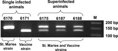
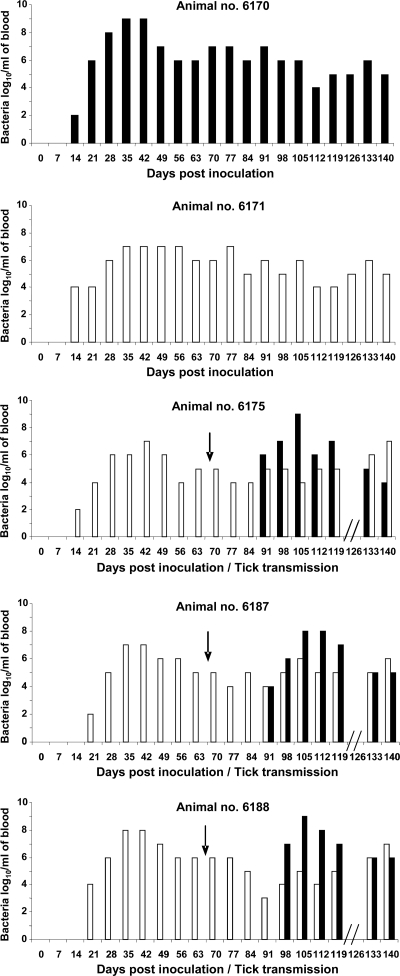
Calves persistently infected with the A. marginale subsp. centrale Israel vaccine strain became superinfected upon transmission feeding of ticks infected with the St. Maries strain of A. marginale. Both strains were detected in the three challenged calves (no. 6175, 6187, and 6188) and persisted (Fig. 1). The calves maintained as single-strain-infected controls, no. 6170 and 6171, contained only the single strain (the St. Maries strain and the vaccine strain, respectively) (Fig. 1). Quantitative tracking using strain-specific real-time PCR revealed an initially higher level of the St. Maries strain following superinfection; however, this was not maintained throughout superinfection (Fig. 2). There was no statistically significant difference in the levels of the vaccine strain between the superinfected animals and the single-strain-infected control over the same time period postchallenge (Fig. 2).
FIG. 1.
Detection of St. Maries strain superinfection. Results are shown for strain-specific PCR amplification of the primary vaccine strain (122 bp) and superinfection of the St. Maries strain (155 bp) in animals 6175, 6187, and 6188. The single-strain-infection controls were animals 6170 (St. Maries strain) and 6171 (vaccine strain).
FIG. 2.
Bacteremia levels in single-strain-infected and superinfected animals. Bacteremia levels are shown for the vaccine strain (white bars) following primary infection and for the St. Maries strain (black bars) following superinfection of animals 6175, 6187, and 6188. Animals 6170 and 6171 represent single-strain-infection controls for the St. Maries and vaccine strains, respectively. The arrows indicate the times of tick-borne transmission of the St. Maries strain.
Tick infection following acquisition feeding on superinfected animals.
Uninfected adult male D. andersoni ticks were acquisition fed on the superinfected calves and the single-strain-infected control calves. During the 7-day acquisition feeding, the mean levels were within the range of 105 to 107 bacteria per ml of blood, with statistically significantly (P = 0.02) higher levels of the vaccine strain than of the St. Maries strain in the superinfected animals (Table 1). Following acquisition feeding, the ticks were incubated at 26°C for an additional 7 days to ensure complete digestion of the blood meal. Separate cohorts of ticks fed on each individual calf were then dissected, and the presence and level of each pathogen strain in the tick salivary gland were determined. Ticks fed on the animals infected with a single strain, no. 6170 and 6171, were infected with only the expected strain (the St. Maries strain and the vaccine strain, respectively) (Table 2). The higher infection rate (percentage of fed ticks that acquired infection) for the St. Maries strain than for the vaccine strain (Table 2) is consistent with previous studies examining acquisition of these two strains from single-strain infections (26, 28, 35, 36). The ticks acquisition fed on superinfected calves represented all four possible classes of infection status: those infected with only the initial vaccine strain, those infected with only the superinfecting St. Maries strain, those coinfected with both strains, and those uninfected (Fig. 3). Analyzed as a group, 36% of the ticks acquisition fed on the three superinfected animals (n = 353 ticks) were found to be coinfected with the two strains. The levels of each strain within individual coinfected ticks were then quantified and used to test whether one strain had a competitive advantage. By quantitative competition index analysis (where the competition index is the number of St. Maries strain organisms minus the number of vaccine strain organisms, divided by the total number for the two strains combined), the St. Maries strain significantly predominated (P < 0.001; Wilcoxon rank sum test). This competitive advantage occurred despite the presence of significantly lower bacteremia levels of the St. Maries strain than of the vaccine strain in the superinfected animals during acquisition feeding (Table 1).
TABLE 1.
Anaplasma marginale strain-specific bacteremia during tick acquisition feeding
| Strain | No. of bacteria (mean ± SE)/ml blood during 7-day feed
|
||||
|---|---|---|---|---|---|
| Single-strain-infected animals
|
Superinfected animalsa
|
||||
| 6170 | 6171 | 6175 | 6187 | 6188 | |
| Vaccine | 106.3 ± 0.4 | 106.8 ± 0.6 | 107.2 ± 0.05 | 107.1 ± 0.4 | |
| St. Maries | 107.0 ± 0.1 | 105.2 ± 0.2 | 105.3 ± 0.05 | 106.0 ± 0.05 | |
Strain superinfections were established by first infecting animals with the vaccine strain, followed by tick-borne transmission of the St. Maries strain. There were significantly higher levels of the vaccine strain than of the St. Maries strain in the superinfected animals during the 7-day tick acquisition feeding period (P = 0.02; analysis of variance for data with equal variances, as determined using the modified Levene equal-variance test).
TABLE 2.
Strain-specific infection rates in Dermacentor andersoni ticks fed on superinfected or single-strain-infected Anaplasma marginale hosts
| Infection group | % of infected ticksa (no. infected/total no.)
|
||||
|---|---|---|---|---|---|
| Single-strain-infected animals
|
Superinfected animals
|
||||
| 6170 | 6171 | 6175 | 6187 | 6188 | |
| Vaccine strain | |||||
| Single-strain infection | 45 (54/120) | 30 (36/119) | 11 (13/118) | 13 (15/116) | |
| Coinfection | 27 (32/119) | 64 (75/118) | 17 (20/116) | ||
| Total | 45 (54/120) | 57 (68/119) | 75 (88/118) | 30 (35/116) | |
| St. Maries strain | |||||
| Single-strain infection | 80 (38/48) | 13 (15/119) | 17 (20/118) | 23 (27/116) | |
| Coinfection | 27 (32/119) | 64 (75/118) | 17 (20/116) | ||
| Total | 80 (38/48) | 40 (47/119) | 81 (95/118) | 40 (47/116) | |
Percentage of fed ticks that acquired infection.
FIG. 3.
Detection of single-strain-infected and coinfected tick salivary glands following acquisition feeding on a superinfected reservoir host. DNA from salivary gland pairs isolated from single ticks was amplified using strain-specific primers and detected using strain-specific probes (upper, St. Maries strain; lower, vaccine strain) by Southern hybridization. Lanes 1, 5, 9 and 10 represent coinfected ticks, lanes 6 and 7 represent uninfected ticks, lanes 2, 4, and 8 represent single St. Maries strain infection, and lane 3 represents single vaccine strain infection.
Transmission to naïve animals following acquisition feeding on superinfected animals.
The remaining cohorts of ticks acquisition fed on individual superinfected calves were then transmission fed on individual naïve calves (n = 70 ticks/animal). Ticks initially acquisition fed on calves infected with a single strain were handled identically for transmission feeding on naïve calves. The St. Maries strain was transmitted using ticks acquisition fed on each of the three superinfected calves and using ticks acquisition fed on the calf infected with only the St. Maries strain (Fig. 4). The St. Maries strain infection rates for the cohorts of transmission-fed ticks ranged from 60 to 94%. The intervals from tick-borne transmission by feeding until first detection of the St. Maries strain were similar among animals (range of 19 to 24 days after initiation of tick feeding), all animals seroconverted within 30 days, and all animals developed a peak acute bacteremia of >108 organisms per ml. In contrast, there was no transmission of the vaccine strain by ticks acquisition fed on either the superinfected animals or the single-strain-infected animal (Fig. 4), consistent with a lack of marked enhancement of efficiency of transmission of the vaccine strain in coinfected ticks. The vaccine strain infection rates for the cohorts of transmission-fed ticks ranged from 36 to 95%. All animals remained PCR negative for the vaccine strain throughout the 80-day observation period, representing >3 standard deviations beyond the mean interval for detection of St. Maries strain transmission. The identities of the strain-specific amplicons detected in the calves used for acquisition feeding, in the ticks fed on either superinfected or single-strain-infected calves, and in the calves following transmission were confirmed by sequencing (data not shown).
FIG. 4.
Transmission of the St. Maries strain but not the vaccine strain from coinfected tick populations. Ticks acquisition fed on superinfected animals were subsequently transmission fed on naïve hosts, and infection was detected by duplex PCR amplification using primer sets specific for both strains. “AF animal (6175)” designates the amplicon generated from a superinfected animal used for acquisition tick feeding and represents a positive control for detection of both strains. “TF animals” designates the amplicons generated from the animals following transmission feeding with cohorts of ticks infected with only the St. Maries strain (no. 32004), infected with only the vaccine strain (no. 32023), or coinfected (no. 32001, 31991, and 32008).
DISCUSSION
The establishment of strain superinfection following tick-borne transmission of the St. Maries strain to animals already persistently infected with the vaccine strain is consistent with the model proposed by Rodriguez et al. (25) and experimentally shown by Futse et al. (8), in which A. marginale superinfection occurs when two strains carry unique msp2 allelic repertoires. Although the complete msp2 allelic repertoire for the vaccine strain has not yet been reported, there are clearly marked differences in this repertoire, as indicated by sequencing of the expression site msp2 in the vaccine strain (30) and comparison to the full msp2 repertoire available in the complete genome sequence of the St. Maries strain (2). This is in contrast to the lack of superinfection observed among closely related strains with shared msp2 allelic repertoires (22, 23, 25).
Superinfection of natural reservoir hosts by strains with significantly different transmission phenotypes permitted testing of the interaction among strains during acquisition by the tick and subsequent replication. We reject the hypothesis that exposure to two strains during tick acquisition feeding significantly affects development within the vector: the results indicated that the efficient-transmission phenotype of the St. Maries strain was unaffected by the presence of a second, less efficiently transmitted strain. Although the overall tick infection rate for the St. Maries strain (including ticks that acquired only the St. Maries strain and coinfected ticks) was lower for cohorts of ticks fed on two of the superinfected animals than for those fed on a host with the single St. Maries strain infection, this observation was not consistent among all the superinfected animals (Table 2) and the difference was not statistically significant. In addition, there were no significant differences in salivary gland levels of the St. Maries strain between cohorts of ticks fed on superinfected animals and those fed on single-strain-infected animals. Most importantly, there was no marked loss of infectivity: cohorts of 70 ticks successfully transmitted the St. Maries strain regardless of whether they were initially acquisition fed on reservoir hosts infected with a single strain or superinfected. Similarly, there was no evidence that the presence of the St. Maries strain enhanced the tick infection rate, salivary gland levels, or low-efficiency-transmission phenotype of the vaccine strain.
These results are consistent with attributing the observed predominance of a specific strain in a naturally infected reservoir host population (22, 23) to intrinsic transmission efficiency. There are two important caveats to this conclusion. The first is that the vaccine strain may not faithfully represent transmission phenotypes of currently circulating wild-type sensu stricto A. marginale strains. However, multiple A. marginale strains that have dramatically reduced efficiencies of transmission by Dermacentor spp. compared to the levels for the St. Maries strain have been isolated (27, 28, 33, 35, 38). Importantly, these strain-specific differences in transmission efficiency are not linked to in vivo or in vitro passage history. Thus, there is compelling evidence that there is a spectrum of transmission phenotypes among circulating sensu stricto strains. The second caveat is that transmission of the St. Maries strain by ticks initially acquisition fed on superinfected animals used a population of ticks that included both coinfected ticks and ticks infected with only the St. Maries strain. Thus, the possibility that only the singly infected ticks were responsible for transmission cannot be excluded. However, this seems unlikely, given that there was no significant difference in the numbers of organisms of the St. Maries strain in the salivary gland between coinfected and singly infected ticks at the time of transmission feeding. Furthermore, the overall transmission phenotypes at the population level were unchanged when numbers of ticks above the threshold for the St. Maries strain and below the threshold for vaccine strain transmission were used. The number of ticks used (n = 70 per animal) would have allowed detection of phenotypic change within a 10-fold range, a narrower range than that of differences between sensu stricto strains (27, 28), for either loss of infectivity for the St. Maries strain (transmitted by ≤10 ticks) or enhancement of infectivity for the vaccine strain (transmitted by >100 ticks) (26, 28, 35, 36).
The ability of ticks to acquire more than a single A. marginale strain has been controversial. The concept of “infection exclusion” within the tick has been evoked to explain why a single strain predominates in the reservoir host population during natural transmission (5, 14). Two distinct mechanisms in the tick could underlie this concept. The first is that there is a limited capacity either in terms of the number of permissive cells for pathogen invasion or in terms of support for subsequent replication. As the pathogen load in superinfected animals is additive (8, 18), this increased load could exceed the threshold capacity and permit tick infection and replication by one strain to the exclusion of the second. Our results argue that this does not occur and that two strains with differing transmission phenotypes may establish infection simultaneously and essentially independently. This is also supported by a study in which ticks simultaneously acquired and transmitted two A. marginale strains with highly transmissible phenotypes (18). A second exclusion mechanism is the induction of the tick's innate defense responses following invasion of the first strain, which would then prevent colonization by the second strain (3, 4, 31, 32). How this would manifest temporally when ticks simultaneously acquire two strains while feeding on a superinfected animal is unclear. However, this mechanism could come into play if ticks acquired the first strain feeding on one infected animal host and, following interhost transfer, subsequently fed on a second animal infected with a distinct strain (13, 15-17, 39). This is compatible with the intermittent feeding and mate-seeking behavior of adult male D. andersoni ticks (11, 29); however, its relevance to strain predominance under conditions of natural transmission remains unexplored.
Strain superinfection introduces a new strain into the reservoir host population. The consequences of this for disease epidemiology depend not only on the onward transmission fitness of the newly introduced strain but also on critical determinants responsible for virulence, immunogenicity, and host range, among others. Improved understanding of shifts in pathogen strain structure within reservoir hosts will likely prove essential to better prediction and early control of new disease phenotypes.
Acknowledgments
We thank Ralph Horn, Beverly Hunter, Duayne Chandler, and Amy Hetrick for technical assistance.
This work was supported by National Institutes of Health grant AI44005, Wellcome Trust grant GR075800M, and U.S. Department of Agriculture Agricultural Research Service grant 5348-32000-027-00D/-01S. M.W.U. was supported by National Institutes of Health grant T32 AI007025.
Editor: R. P. Morrison
Footnotes
Published ahead of print on 2 February 2009.
REFERENCES
- 1.Blackard, J. T., and K. E. Sherman. 2007. Hepatitis C virus coinfection and superinfection. J. Infect. Dis. 195519-524. [DOI] [PubMed] [Google Scholar]
- 2.Brayton, K. A., L. S. Kappmeyer, D. R. Herndon, M. J. Dark, D. L. Tibbals, G. H. Palmer, T. C. McGuire, and D. P. Knowles, Jr. 2005. Complete genome sequencing of Anaplasma marginale reveals that the surface is skewed to two superfamilies of outer membrane proteins. Proc. Natl. Acad. Sci. USA 102844-849. [DOI] [PMC free article] [PubMed] [Google Scholar]
- 3.Ceraul, S. M., S. M. Dreher-Lesnick, J. J. Gillespie, M. S. Rahman, and A. F. Azad. 2007. New tick defensin isoform and antimicrobial gene expression in response to Rickettsia montanensis challenge. Infect. Immun. 751973-1983. [DOI] [PMC free article] [PubMed] [Google Scholar]
- 4.Ceraul, S. M., S. M. Dreher-Lesnick, A. Mulenga, M. S. Rahman, and A. F. Azad. 2008. Functional characterization and novel rickettsiostatic effects of a Kunitz-type serine protease inhibitor from the tick Dermacentor variabilis. Infect. Immun. 765429-5455. [DOI] [PMC free article] [PubMed] [Google Scholar]
- 5.de la Fuente, J., E. F. Blouin, and K. M. Kocan. 2003. Infection exclusion of the rickettsial pathogen Anaplasma marginale in the tick vector Dermacentor variabilis. Clin. Diagn. Lab. Immunol. 10182-184. [DOI] [PMC free article] [PubMed] [Google Scholar]
- 6.de la Fuente, J., A. Lew, H. Lutz, M. L. Meli, R. Hofmann-Lehmann, V. Shkap, T. Molad, A. J. Mangold, C. Almazan, V. Naranjo, C. Gortazar, A. Torina, S. Caracappa, A. L. Garcia-Perez, M. Barral, B. Oporto, L. Ceci, G. Carelli, E. F. Blouin, and K. M. Kocan. 2005. Genetic diversity of Anaplasma species major surface proteins and implications for anaplasmosis serodiagnosis and vaccine development. Anim. Health Res. Rev. 675-89. [DOI] [PubMed] [Google Scholar]
- 7.Eriks, I. S., D. Stiller, W. L. Goff, M. Panton, S. M. Parish, T. F. McElwain, and G. H. Palmer. 1994. Molecular and biological characterization of a newly isolated Anaplasma marginale strain. J. Vet. Diagn. Investig. 6435-441. [DOI] [PubMed] [Google Scholar]
- 8.Futse, J. E., K. A. Brayton, M. J. Dark, D. P. Knowles, Jr., and G. H. Palmer. 2008. Superinfection as a driver of genomic diversification in antigenically variant pathogens. Proc. Natl. Acad. Sci. USA 1052123-2127. [DOI] [PMC free article] [PubMed] [Google Scholar]
- 9.Futse, J. E., M. W. Ueti, D. P. Knowles, Jr., and G. H. Palmer. 2003. Transmission of Anaplasma marginale by Boophilus microplus: retention of vector competence in the absence of vector-pathogen interaction. J. Clin. Microbiol. 413829-3834. [DOI] [PMC free article] [PubMed] [Google Scholar]
- 10.Herring, B. L., K. Page-Shafer, L. H. Tobler, and E. L. Delwart. 2004. Frequent hepatitis C virus superinfection in injection drug users. J. Infect. Dis. 1901396-1403. [DOI] [PubMed] [Google Scholar]
- 11.Homsher, P. J., and D. E. Sonenshine. 1976. The effect of presence of females on spermatogenesis and early mate seeking behavior in two species of Dermacentor ticks (Acari: Ixodidae). Acarologia 18226-233. [PubMed] [Google Scholar]
- 12.Hutchinson, O. C., K. Picozzi, N. G. Jones, H. Mott, R. Sharma, S. C. Welburn, and M. Carrington. 2007. Variant surface glycoprotein gene repertoires in Trypanosoma brucei have diverged to become strain-specific. BMC Genomics 8234. [DOI] [PMC free article] [PubMed] [Google Scholar]
- 13.Kocan, K. M. 1986. Development of Anaplasma marginale in ixodid ticks: coordinated development of a rickettsial organism and its tick host, p. 472-505. In J. R. Sauer and J. A. Hair (ed.), Morphology, physiology, and behavioral ecology of ticks. Ellis Horwood, Ltd., Chichester, United Kingdom.
- 14.Kocan, K. M., J. de la Fuente, E. F. Blouin, and J. C. Garcia-Garcia. 2004. Anaplasma marginale (Rickettsiales: Anaplasmataceae): recent advances in defining host-pathogen adaptations of a tick-borne rickettsia. Parasitology 129(Suppl.)S285-S300. [DOI] [PubMed] [Google Scholar]
- 15.Kocan, K. M., W. L. Goff, D. Stiller, P. L. Claypool, W. Edwards, S. A. Ewing, J. A. Hair, and S. J. Barron. 1992. Persistence of Anaplasma marginale (Rickettsiales: Anaplasmataceae) in male Dermacentor andersoni (Acari: Ixodidae) transferred successively from infected to susceptible calves. J. Med. Entomol. 29657-668. [DOI] [PubMed] [Google Scholar]
- 16.Kocan, K. M., W. L. Goff, D. Stiller, W. Edwards, S. A. Ewing, P. L. Claypool, T. C. McGuire, J. A. Hair, and S. J. Barron. 1993. Development of Anaplasma marginale in salivary glands of male Dermacentor andersoni. Am. J. Vet. Res. 54107-112. [PubMed] [Google Scholar]
- 17.Kocan, K. M., D. Stiller, W. L. Goff, P. L. Claypool, W. Edwards, S. A. Ewing, T. C. McGuire, J. A. Hair, and S. J. Barron. 1992. Development of Anaplasma marginale in male Dermacentor andersoni transferred from parasitemic to susceptible cattle. Am. J. Vet. Res. 53499-507. [PubMed] [Google Scholar]
- 18.Leverich, C. K., G. H. Palmer, D. P. Knowles, Jr., and K. A. Brayton. 2008. Tick-borne transmission of two genetically distinct Anaplasma marginale strains following superinfection of the mammalian reservoir host. Infect. Immun. 764066-4070. [DOI] [PMC free article] [PubMed] [Google Scholar]
- 19.Lew, A. E., R. E. Bock, C. M. Minchin, and S. Masaka. 2002. A msp1alpha polymerase chain reaction assay for specific detection and differentiation of Anaplasma marginale isolates. Vet. Microbiol. 86325-335. [DOI] [PubMed] [Google Scholar]
- 20.Marcello, L., and J. D. Barry. 2007. From silent genes to noisy populations-dialogue between the genotype and phenotypes of antigenic variation. J. Eukaryot. Microbiol. 5414-17. [DOI] [PMC free article] [PubMed] [Google Scholar]
- 21.Palmer, G. H., W. C. Brown, and F. R. Rurangirwa. 2000. Antigenic variation in the persistence and transmission of the ehrlichia Anaplasma marginale. Microbes Infect. 2167-176. [DOI] [PubMed] [Google Scholar]
- 22.Palmer, G. H., D. P. Knowles, Jr., J. L. Rodriguez, D. P. Gnad, L. C. Hollis, T. Marston, and K. A. Brayton. 2004. Stochastic transmission of multiple genotypically distinct Anaplasma marginale strains in a herd with high prevalence of Anaplasma infection. J. Clin. Microbiol. 425381-5384. [DOI] [PMC free article] [PubMed] [Google Scholar]
- 23.Palmer, G. H., F. R. Rurangirwa, and T. F. McElwain. 2001. Strain composition of the ehrlichia Anaplasma marginale within persistently infected cattle, a mammalian reservoir for tick transmission. J. Clin. Microbiol. 39631-635. [DOI] [PMC free article] [PubMed] [Google Scholar]
- 24.Piantadosi, A., B. Chohan, V. Chohan, R. S. McClelland, and J. Overbaugh. 2007. Chronic HIV-1 infection frequently fails to protect against superinfection. PLoS Pathog. 3e177. [DOI] [PMC free article] [PubMed] [Google Scholar]
- 25.Rodriguez, J. L., G. H. Palmer, D. P. Knowles, Jr., and K. A. Brayton. 2005. Distinctly different msp2 pseudogene repertoires in Anaplasma marginale strains that are capable of superinfection. Gene 361127-132. [DOI] [PubMed] [Google Scholar]
- 26.Scoles, G. A., A. B. Broce, T. J. Lysyk, and G. H. Palmer. 2005. Relative efficiency of biological transmission of Anaplasma marginale (Rickettsiales: Anaplasmataceae) by Dermacentor andersoni (Acari: Ixodidae) compared with mechanical transmission by Stomoxys calcitrans (Diptera: Muscidae). J. Med. Entomol. 42668-675. [DOI] [PubMed] [Google Scholar]
- 27.Scoles, G. A., T. F. McElwain, F. R. Rurangirwa, D. P. Knowles, and T. J. Lysyk. 2006. A Canadian bison isolate of Anaplasma marginale (Rickettsiales: Anaplasmataceae) is not transmissible by Dermacentor andersoni (Acari: Ixodidae), whereas ticks from two Canadian D. andersoni populations are competent vectors of a U.S. strain. J. Med. Entomol. 43971-975. [DOI] [PubMed] [Google Scholar]
- 28.Scoles, G. A., M. W. Ueti, S. M. Noh, D. P. Knowles, and G. H. Palmer. 2007. Conservation of transmission phenotype of Anaplasma marginale (Rickettsiales: Anaplasmataceae) strains among Dermacentor and Rhipicephalus ticks (Acari: Ixodidae). J. Med. Entomol. 44484-491. [DOI] [PubMed] [Google Scholar]
- 29.Scoles, G. A., M. W. Ueti, and G. H. Palmer. 2005. Variation among geographically separated populations of Dermacentor andersoni (Acari: Ixodidae) in midgut susceptibility to Anaplasma marginale (Rickettsiales: Anaplasmataceae). J. Med. Entomol. 42153-162. [DOI] [PubMed] [Google Scholar]
- 30.Shkap, V., T. Molad, K. A. Brayton, W. C. Brown, and G. H. Palmer. 2002. Expression of major surface protein 2 variants with conserved T-cell epitopes in Anaplasma centrale vaccinates. Infect. Immun. 70642-648. [DOI] [PMC free article] [PubMed] [Google Scholar]
- 31.Simser, J. A., K. R. Macaluso, A. Mulenga, and A. F. Azad. 2004. Immune-responsive lysozymes from hemocytes of the American dog tick, Dermacentor variabilis and an embryonic cell line of the Rocky Mountain wood tick, D. andersoni. Insect Biochem. Mol. Biol. 341235-1246. [DOI] [PubMed] [Google Scholar]
- 32.Simser, J. A., A. Mulenga, K. R. Macaluso, and A. F. Azad. 2004. An immune responsive factor D-like serine proteinase homologue identified from the American dog tick, Dermacentor variabilis. Insect Mol. Biol. 1325-35. [DOI] [PubMed] [Google Scholar]
- 33.Smith, R. D., M. G. Levy, M. S. Kuhlenschmidt, J. H. Adams, D. L. Rzechula, T. A. Hardt, and K. M. Kocan. 1986. Isolate of Anaplasma marginale not transmitted by ticks. Am. J. Vet. Res. 47127-129. [PubMed] [Google Scholar]
- 34.Torioni de Echaide, S., D. P. Knowles, T. C. McGuire, G. H. Palmer, C. E. Suarez, and T. F. McElwain. 1998. Detection of cattle naturally infected with Anaplasma marginale in a region of endemicity by nested PCR and a competitive enzyme-linked immunosorbent assay using recombinant major surface protein 5. J. Clin. Microbiol. 36777-782. [DOI] [PMC free article] [PubMed] [Google Scholar]
- 35.Ueti, M. W., J. O. Reagan, Jr., D. P. Knowles, Jr., G. A. Scoles, V. Shkap, and G. H. Palmer. 2007. Identification of midgut and salivary glands as specific and distinct barriers to efficient tick-borne transmission of Anaplasma marginale. Infect. Immun. 752959-2964. [DOI] [PMC free article] [PubMed] [Google Scholar]
- 36.Ueti, M. W., D. P. Knowles, C. M. Davitt, G. A. Scoles, T. V. Baszler, and G. H. Palmer. 2009. Quantitative differences in salivary pathogen load during tick transmission underlie strain-specific variation in transmission efficiency of Anaplasma marginale. Infect. Immun. 7770-75. [DOI] [PMC free article] [PubMed] [Google Scholar]
- 37.van der Kuyl, A. C., and M. Cornelissen. 2007. Identifying HIV-1 dual infections. Retrovirology 467-79. [DOI] [PMC free article] [PubMed] [Google Scholar]
- 38.Wickwire, K. B., K. M. Kocan, S. J. Barron, S. A. Ewing, R. D. Smith, and J. A. Hair. 1987. Infectivity of three Anaplasma marginale isolates for Dermacentor andersoni. Am. J. Vet. Res. 4896-99. [PubMed] [Google Scholar]
- 39.Zaugg, J. L., D. Stiller, M. E. Coan, and S. D. Lincoln. 1986. Transmission of Anaplasma marginale Theiler by males of Dermacentor andersoni Stiles fed on an Idaho field-infected, chronic carrier cow. Am. J. Vet. Res. 472269-2271. [PubMed] [Google Scholar]


