Abstract
Although the Ponseti method has been effective in patients up to 2 years old, limited information is available on the use of this method in older patients. We retrospectively reviewed the records of 171 patients (260 feet) to determine whether initial correction of the deformity (a plantigrade foot) could be achieved using the Ponseti method in untreated idiopathic clubfeet in patients presenting between the ages of 1 and 6 years. A mean of seven casts was required, and there were no differences in the number of casts between the different age groups. Two hundred fifty (95%) of the 260 feet were treated surgically for residual equinus after a plateau in casting, and procedures included percutaneous tendo-Achilles release (n = 205 [79%]), open tendo-Achilles lengthening (n = 8 [3%]), posterior release (n = 21 [8%]), and extensive soft tissue release (posteromedial release, n = 16 [6%]). The mean dorsiflexion after removal of the last cast was 12.5° for the entire group and was greater in 1 year olds compared with 3 year olds. Although all patients achieved a plantigrade foot, the importance of the mild loss of passive dorsiflexion remains to be determined. An extensive soft tissue release was avoided in 94% of patients using the Ponseti method. We intend a followup study to ascertain whether the correction is maintained.
Level of Evidence: Level III, therapeutic study. See the Guidelines for Authors for a complete description of levels of evidence.
Introduction
With the perception that the treatment of idiopathic clubfoot by extensive soft tissue release is often complicated by stiffness, recurrence, and the need for additional procedures, many treatment centers have adopted the minimally invasive Ponseti method, which has achieved excellent results in both economically developed [1–4, 6–8, 10, 11, 13, 14, 16, 18, 19, 21–26] and underdeveloped regions [9, 12, 17, 28]. Although the majority of patients in these studies have been treated in infancy, recent evidence suggests the method may be appropriate for patients of walking age [9, 17, 28], and the upper age limit remains to be established. In Malawi, for example, adequate correction (plantigrade foot) was achieved in 98% of feet, and 25% of patients presented between 18 and 48 months of age [28]. In Brazil, the Ponseti method (with slight modifications) was successful in 16 of 24 feet (1–9 years of age), and failure was defined as the need for a posterior or an extensive soft tissue release to correct residual equinus after casting [17]. The Ponseti casting method, supplemented by a tibialis anterior tendon transfer, was also successful in correcting 17 feet (1–8 years of age) previously treated by an extensive soft tissue release [9].
The goal of this retrospective case series was to determine whether the Ponseti method could achieve initial correction (a plantigrade foot) of untreated idiopathic clubfeet in patients presenting between 1 and 6 years of age. We also sought to determine whether there were any differences when comparing selected treatment variables (number of casts, Pirani scores pre- and postcasting, and degree of dorsiflexion posttreatment) between different age groups.
Materials and Methods
Since July 2004, all patients with an untreated idiopathic clubfoot presenting to the Hospital and Rehabilitation Centre for Disabled Children between birth and 6 years of age have been treated by the Ponseti method [22]. Of 375 consecutive patients with clubfoot treated July 2004 through December 2005, 185 patients (292) met our inclusion criteria, namely of an untreated idiopathic clubfoot in a child presenting between 1 and 6 years of age. A standardized data collection sheet was used to evaluate and follow each patient since inception of the program. We excluded 190 patients for the following reasons: nonidiopathic clubfoot (syndromic, neuromuscular, other), previous treatment, or age younger than 1 year or older than 6 years. We located data for 171 patients (260 feet), 89% of those meeting our inclusion criteria and 45% of the total patients (Table 1). The number of feet in each age group was: 1 to 2 years old, 128 feet; 2 to 3 years old, 74 feet; 3 to 4 years old, 21 feet; 4 to 5 years old, 18 feet; and 5 to 6 years old, 19 feet. Seventy percent were boys, and the clubfoot was bilateral in 73%. One hundred sixteen of the 171 (68%) patients were admitted to the hospital for a mean duration of 38 days (range, 19–60 days). We reviewed the records do ascertain in the likelihood of correction with initial treatment; no followup data is reported to determine whether that correction was maintained. Institutional Review Board approval was obtained for this retrospective review.
Table 1.
Characteristics of the study sample*
| Variable | Age | |||||
|---|---|---|---|---|---|---|
| Total (n = 260) | 1 year old (n = 128) | 2 years old (n = 74) | 3 years old (n = 21) | 4 years old (n = 18) | 5 years old (n = 19) | |
| Gender | ||||||
| Female | 30% | 30% | 28% | 33% | 33% | 26% |
| Male | 70% | 70% | 72% | 67% | 67% | 74% |
| Patient category | ||||||
| Bilateral | 73% | 73% | 76% | 57% | 67% | 84% |
| Unilateral | 27% | 27% | 24% | 43% | 33% | 16% |
| Side | ||||||
| Left | 49% | 49% | 47% | 48% | 56% | 53% |
| Right | 51% | 51% | 53% | 52% | 44% | 47% |
| Admit | ||||||
| No | 32% | 33% | 43% | 19% | 22% | 11% |
| Yes | 68% | 67% | 57% | 81% | 78% | 90% |
| Number of casts (mean, SD) | 7 (1) | 6 (2) | 6 (1) | 7 (1) | 7 (2) | 7 (1) |
| Pre-Pirani score (mean, SD) | 5.15 (0.92) | 5.17 (0.94) | 5.23 (0.90) | 5.55 (0.59) | 4.86 (0.64) | 4.55 (1.08) |
| Post-Pirani score (mean, SD) | 2.07 (1.03) | 2.02 (0.94) | 2.10 (1.28) | 1.98 (0.87) | 2.50 (1) | 2.05 (0.52) |
| Degree of dorsiflexion | 12.49 (5.91) | 13.54 (5.62) | 12.54 (5.94) | 9.76 (6.22) | 10.63 (5.44) | 10.00 (6.24) |
| Surgery | ||||||
| HCR | 79% | 90% | 81% | 71% | 39% | 47% |
| PMR | 6% | 3% | 9% | 0% | 22% | 0% |
| PR | 8% | 0% | 1% | 29% | 39% | 37% |
| TAL | 3% | 1% | 4% | 0% | 0% | 16% |
| None | 5% | 6% | 5% | 0% | 0% | 0% |
| Treatment outcome | ||||||
| Success | 94% | 97% | 92% | 100% | 78% | 100% |
| Failure (PMR needed) | 6% | 3% | 8% | 0% | 22% | 0% |
* Data represent percentages of feet in each column, unless otherwise specified; percentages may not sum to 100 as a result of rounding; sample n = 260 for all variables except degree of dorsiflexion (n = 247) and admit (n = 258); SD = standard deviation; HCR = percutaneous tendo-Achilles release; PMR = ; PR = posterior release; TAL = tendo-Achilles release.
Physiotherapists were trained to perform the casting by the attending staff and residents, and since the first 6 months of the program, they have been responsible for the majority of casting. When needed, surgery was performed by orthopaedic residents supervised by the attending staff. Although patients who could travel to and from the hospital were treated as outpatients (cast changes every 7 days), those coming from more remote regions were admitted to our rehabilitation unit for an “accelerated” Ponseti protocol (cast changes every 5 days) [18]. The majority of patients treated using the Ponseti method require surgery for residual equinus after a plateau is reached in casting, typically a percutaneous tendo-Achilles release (HCR). With no published guidelines on the upper age limit for performing a percutaneous release of the tendo-Achilles, the type of surgery required to correct residual equinus was determined by the attending surgeon based on an examination under anesthesia. Although 205 clubfeet were treated by a HCR in our minor surgical room under ketamine anesthesia, 29 were treated by either open tendo-Achilles release or posterior release. Patients were then placed in a long leg cast in maximal abduction and 10° to 15° dorsiflexion for 3 weeks.
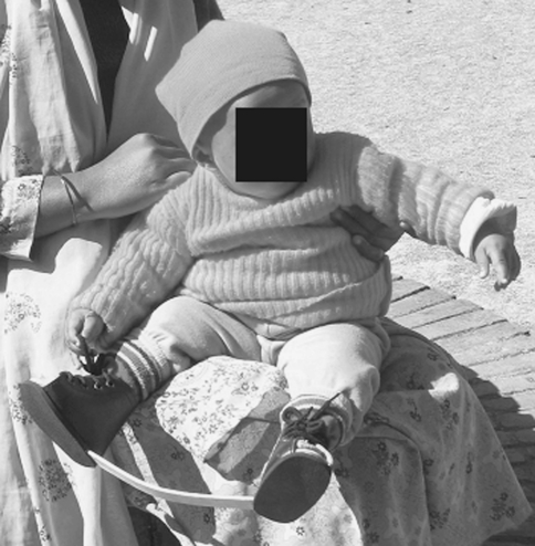
As a result of the inability to enforce full-time use of the foot abduction orthosis in ambulatory patients, our patients were transitioned to nighttime use of a foot abduction orthosis after the final cast was removed (Fig. 1). We have advised splinting until 5 years of age. Further study of this cohort will be required to document what percentage of patients has worn the splint as well as the rates of recurrence.
Fig. 1.
The foot abduction orthosis is crafted in our surgical workshop.
We recorded age in months, whether the patient was treated as an outpatient or inpatient (and number of days in the hospital), Pirani score [7, 20] (pretreatment, after casting), number of casts, type of surgical procedure (if required), complications, passive dorsiflexion after treatment (measured with the knee extended using a goniometer by either a physiotherapist or orthopaedic resident), and the ability to squat. The Pirani system assigns a score (0 = normal, 0.5 = moderate abnormality, 1 = severe abnormality) to each of six signs. Three of these involve the midfoot (curvature of the lateral border, position of the lateral head of the talus, severity of the medial crease) and three involve the hindfoot (rigidity of equinus, emptiness of the heel pad, and depth or severity of the posterior crease) [7, 20].
We defined a successful outcome as achieving a plantigrade foot without the need for an extensive soft tissue release. The need for an extensive soft tissue release (posteromedial release) was considered a failure of treatment. We used descriptive analysis for all numeric data. We compared pre- and post-Pirani scores for each foot using paired-samples t-tests. One-way analysis of variance was used to evaluate differences between arbitrarily selected age groups (1–2 years, 2–3 years, 3–4 years, 4–5 years, 5–6 years) and the average number of casts required, average pre-Pirani scores, average post-Pirani scores, and the average degree of dorsiflexion achieved when the final cast was removed. When we identified differences (p < 0.05) in the analysis of variance, those differences were further analyzed using Tukey’s honestly significant difference test.
Results
A mean of seven casts was required (range, 4–14; standard deviation [SD], 1), and the number of casts required was similar (p = 0.26) between the age groups. The Pirani score for the entire group improved (p < 0.001) after casting (mean of 5.2 ± 0.92 before casting and 2.1 ± 1.03 after casting). Pre-Pirani scores were lowest among the 5-year-old feet when compared with 2-year-old feet (p = 0.03), 3-year-old feet (p = 0.01), and 1-year-old feet (p = 0.05). Age did not influence (p = 0.44) the post-Pirani scores. The average dorsiflexion after treatment was 12.5° (9.8°–13.5°; SD, 5.91) for the group as a whole. The feet of 1-year-old children achieved a marginally greater (p = 0.05) average dorsiflexion than those of 3-year-old children, but there were no other differences by age group. Eighty-four percent of patients (data available for 126 of the 171 patients) were able to squat.
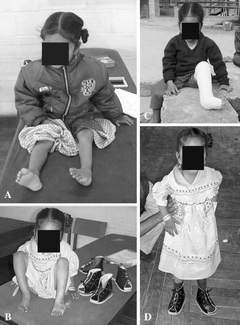
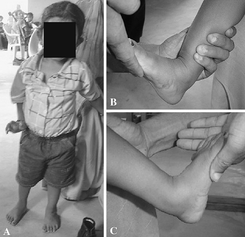
We performed surgery of some sort in 248 of the 260 feet (95%), the majority (94%) for residual equinus (234 of 248). The procedures included percutaneous tendo-Achilles release in 205 (83%), open tendo-Achilles lengthening in eight (3%), posterior release in 21 (8%), and extensive soft tissue release (posteromedial release) in 16 (6%). A plantigrade foot was achieved in 94% of cases without an extensive soft tissue release (Figs. 2A–C, 3A–C).
Fig. 2A–D.
A patient with a clubfoot on the left is shown before and after Ponseti treatment. (A) This 4-year-old girl has had her first cast applied. Note the position of the patella and the large callus over the lateral aspect of her calcaneus. (B) During the casting phase, her foot was progressively abducted. (C) Her last long leg cast is shown. (D) She is now able to walk with a plantigrade foot. We initially recommended wearing high top, reverse last shoes for several months after casting, but have since abandoned this practice.
Fig. 3A–C.
A patient treated by the Ponseti method for bilateral idiopathic clubfoot is shown with followup photograph (A) standing and (B–C) with maximal passive dorsiflexion. (A) This boy has just completed the Ponseti method for a bilateral clubfoot deformity and has achieved a heel-toe gait. (B) Maximum passive dorsiflexion on the right side. (C) Maximum passive dorsiflexion on the left side.
Complications included wound dehiscence (n = 5) after a posterior release. All healed with local wound care. Prolonged bleeding was seen in one case after percutaneous tenotomy and was controlled with local pressure. Two cases of tibial bowing during casting were observed, and both resolved after casting.
Discussion
The treatment of congenital clubfoot has evolved over the past few decades. Recently, with the perception that extensive soft tissue releases are often complicated by stiffness and residual or recurrent deformities at long-term followup, there has been considerable interest in the minimally invasive Ponseti method. Dobbs et al. [5] reported poor results in nearly 50% of patients treated by an extensive soft tissue release at 25 years followup, mainly as a result of stiffness. An average of 2.3 surgeries was performed per patient, and nearly all had a weakness in pushoff [5]. The Ponseti method has dramatically reduced the number of extensive soft tissue releases performed in selected centers in both economically developed and underdeveloped regions [1–4, 6–14, 16–19, 28]. Before 2004, our approach involved serial casting using the Kite method [15]; however, the majority of patients required a posteromedial release. At the time when this treatment protocol was established at our institution, the upper age limit for the Ponseti method (in published series) was 2 years. We therefore asked whether the Ponseti method could achieve initial correction (a plantigrade foot) of untreated idiopathic clubfeet in patients presenting between 1 and 6 years of age, and the results of our study suggest initial correction of the deformity can be achieved without the need for an extensive soft tissue release (posteromedial release or other) in 94% of such cases (Figs. 1, 2). The upper age limit for this method remains to be determined.
There are several limitations to this study that must be mentioned. Despite the fact that data were collected in a prospective manner, we were only able to locate the medical records for 89% of this initial cohort at the time of review. This reflects a deficiency in recordkeeping at the hospital, which we hope to improve with a computerized database. In addition, although the percentage of patients requiring surgical treatment for residual equinus after casting (95%) is comparable to published reports in patients younger than 2 years of age, we did not standardize the type of procedure used for correction of equinus. Without published guidelines on an upper age limit for a percutaneous tenotomy of the tendo-Achilles (standard component of Ponseti method), the decision was made by the attending surgeon based on an examination under anesthesia. Although the most frequent procedure selected was a percutaneous tenotomy (79%), a smaller number were treated by either open tendo-Achilles lengthening (3%) or posterior release (8%). We are unable to draw any conclusions regarding the most appropriate surgical technique for residual equinus after casting in these older patients. Recent studies have indicated a percutaneous release may be performed up to age 9 years [9, 17, 28]. If we were to consider the use of a posterior release to achieve a plantigrade foot as a treatment failure, then the Ponseti protocol failed in an additional 8% of cases (14% were treated by posterior release or posteromedial release), and the majority of patients were older than 3 years of age. The attending surgeons elected to perform a posterior release in 29% to 39% of patients between 3 and 6 years of age (only 1% in patients younger than 3 years of age), which may reflect the perception that the equinus was more rigid in these older patients. Second, the ultimate success of any method of treatment for clubfoot relies not only on the ability to achieve initial correction of the deformity, but also the ability to maintain that correction. Although this report focuses on the ability to achieve correction, we need to follow this initial cohort to determine whether the correction can be maintained. Interestingly, the initial Pirani score (before treatment) was lower in 5 year olds compared with the 1-, 2-, and 3-year-old feet. Although it is possible that clubfeet in these older patients may be more supple as a result of weightbearing forces or other factors, our impression is this scoring system may be less reliable in the older age groups, as the medial and posterior creases gradually disappear, and the “empty” heel pad may decrease with the normal loss of subcutaneous fat as a child grows. Although we do not believe this detracts from the observations and conclusions of the study, we plan to consider another means to grade the initial deformity in the older patients.
For a variety of reasons, many of our patients present at a later stage in their disease process, and clubfoot is no exception. Although most of the literature on the Ponseti method has concerned infants presenting younger than 1 year of age, several recent studies have investigated the use of this method in patients of walking age [9, 17, 28]. Tindall et al. [28] reviewed 100 clubfeet treated by the orthopaedic clinical officers in Malawi (25% between 18 and 48 months of age) and reported only 2% required a posteromedial release. An average of five casts was required, and 59% of feet did not require any surgical intervention. Forty-one percent were treated by a percutaneous tenotomy after a plateau in casting. Success was defined as a plantigrade foot, and the final degree of dorsiflexion was not reported. Followup was impossible as a result of social and economic factors. Lourenco and Morcuende studied 24 feet in patients from 1 to 9 years of age [17]. The Ponseti protocol was modified for these older patients; each cast was left in place for 2 weeks, and an ankle-foot orthosis was worn full-time for 11 months after the initial correction was achieved (adherence to nighttime abduction splinting could not be achieved). Additional surgery was required in nine of 17 feet to achieve a plantigrade foot, including repeat percutaneous tenotomy (four) and posterior release (five). At a mean of more than 3 years followup, all patients had a successful outcome, defined as the absence of a limp, the ability to wear standard shoes, and the ability to participate in regular activities of daily living.
We found no differences in the number of casts required when comparing the different age groups, and our average of seven casts per patient compares favorably with data from Malawi (five) [28] and Brazil (nine) [17]. We elected to change the casts weekly or every 5 days for those patients who were admitted (standard Ponseti protocol); in contrast, Lourenco and Morcuende recommend leaving each cast for 2 weeks in older patients to allow more time for relaxation of soft tissues (and presumably chondro-osseous remodeling) [17]. The mean dorsiflexion after treatment was 12.5° (range, 10°–14°), and there was a marginal difference in which 1 year olds had more dorsiflexion than 3 year olds. Because full passive dorsiflexion was not obtained in the majority of patients, longer-term followup will be required to determine rates of recurrence and whether any functional consequences will be observed. Lourenco and Morcuende reported an average dorsiflexion of 5° (range, 0°–10°) at a mean followup of 3.1 years and reported no limitation in activities of daily living.
Another issue is the role of abduction splinting in patients of walking age. Abduction splinting is an essential component of the Ponseti method, and relapse rates of up to 70% may be expected when the abduction splint is not worn [6, 10, 11, 27]. Reasons for a lack of adherence to the splinting program may include noncompliance (patient or family chooses not to wear the splint) and brace intolerance (discomfort from skin irritation or other cause). In the United States, Dobbs et al. [6] identified a lack of adherence to the splinting program and the level of education of the parents (high school or less) as important factors leading to relapse. Whether the same rates of recurrence accompany nonadherence with abduction bracing in the older patients remains to be determined. In addition, followup will be required to whether the initial correction has been maintained and to determine the rate of recurrent deformities such as equinus, supination, or varus. As of now, longer-term followup of a cohort of patients of similar age treated by the Ponseti method is unavailable. In resource-challenged environments such as Nepal, obtaining adequate followup is often difficult or unachievable as a result of economic and social factors. Many patients have to travel for days to reach the hospital, and those from remote villages are often unable to return for routine followup. We must also assess whether patient function is satisfactory using an outcome measure appropriate to our environment. Such a followup is currently being planned using a community-based rehabilitation (CBR) network.
From a population-based (rather than a patient-based) perspective, future challenges include how to reduce the number of neglected cases (improve case identification through a mechanism for screening and timely referral) and how to effectively decentralize the delivery of services so that patients can be treated in proximity to their home villages. In environments such as Nepal, barriers to timely and effective diagnosis and treatment include geographic constraints (including lack of roads and the mountainous terrain), insufficient physical resources and/or trained healthcare providers in more rural communities, and often a lack of awareness that treatment is available. Earlier case identification may be achieved by promoting community awareness, and screening may be carried out by local health professionals in collaboration with our CBR workers. Because nearly 70% of our patients required inpatient services (for up to 60 days), models to decentralize the delivery of services must be explored. The economic and social consequences of time away from home must be recognized in a society where subsistence agriculture is the principle means of support. Realistically, this will involve the training of health professionals other than orthopaedic surgeons, and our experience suggests paraprofessionals may effectively administer the casting as shown in the United Kingdom (physiotherapists) [25] and Malawi (orthopaedic clinical officers) [27]. Such nonconventional models must be explored if clubfoot care is to be delivered at the population level in low-income countries.
The Ponseti method has been effective in achieving correction in patients with idiopathic clubfoot up to 2 years of age, and the upper age limit remains to be established. Our results suggest initial correction (a plantigrade foot) of an untreated idiopathic clubfoot may be achieved in the majority of patients up to 6 years of age. The results of several recent studies also suggest a role for this method in patients of walking age. Long-term followup will be required to assess adherence to abduction splinting, to define rates of recurrence, and to evaluate the functional outcome.
Acknowledgments
We thank Seema Sonnad and Meredith Bergey for their help in performing the statistical analysis.
Footnotes
Study conducted at the Hospital and Rehabilitation Centre for Disabled Children, Banepa, Nepal.
Each author certifies that he or she has no commercial associations (eg, consultancies, stock ownership, equity interest, patent/licensing arrangements, etc) that might pose a conflict of interest in connection with the submitted article.
Each author certifies that his or her institution has approved or waived approval for the human protocol for this investigation and that all investigations were conducted in conformity with ethical principles of research.
References
- 1.Bor N, Herzenberg JE, Frick SL. Ponseti management of clubfoot in older infants. Clin Orthop Relat Res. 2006;444:224–228. doi: 10.1097/01.blo.0000201147.12292.6b. [DOI] [PubMed] [Google Scholar]
- 2.Chotel F, Parot R, Durand JM, Garnier E, Hodgkinson I, Bérard J. Initial management of congenital varus equinus clubfoot by Ponseti’s method [in French] Rev Chir Orthop Reparatrice Appar Mot. 2002;88:710–717. [PubMed] [Google Scholar]
- 3.Colburn M, Williams M. Evaluation of the treatment of idiopathic clubfoot using the Ponseti method. J Foot Ankle Surg. 2003;42:259–267. doi: 10.1016/S1067-2516(03)00312-0. [DOI] [PubMed] [Google Scholar]
- 4.Cooper DM, Dietz FR. Treatment of idiopathic clubfoot. A thirty-year follow-up note. J Bone Joint Surg Am. 1995;77:1477–1489. doi: 10.2106/00004623-199510000-00002. [DOI] [PubMed] [Google Scholar]
- 5.Dobbs MB, Nunley R, Schoenecker PL. Long-term follow-up of patients with clubfeet treated with extensive soft-tissue release. J Bone Joint Surg Am. 2006;88:986–996. doi: 10.2106/JBJS.E.00114. [DOI] [PubMed] [Google Scholar]
- 6.Dobbs MB, Rudzki JR, Purcell DB, Walton T, Porter KR, Gurnett CA. Factors predictive of outcome after use of the Ponseti method for the treatment of idiopathic clubfeet. J Bone Joint Surg Am. 2004;86:22–27. doi: 10.2106/00004623-200401000-00005. [DOI] [PubMed] [Google Scholar]
- 7.Dyer PJ, Davis N. The role of the Pirani scoring system in the management of club foot by the Ponseti method. J Bone Joint Surg Br. 2006;88:1082–1084. doi: 10.1302/0301-620X.88B8.17482. [DOI] [PubMed] [Google Scholar]
- 8.Eberhardt O, Schelling K, Parsch K, Wirth T. Treatment of congenital clubfoot with the Ponseti method. Z Orthop Ihre Grenzgeb. 2006;144:497–501. doi: 10.1055/s-2006-942239. [DOI] [PubMed] [Google Scholar]
- 9.Garg S, Dobbs MB. Use of the Ponseti method for recurrent clubfoot following posteromedial release. Indian J Orthop. 2008;42:68–72. doi: 10.4103/0019-5413.38584. [DOI] [PMC free article] [PubMed] [Google Scholar]
- 10.Goksan SB. Treatment of congenital clubfoot with the Ponseti method. Acta Orthop Traumatol Turc. 2002;36:281–287. [PubMed] [Google Scholar]
- 11.Goksan SB, Bursali A, Bilgili F, Sivacioglu S, Ayanoglu S. Ponseti technique for the correction of idiopathic clubfeet presenting up to 1 year of age. A preliminary study in children with untreated or complex deformities. Arch Orthop Trauma Surg. 2006;126:15–21. doi: 10.1007/s00402-005-0070-9. [DOI] [PubMed] [Google Scholar]
- 12.Gupta A, Singh S, Patel P, Patel J, Varshney MK. Evaluation of the utility of the Ponseti method of correction of clubfoot deformity in a developing nation. Int Orthop. 2008;32:75–79. doi: 10.1007/s00264-006-0284-7. [DOI] [PMC free article] [PubMed] [Google Scholar]
- 13.Herzenberg JE, Radler C, Bor N. Ponseti versus traditional methods of casting for idiopathic clubfoot. J Pediatr Orthop. 2002;22:517–521. doi: 10.1097/00004694-200207000-00019. [DOI] [PubMed] [Google Scholar]
- 14.Ippolito E, Farsetti P, Caterini R, Tudisco C. Long-term comparative results in patients with congenital clubfoot treated with two different protocols. J Bone Joint Surg Am. 2003;85:1286–1294. doi: 10.2106/00004623-200307000-00015. [DOI] [PubMed] [Google Scholar]
- 15.Kite JH. Some suggestions on the treatment of clubfoot by casts. J Bone Joint Surg Am. 1963;45:406–412. [PubMed] [Google Scholar]
- 16.Laaveg SJ, Ponseti IV. Long-term results of treatment of congenital club foot. J Bone Joint Surg Am. 1980;62:23–31. [PubMed] [Google Scholar]
- 17.Lourenco AF, Morcuende JA. Correction of neglected idiopathic club foot by the Ponseti method. J Bone Joint Surg Br. 2007;89:378–381. doi: 10.1302/0301-620X.89B3.18313. [DOI] [PubMed] [Google Scholar]
- 18.Morcuende JA, Abbasi D, Dolan LA, Ponseti IV. Results of an accelerated Ponseti protocol for clubfoot. J Pediatr Orthop. 2005;25:623–626. doi: 10.1097/01.bpo.0000162015.44865.5e. [DOI] [PubMed] [Google Scholar]
- 19.Morcuende JA, Dolan LA, Dietz FR, Ponseti IV. Radical reduction in the rate of extensive corrective surgery for clubfoot using the Ponseti method. Pediatrics. 2004;113:376–380. doi: 10.1542/peds.113.2.376. [DOI] [PubMed] [Google Scholar]
- 20.Pirani S, Outerbridge HK, Sawatzky B, Stothers K. A reliable method of clinically evaluating a virgin clubfoot evaluation. 21st SICOT conference, 1999.
- 21.Ponseti IV. Treatment of congenital clubfoot. J Bone Joint Surg Am. 1992;74:448–454. [PubMed] [Google Scholar]
- 22.Ponseti IV. Congenital Clubfoot: Fundamentals of Treatment. New York, NY: Oxford University Press; 1996. [Google Scholar]
- 23.Ponseti IV, Smoley EN. Congenital club foot: the results of treatment. J Bone Joint Surg Am. 1963;45:261–266. [Google Scholar]
- 24.Radler C, Suda R, Manner HM, Grill F. Early results of the Ponseti method for the treatment of idiopathic clubfoot. Z Orthop Ihre Grenzgeb. 2006;144:80–86. doi: 10.1055/s-2006-921413. [DOI] [PubMed] [Google Scholar]
- 25.Segev E, Keret D, Lokiec F, Yavor A, Wientraub S, Ezra E, Hayek S. Early experience with the Ponseti method for the treatment of congenital idiopathic clubfoot. Isr Med Assoc J. 2005;7:307–310. [PubMed] [Google Scholar]
- 26.Shack N, Eastwood DM. Early results of a physiotherapist-delivered Ponseti service for the management of idiopathic congenital talipes equinovarus foot deformity. J Bone Joint Surg Br. 2006;88:1085–1089. doi: 10.1302/0301-620X.88B8.17919. [DOI] [PubMed] [Google Scholar]
- 27.Thacker MM, Scher DM, Sala DA, Bosse HJ, Feldman DS, Lehman WB. Use of the foot abduction orthosis following Ponseti casts: is it essential? J Pediatr Orthop. 2005;25:225–228. doi: 10.1097/01.bpo.0000150814.56790.f9. [DOI] [PubMed] [Google Scholar]
- 28.Tindall AJ, Steinlechner CWB, Lavy CBD, Mannion S, Mkandawire N. Results of manipulation of idiopathic clubfoot deformity in Malawi by orthopaedic clinical officers using the Ponseti method: a realistic alternative for the developing world? J Pediatr Orthop. 2005;25:627–629. doi: 10.1097/01.bpo.0000164876.97949.6b. [DOI] [PubMed] [Google Scholar]


