Abstract
In congenital clubfoot, residual deformities are not well-documented and they may change depending on different treatments. To identify the treatment that provides better outcome at maturity, we studied the computed tomography of two cohorts of patients affected with congenital clubfoot who were treated using two distinct protocols. Forty-seven clubfeet were treated according to the traditional protocol of our hospital and 61 were treated according to the Ponseti technique. The normal feet of the unilateral deformities served as controls. All patients were followed to skeletal maturity. The ankle torsion angle and the declination angle of the neck of the talus were higher than normal but different only in patients treated with the traditional method. The calcaneocuboid angle was lower but only in patients treated with the Ponseti method. The shape of the talar joints was altered in many feet regardless of protocol. The CT images suggest the modifications of the torsion angle of the ankle, the declination angle of the neck of the talus, and the calcaneocuboid angle at maturity are related to the treatment protocol followed. The Ponseti manipulative technique provided better anatomical results in comparison to our traditional technique.
Introduction
Abnormalities in both size and shape of the tarsal bones, as well as alterations of their relationships, have been reported in several pathologic studies on aborted fetuses and stillborn infants affected with idiopathic congenital clubfoot [3, 9, 10, 16, 19, 21]. Similar abnormalities have been described in radiographic and computed tomography studies conducted at skeletal maturity in treated congenital clubfeet with various residual foot deformities [5, 7, 12, 17]. The talus and the calcaneus are smaller and morphologically distinct from those in normal feet: the neck of the talus is medially angulated and the navicular is wedge-shaped. The relationships between the tarsal bones are abnormal. The residual clubfoot deformities at maturity seem related to the severity of the pathologic abnormalities present at birth as well as to the treatment performed [17]. However, whether the residual abnormalities relate to choice of treatment is not well-documented [4], and they may differ depending on the choice of treatment protocol. To select the most appropriate treatment, we believe it important to know which treatment provides the best anatomical results at maturity in idiopathic congenital clubfoot.
We therefore evaluated at maturity the torsion angle of the ankle mortise, the declination angle of the neck of the talus (talar body-neck angle), the calcaneocuboid angle, and the shape of the tarsal joints (subtalar joint, calcaneocuboid joint, and talonavicular joint) in two groups of patients with idiopathic clubfoot treated by either Marino-Zuco technique or the Ponseti technique.
Materials and Methods
In 2003, we published the long-term comparative results in two series of patients with idiopathic congenital clubfoot treated with two different protocols [4]. The two series comprised 32 patients each; the first series, treated between 1973 and 1977 using a traditional approach [14], included 47 clubfeet, whereas the second one, treated between 1979 and 1984 using the Ponseti method [16], was made up of 49 clubfeet. Since 2003, eight more patients (12 clubfeet) of the second series were added to the original cohort because they reached skeletal maturity. Therefore, in this study, the second series included 40 patients with 61 clubfeet. All patients were younger than 3 weeks of age at the start of treatment and all their clubfeet were severe, graded as Group 3 according to the Manes classification [13]. No patient had previous treatment. All the patients were followed up at the end of skeletal growth. In the first series, 24 patients were male and eight female; the deformity was bilateral in 15 patients and unilateral in 17. In the second series, 28 patients were male and 12 female; the clubfoot was bilateral in 21 patients and unilateral in 19. The minimum followup in the first group was 24 years (mean, 25 years; range, 24–28 years), whereas the minimum followup in the second group was 17 years (mean, 18.8 years; range, 17–22 years). The study was approved by the Ethics Committee of our University Hospital, and informed consent was obtained from all the patients included in the study.
The patients of the first series were treated with manipulation and casting according to the Marino-Zuco technique [14] up to 5 to 7 months of age (average, 16 casts) followed in 41 resistant cases by a posteromedial release according to Codivilla [1], later modified by Turco [20], performed between 5 and 8 months of age. The manipulation technique described by Marino-Zuco was based on abduction and pronation of the forefoot with counterpressure applied at the anterior tuberosity of the calcaneus grasped with the other hand [14]. In 11 feet, the Codivilla operation was performed with two separate incisions. An aluminum brace extending proximally to the knee was applied at night until 3 years of age, whereas high-top reverse-last shoes were used until the child was 5 years old.
The patients in the second series were treated with manipulation and casting according to the Ponseti technique [18] up to 2 to 3 months of age (average, five casts). In the resistant cases the manipulations and castings were followed by limited posterior release at 3 to 4 months of age instead of the subcutaneous tenotomy recommended by Ponseti. The manipulation technique described by Ponseti was based on abduction of the foot in supination under the talus with counterpressure applied at the lateral aspect of the head of the talus. An aluminum brace with the knee flexed 90° was worn until 4 years of age. Relapsing feet, passively correctable, were treated with transfer of the anterior tibial tendon on the third cuneiform [2], whereas relapsed stiff feet, observed only in the first series, were treated with a second posteromedial release. The most important differences between the two manipulation techniques are that in the Marino-Zuco technique, the foot is abducted and pronated, whereas in the Ponseti technique, the foot is abducted in supination; therefore, in the traditional technique, counterpressure is applied at the level of the calcaneus, whereas in the Ponseti technique, it is applied at the level of the head of the talus.
All the clubfeet were evaluated at followup using a CT scan (LightSpeed Series 5.X; GE Healthcare, Chalfont St Giles, UK) with 3-D reconstruction. The patient was positioned supine on the machine platform with the lower limbs parallel and the knees straight. The feet were locked parallel in a radiolucent support by a belt, in a neutral position with the ankles at 90°, to simulate a standing view. Scans 1.25-mm thick were taken on the three different spatial planes.
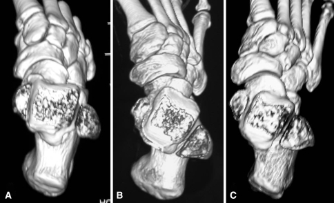
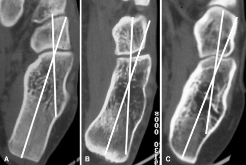
The images to determine the torsion angle of the ankle mortise, the declination angle of the neck of the talus, and the calcaneocuboid angle were evaluated by a radiologist (LR), who used a computer program to draw the lines representing the different angles. The torsion angle of the ankle mortise was evaluated through a 3-D CT scan of the lower limb with axial sections obtained at the levels of the proximal juxta-articular epiphysis of the tibia and of the ankle mortise. The angle was measured between the two lines of the axis of the proximal tibial epiphysis and the bimalleolar axis according to the method described by Jacob et al in 1980 [11] (Fig. 1A–C). The declination angle of the neck of the talus was evaluated in a 3-D CT scan reconstruction obtained at the level of the talar bone as the angle formed by the axes of the body and the neck of the talus [6, 7] (Fig. 1A–C). The calcaneocuboid angle was evaluated in a CT scan obtained on the plantar plane as the angle formed by the longitudinal axes of the calcaneus and the cuboid [6, 7] (Fig. 2A–C).
Fig. 1A–C.
Three-dimensional computed tomography scan reconstruction of the foot in the transverse plane at the level of the ankle mortise in (A) a normal foot, (B) a clubfoot of the first series, and (C) a clubfoot of the second series. The torsion angle of the ankle measured 19° in the normal foot, 30.5° in the clubfoot of the first series, and 26.5° in the clubfoot of the second series. The declination angle of the neck of the talus measured 18° in the normal foot, 31° in the first series clubfoot, and 23.5 in the second series clubfoot. A residual varus deformity of the calcaneus is present in the clubfoot of the first series. The navicular tuberosity is very close to the medial malleolus in the clubfoot of the second series, but the cuneiforms and the cuboid are shifted laterally.
Fig. 2A–C.
Computed tomography image of the calcaneocuboid joint (A) in a normal foot, (B) in a clubfoot of the first series, in which the joint appears inverted, and (C) in a clubfoot of the second series, in which it appears flat. The calcaneocuboid angle measured 19.5° in the normal foot, 15.5° in the clubfoot of the first series, and 13° in the clubfoot of the second series.
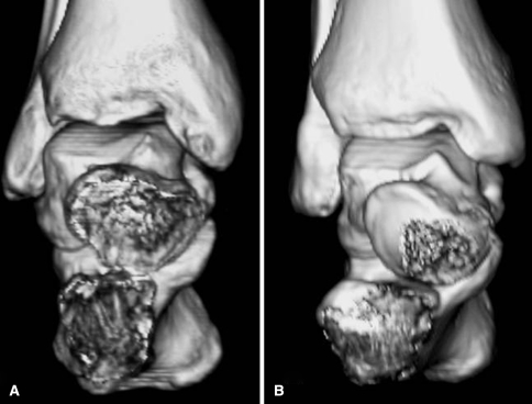
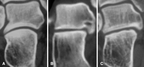
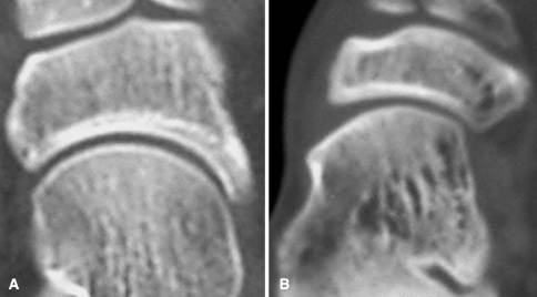
We also determined the shape of the subtalar joint (Fig. 3A–B), of the calcaneocuboid joint, and of the talonavicular joint. The normal feet of the unilateral deformities served as controls. The subtalar joint was evaluated on the coronal plane at the level of the posterior articulation (Fig. 4A–C). The calcaneocuboid joint was evaluated on the plantar plane (Fig. 2A–C). The talonavicular joint was evaluated on the plantar plane (Fig. 5A–B). To evaluate the joint shapes, we have selected the CT scan at the level of the middle part of each joint.
Fig. 3A–B.
Three-dimensional computed tomography scan reconstruction of the hindfoot in the coronal plane in (A) a clubfoot of the first series and (B) a clubfoot of the second series. Talus and calcaneus are coincident in the clubfoot of the first series and divergent in the clubfoot of the second series.
Fig. 4A–C.
Computed tomography image of the subtalar joint at the level of the posterior articulation in (A) a normal foot, (B) a clubfoot of the second series, in which it appears flat, and (C) a clubfoot of the first series, in which the joint appears slanted.
Fig. 5A–B.
Computed tomography image of the talonavicular joint in (A) a normal foot and (B) in a clubfoot of the second series, in which it is medially subluxated.
We determined differences in the values of the torsion angle of the ankle, of the declination angle of the neck of the talus, and of the calcaneocuboid angle (dependent variables) between the two series of clubfeet and normal feet (independent variables) using the t-test with a Bonferroni post hoc correction for multiple tests.
Results
The average value of the torsion angle of the mortise was greater (p = 0.002) in the first than in the second series (average, 29.9° versus 23.7°, respectively) and greater (p = 0.0001) in the first than in the normal feet (average, 21.4°) (Table 1; Fig. 1A–C). The average value of the torsion angle of the mortise measured 29.9° in the first series (range, 22.4°–37.5°), 23.7° in the second series (range, 16.2°–31.2°), and 21.4° in the normal feet (range, 13.9°–29°).
Table 1.
Tarsal angles, together with statistical significance, in the two series of clubfeet and in normal feet measured at the CT scan examination
| Variable | Torsion angle of ankle | Talar body-neck angle | Calcaneocuboid angle |
|---|---|---|---|
| Congenital clubfeet of the first series (Marino-Zuco technique) | 29.9° (22.4°–37.5°) | 30.3° (23°–37.6°) | 7.7° (–9.1°–24.6°) |
| Congenital clubfeet of the second series (Ponseti technique) | 23.7° (16.2°–31.2°) | 22.9° (14.8°–31.8°) | 2.5° (–14.9°–19.3°) |
| Normal feet | 21.4° (13.9°–29°) | 17.8° (12.3°–23.3°) | 13.8° (6.2°–21.5°) |
| Statistical significance | 1st series > 2nd series p = 0.002 1st series > normal feet p = 0.0001 2nd series > normal feet not significant |
1st series > 2nd series p = 0.001 1st series > normal feet p = 0.0001 2nd series > normal feet not significant |
2nd series < normal feet p = 0.001 1st series < normal feet not significant 1st series > 2nd series not significant |
The average value of the declination angle of the neck of the talus was greater (p = 0.001) in the first than in the second series (average, 30.3 versus 22.9, respectively) and greater (p = 0.0001) in the first series than in the normal feet (average, 17.8°) (Table 1; Fig. 1A–C). The average value of the declination angle of the neck of the talus measured 30.3° in the first series (range, 23°–37.6°), 22.9° in the second series (range, 14.8°–31.8°), and 17.8° in the normal feet (range, 12.3°–23.3°).
The average value of the calcaneocuboid angle was less (p = 0.001) in the second series than in the normal feet (average, 2.5° versus 13.8°, respectively). The average value of the calcaneocuboid angle was 7.7° in the first series (range, −9.1–24.6°), 2.5° in the second series (range, −14.9–19.3°), and 13.8° (range, 6.2°–21.5°) in the normal feet (Table 1; Fig. 2A–C).
The shapes of the subtalar joint were similarly distributed in the two series (Table 2; Fig. 4A–C). In the first series, the subtalar joint was normal in five feet, presented less curvature than normal in 17 feet, was flat in 18 feet, and was slanted in seven feet. For the second series, the subtalar joint was normal in seven feet, presented less curvature than normal in 25 feet, was flat in 22 feet, and was slanted in seven feet.
Table 2.
Shape of the tarsal joints in the two series of clubfeet and in normal feet
| Clubfeet studied | Subtalar joint | Calcaneocuboid joint | Talonavicular joint |
|---|---|---|---|
| Congenital clubfeet of the first series (Marino-Zuco technique) | Normal: 5 feet Lesser than normal: 17 feet Flat: 18 feet Slanted: 7 feet |
Normal: 6 feet Inverted: 9 feet Wave-shaped: 14 feet Flat: 9 feet Medially subluxated: 9 feet |
Normal: 12 feet Medially subluxated: 35 feet |
| Congenital clubfeet of the second series (Ponseti technique) | Normal: 7 feet Lesser than normal: 25 feet Flat: 22 feet Slanted: 7 feet |
Normal: 5 feet Inverted: 14 feet Wave-shaped: 19 feet Flat: 23 feet Medially subluxated: none |
Normal: 6 feet Medially subluxated: 55 feet |
The shapes of the calcaneocuboid joint were similarly distributed in the two series (Table 2; Fig. 2A–C). For the first series, the calcaneocuboid joint was normal in six feet, inverted in nine feet, wave-shaped in 14 feet, flat in nine feet, and medially subluxated in nine feet. Of the second series, the calcaneocuboid joint was normal in five feet, inverted in 14 feet, wave-shaped in 19 feet, and flat in 23 feet. No clubfoot of the second series showed medial subluxation of this joint.
The shapes of the talonavicular joint were similarly distributed in the two series (Table 2; Fig. 5A–B). Of the first series, the talonavicular joint was normal in 12 feet, whereas it was medially subluxated in the remaining 35 feet. For the second series, the talonavicular joint was normal in six feet and medially subluxated in 55.
Discussion
The long-term followup studies published on idiopathic clubfoot that report the final outcomes evaluated by conventional radiographic methods often do not show tarsal bones and joints from a morphological point of view [4, 17, 20]. CT scan examination, performed at the end of skeletal growth, allows better evaluation of the actual final shapes of the tarsal bones, their relationships, and any residual deformity. In congenital clubfoot, these deformities are not well-studied, and may be related to the treatment protocol used. Using CT scans, we therefore evaluated two series of patients affected by idiopathic clubfoot treated with two distinct protocols to assess their influence on the shapes and relationships of the hindfoot bones at the end of skeletal growth. We specifically investigated six variables, three regarding the tarsal angles (torsion angle of the ankle, talar body-neck angle, and calcaneocuboid angle) and three regarding the shape of the tarsal joints (subtalar, calcaneocuboid, and talonavicular).
A limitation of this study is that the two series of patients had not been randomized, because they were treated in different periods of time: the first series between 1973 and 1977 and the second between 1979 and 1984. Another limitation of the study is that the patients included in the Ponseti series were surgically treated by a limited posterior release after the manipulations, instead of the subcutaneous tenotomy of the Achilles tendon recommended by Ponseti [16].
In a pathologic study published in 1993 [3], Howard and Benson demonstrated the torsion angle of the ankle mortise in stillborn infants with congenital clubfoot is normal. We observed an increase of the average value of this angle in both series of patients reported, although we found a greater increase in the first series. The average angle was greater than that of normal in the first series but not in the second. We believe the external torsion of the ankle is related to the manipulative treatment performed, in which ankle torsion compensates for the lack of eversion and abduction of the calcaneus and the increased declination angle of the neck of the talus (Figs. 1A–C and 3A–B). In fact, the Ponseti manipulative technique allows calcaneus eversion in the subtalar joint according to its shape, whereas our traditional technique blocks the calcaneus underneath the talus.
In their histologic study published in 1980, Ippolito and Ponseti [9] reported an increase of the declination angle of the neck of the talus in a series of histologic sections cut in the transverse plane in fetuses affected by congenital clubfoot at the level of the talar bone. Howard and Benson [3] reported this angle was greater than normal in fetuses with congenital clubfoot and that in the most severely deformed specimens, the body-neck angle measured as much as 90°. We observed an increase of the average value of this angle in our two series of patients, although we found a greater increase in the first series. Therefore, the declination angle of the neck of the talus was better corrected in our second series treated with the Ponseti method. We observed a difference between the two series and between the first series and the normal feet. A recent MRI study showed an improvement in the declination angle of the neck of the talus in infant clubfeet treated with the Ponseti method [15]. It is likely the Ponseti manipulative technique may straighten the angle of the neck of the talus by the pressure exerted by the laterally stretched navicular on the head and the neck of the talus (Figs. 1A–C and 3A–B).
In both series of patients, we observed a decrease of the calcaneocuboid angle in comparison to the normal feet. We believe the angle value decrease is related to the everting force caused by the manipulative technique performed in both series. However, we suspect the everting force produced by the Ponseti technique is stronger than that produced by the Marino-Zuco technique. In the relapsed clubfeet, treated with transfer of the anterior tibial tendon to the third cuneiform, we observed the lowest values of the calcaneocuboid angle, probably as a result of the abducting-everting force of the transferred tendon that was able to shift the cuneiforms, the cuboid, and the whole forefoot more laterally [2].
Some anatomic studies demonstrate abnormality of the subtalar joint in fetuses and stillborns with congenital clubfoot from the early stages of foot development [3, 9, 16]. We found a normal subtalar joint in only five feet of the first series (10.2%) and in seven feet of the second series (11.4%), whereas in most cases, the CT scan showed various anatomic abnormalities in the subtalar joint of the treated clubfeet, although the distribution of the abnormalities was similar in the two series. The abnormalities in the shape of the subtalar joint, present at birth, appear not to be correctable with either manipulative or surgical treatment. Therefore, these abnormalities may affect the final clinical result; in fact, at followup, the clubfeet with the described abnormalities showed residual heel varus deformity as well as limitation of the hindfoot movements [8].
The calcaneocuboid joint presented various anatomic abnormalities in 41 feet of the first series (87.2%) and in 55 feet of the second series (90.1%) with similar distributions in the two series. We found the manipulative techniques used in our two series of patients are able to obtain a satisfactory reduction of the medial subluxation of the calcaneocuboid joint or even an overreduction, especially in the second series of patients, but the manipulative techniques are not able to modify the shape of the calcaneocuboid joint that in most cases remains abnormal.
The talonavicular joint was medially subluxated in 35 feet of the first series (74.5%) and 55 feet of the second series (90.2%). This difference could be related to the medial release performed in our first series of patients. In fact, posteromedial release seems to better reduce the medial subluxation of the navicular; however, the better reduction of the talonavicular joint, observed in approximately one-fourth of the patients of the first series, did not influence the final functional results that were much better in the second series than in the first [4].
The Ponseti manipulative technique provides good and permanent correction of clubfoot deformities. In particular, the torsion angle of the ankle and the talar body-neck angle appear normal, whereas the decrease of the calcaneocuboid angle seems compensatory for the lack of correction of the relationship of the talus and calcaneus resulting from the abnormality of the subtalar joint observed in most cases. Moreover, clinical results obtained with the Ponseti technique were better than those obtained with our traditional method as reported in a previous paper [4].
Acknowledgments
We thank Dr Giovanna Brancato of the Italian Institute of Statistics (ISTAT) for the statistical analysis.
Footnotes
Each author certifies that he or she has no commercial associations (eg, consultancies, stock ownership, equity interest, patent/licensing arrangements, etc) that might pose a conflict of interest in connection with the submitted article.
Each author certifies that his or her institution has approved the human protocol for this investigation, that all investigations were conducted in conformity with ethical principles of research, and that informed consent for participation in the study was obtained.
References
- 1.Codivilla A. Sulla cura del piede equino varo congenito: nuovo metodo di cura cruenta [Congenital talipes equinovarus: a new method of surgical treatment] Arch Chir Orthop. 1906;23:245–258. [Google Scholar]
- 2.Farsetti P, Caterini R, Mancini F, Potenza V, Ippolito E. Anterior tibial tendon transfer in relapsing congenital clubfoot. J. Pediatr. Orthop. 2006;26:83–90. doi: 10.1097/01.bpo.0000188999.21427.fb. [DOI] [PubMed] [Google Scholar]
- 3.Howard CB, Benson MKD. Clubfoot: its pathological anatomy. J. Pediatr. Orthop. 1993;13:654–659. [PubMed] [Google Scholar]
- 4.Ippolito E, Farsetti P, Caterini R, Tudisco C. Long-term comparative results in patients with congenital clubfoot treated with two different protocols. J. Bone Joint Surg. Am. 2003;85:1286–1294. doi: 10.2106/00004623-200307000-00015. [DOI] [PubMed] [Google Scholar]
- 5.Ippolito E, Fraracci L, Caterini R, Di Mario M, Farsetti P. A radiographic comparative study of two series of skeletally mature clubfeet treated by two different protocols. Skeletal Radiol. 2003;32:446–453. doi: 10.1007/s00256-003-0620-6. [DOI] [PubMed] [Google Scholar]
- 6.Ippolito E, Fraracci L, Farsetti P, Maio F. Validity of the anteroposterior talocalcaneal angle to assess congenital clubfoot correction. AJR Am. J. Roentgenol. 2004;182:1279–1282. doi: 10.2214/ajr.182.5.1821279. [DOI] [PubMed] [Google Scholar]
- 7.Ippolito E, Fraracci L, Farsetti P, Di Mario M, Caterini R. The influence of treatment on skeletal pathology of clubfoot. CT study at maturity. J. Bone Joint Surg. Br. 2004;86:574–580. [PubMed] [Google Scholar]
- 8.Ippolito E, Mancini F, Di Mario M, Farsetti P. A comparison of resultant subtalar joint pathology with functional results in two groups of clubfoot patients treated with two different protocols. J. Pediatr. Orthop. B. 2005;14:358–361. doi: 10.1097/01202412-200509000-00008. [DOI] [PubMed] [Google Scholar]
- 9.Ippolito E, Ponseti IV. Congenital clubfoot in the human fetus: a histological study. J. Bone Joint Surg. Am. 1980;62:8–18. [PubMed] [Google Scholar]
- 10.Irani RN, Sherman MS. The pathological anatomy of clubfoot. J. Bone Joint Surg. Am. 1963;45:45–52. [Google Scholar]
- 11.Jacob RP, Haertel M, Stussi E. Tibial torsion calculated by computerized tomography and compared to other methods of measurements. J. Bone Joint Surg. Br. 1980;62:238–242. doi: 10.1302/0301-620X.62B2.7364840. [DOI] [PubMed] [Google Scholar]
- 12.Johnston CE, 2nd, Hobatho MC, Baker KJ, Baunin C. Three-dimensional analysis of clubfoot deformity by computed tomography. J. Pediatr. Orthop. B. 1995;4:39–48. doi: 10.1097/01202412-199504010-00006. [DOI] [PubMed] [Google Scholar]
- 13.Manes E, Costa CM, Innao V. Treatment of congenital clubfoot during the first years of life. Chir. Organi Mov. 1975;62:301–314. [PubMed] [Google Scholar]
- 14.Marino-Zuco C. Trattamento del piede torto congenito [Treatment of congenital clubfoot]. Roma Arte della Stampa. 1934:9–28.
- 15.Pirani S, Zenznik L, Hodges D. Magnetic resonance imaging study of the congenital clubfoot treated with the Ponseti method. J. Pediatr. Orthop. 2001;21:719–726. doi: 10.1097/00004694-200111000-00004. [DOI] [PubMed] [Google Scholar]
- 16.Ponseti IV. Congenital Clubfoot: Fundamentals and Treatment. New York, NY: Oxford University Press; 1996. [Google Scholar]
- 17.Ponseti IV, El-Khoury GY, Ippolito E, Weinstein SL. A radiographic study of skeletal deformities in treated clubfoot. Clin. Orthop. Relat. Res. 1981;160:30–42. [PubMed] [Google Scholar]
- 18.Ponseti IV, Smoley EN. Congenital clubfoot: the results of treatment. J. Bone Joint Surg. Am. 1963;45:261–275. [Google Scholar]
- 19.Settle GW. The anatomy of congenital talipes equinovarus: sixteen dissected specimens. J. Bone Joint Surg. Am. 1963;45:1341–1354. [PubMed] [Google Scholar]
- 20.Turco VJ. Resistant congenital clubfoot treated by one stage posteromedial release with internal fixation: a follow-up report with a fifteen years experience. J. Bone Joint Surg. Am. 1979;61:805–814. [PubMed] [Google Scholar]
- 21.Waisbrod H. Congenital clubfoot: an anatomical study. J. Bone Joint Surg. Br. 1973;55:796–801. [PubMed] [Google Scholar]


