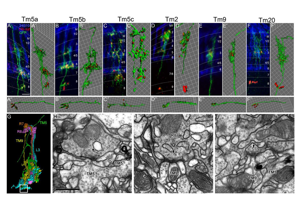
Figure 4. Ort-expressing Tm neurons receive multi-channel inputs in the medulla and are presynaptic at both the medulla and lobula.

(A–F) The distribution of presynaptic terminals of single Ort-expressing Tm neurons was examined in flies carrying ort1–3-Gal4, hs-Flp, TubP->Gal80>, UAS-mCD8GFP (green) and UAS-synaptotagmin-HA (red). Localization of the presynaptic reporter, Synaptotagmin-HA, was visualized using anti-HA antibody. R7 and R8 photoreceptors were visualized using MAb24B10 antibody (blue). Tm cell types are as indicated. IsoSurface representations of medulla arborization (A’–F’) and lobula terminals (A-F”) were generated using Imaris software. Synaptotagmin-HA was localized to the tips of the axon terminals and dendritic arbors, the latter especially in the proximal medulla strata (M7 for Tm5c; M8 for Tm5a, Tm5b, and Tm20; M9 for Tm2).
(G) Profiles of R7, R8, L3, Tm5 and Tm9 reconstructed in three dimensions from a single medulla column. The white square box indicates the contact site between L3 and both Tm5 and Tm9 shown in (J). Although the partially reconstructed profile resembles Tm5a, the subtype reconstructed is still not certain.
(H–J) Synaptic contacts between R7 and Tm5 (H), R8 and Tm9 (I), and L3 and both Tm9 and Tm5 (J) Arrowheads point to T-bar ribbons in presynaptic elements, in the presumed direction of transmission.
Scale bar: in (A), 5 µm (for A–F); in (H), 500nm (for H–J)
