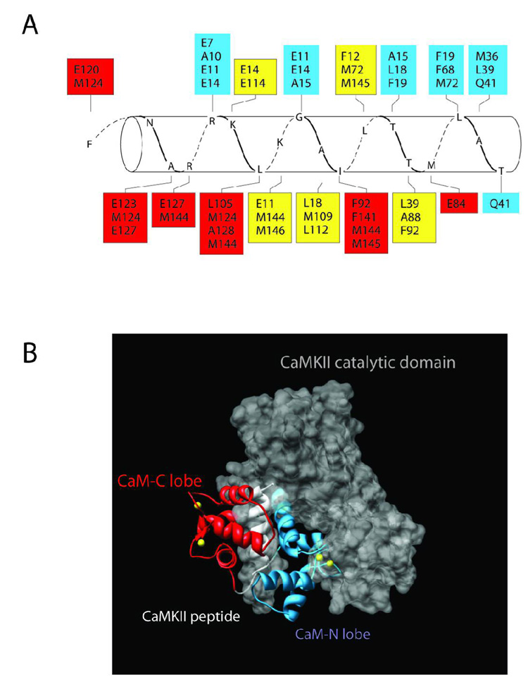
Figure 8.
Structure and Model of Ca2+/CaM bound to CaMKII. A) Amino acids in CaM are shown in boxes that interact with residues of the peptide (CKII 293–314) that mimics the CaM-binding domain of CaMKII. The residues in red are from the C-terminal domain of CaM, blue are from the N-terminal domain and yellow are from both N- and C-domains. The crystal structure that this figure is based upon is derived from PDB # 1CM1 (20). B) The complete crystal structure of PDB #1CM1 was aligned into the crystal structure of the autoinhibited catalytic domain of CaMKII (PDB# 2BDW) (41). The amino acids of the peptide were superimposed on those same residues in the CaM-binding domain of the catalytic domain as a means of constraining the alignment. The backbone of the C-lobe and N-lobes of CaM are shown in red and blue, respectively. The peptide (CKII 293–314) is in white and the catalytic domain is shown as a space filling model in grey. The yellow balls represent Ca2+ ions. Note that the N-lobe lies in a crevice between the autoregulatory domain and the catalytic domain. This figure was created using Chimera (http://www.cgl.ucsf.edu/chimera/).

