Abstract
Although both the H1 and HES2 human embryonic stem cell lines (NIH codes: WA01 and ES02, respectively) are capable of forming all three germ layers and their derivatives, various lines of evidence including the need for using different protocols to induce cardiac differentiation hint that they have distinctive preferences to become chamber-specific heart cells. However, a direct systematic comparison has not been reported. Here we electrophysiologically demonstrated that the distributions of ventricular-, atrial- and pacemaker-like derivatives were indeed different (ratios=39:61:0 and 64:33:3 for H1 and HES2, respectively). Based on these results, we hypothesized the differences in their cardiogenic potentials are imprinted in the proteomes of undifferentiated H1 and HES2. Using the multiplexing, high-resolution 2-D Differential In Gel Electrophoresis (DIGE) to minimize gel-to-gel variations that are common in conventional 2D gels, a total of 2000 individual protein spots were separated. Of which, 55 were >2-fold differentially expressed in H1 and HES2 (p<0.05) and identified by mass spectrometery. Bioinformatic analysis of these protein differences further revealed candidate pathways that contribute to the H1 and HES2 phenotypes. We conclude that H1 and HES2 have predetermined preferences to become ventricular, atrial and pacemaker cells due to discrete differences in their proteomes. These results improve our basic understanding of hESCs and may lead to mechanism-based methods for their directed cardiac differentiation into chamber-specific cardiomyocytes.
Introduction
Normal rhythms originate in the sino-atrial (SA) node, a specialized cardiac tissue consisting of only a few thousands pacemaker cells. The SA node generates spontaneous rhythmic action potentials which subsequently propagate to induce coordinated muscle contractions of the atria and ventricles for effective blood pumping [1; 2]. Since terminally-differentiated adult CMs normally lack the ability to regenerate[3], malfunctions or significant loss of heart cells due to disease or aging can lead to lethal consequences. Human embryonic stem cells (hESCs), isolated from the inner cell mass of blastocysts, possess the ability to remain pluripotent and propagate indefinitely in vitro. When cultured properly, hESCs uniquely maintain their normal karyotype and can differentiate into the derivatives of all three germ layers, including such highly specialized cells as CMs. Thus, hESCs have the potential to act as an unlimited ex vivo source of CMs for transplantation therapies. However, a number of hurdles remain. Particularly, the ability to direct the differentiation of hESC into chamber-specific cell types is crucial for future clinical applications. For instance, while hESC-derived ventricular cardiomyocytes are useful for myocardial repair, nodal pacemaker derivatives can alleviate the need of electronic pacemakers for certain arrhythmias [1; 2; 4–7]. Although several hESC lines are capable of differentiating into CMs [8; 9], various lines of evidence hint that they have distinctive preferences to become chamber-specific pacemaker-, ventricular- or atrial-like cells [9; 10]. For instance, H1 but not HES2 cells (NIH codes: WA01 and ES02, respectively), can form three-dimensional (3-D) embryoid bodies (EBs) that contain CMs when plated and grown in permissive conditions [8; 10]. By contrast, HES2 cells do not form EBs under the same conditions; for forming for spontaneously beating CM-containing outgrowths, they need to be co-cultured with an immortalized endoderm-like derivative of P19 cells (END2) [9]. Interestingly, the same method of END2 co-culturing can also induce H1 to become CMs (unpublished observation, JC Moore and RA Li). Taken together, these observations raise the intriguing possibility that intrinsic differences between the hESC lines, rather than the differentiation methods per se, underlie their different cardiogenic potentials. Understanding the basis of these differences will help develop mechanism-based methods to direct cardiac differentiation into chamber-specific CMs.
The ability to monitor changes in global protein expression and post-translational modifications is a powerful tool to understand stem cell differentiation. The conventional method for identifying quantitative differences in global protein levels involves the use of 2-D gels, which are subject to significant gel-to-gel variability and errors (reviewed in [11]). Since it is often difficult to distinguish between system and biological variations, accurate quantification of differences in the expression levels with statistical confidence can be challenging [12]. In particular, non-abundant proteins important for certain biological processes can be easily masked by others that are highly expressed. These hurdles can be overcome by the use of the multiplexing 2-D Differential In-Gel Electrophoresis (DIGE) technique [12]. DIGE uses size- and charge-matched, spectrally resolvable fluorophores (CyDye) to simultaneously separate up to three samples on a single 2-D gel. Thus, every spot on a gel has its own internal standard. After electrophoresis and scanning on an imager, integrated software can be used to co-detect, locate and analyze protein spots, followed by assigning statistical confidence to each and every difference via a differential analysis algorithm and thereby avoid gel-to-gel variations. For instance, differences as little as 10% can be routinely detected with >95% statistical confidence [13].
Proteomic studies have been done to compare the differentiation profiles of such stem cell types as human mesenchymal stem cells, murine ESCs, neuroblastoma cells, etc [14–17]. However, only two studies analyzing the protein expression profile of hESCs have been reported to date [18; 19]. The first of these studies used mass spectroscopy to identify proteins resolved by conventional SDS-PAGE [18]. Using a subtraction method, van Hoof et al identified several previously unknown factors involved in hESC self-renewal. In addition, they were able to verify the presence of these newly identified factors in 3 undifferentiated hESC lines: EES2, HUES-1 and NL-hESC-03 [18]. The second study used conventional 2-D gels to compare the undifferentiated proteomes of the Royan H2, H3 and H5 hESC lines [19]. In the present study, we hypothesized that the H1 and HES2 lines have distinct preferences to become chamber-specific heart cells, and that their different cardiogenic potentials are predetermined at the pluripotent stage due to discrete differences in their proteomes.
Materials and Methods
Please see Supplemental Material for additional details.
Human ESC Culture and Differentiation
The H1 (WiCell, Madison, WI) [1; 20; 21] and HES2 (ESI, Singapore) [5; 22] lines were cultured and differentiated as described previously.
Electrophysiology
Spontaneously beating HES2- and H1-derived cardiomyocytes were dissected from hESC aggregates 14–21 days after initiating differentiation. After collagenase dissociation, action potential (AP) recordings from single cells were performed using Axopatch 200B, Digitize 1322 and pClamp8 (Axon Burlingame, CA, USA). Patch pipettes typically had resistances of 4–6 M'Ω when filled with an internal solution containing (mM): 110 K+ aspartate, 20 KCl, 1 MgCl2, 0.1 Na-GTP, 5 Mg-ATP, 5 Na2-phospocreatine, 1 EGTA, 10 HEPES, pH adjusted to 7.3 with KOH. The external Tyrode’s bath solution consisted of (mM): 140 NaCl, 5 KCl, 1 CaCl2, 1 MgCl2, 10 glucose, 10 HEPES, pH adjusted to 7.4 with NaOH.
2-D DIGE
Cells from all three different passages were combined and lysed in 2-D lysis buffer. Each sample was labeled with a different CyDye (GE Healthcare/Amersham). CyDye-labeled protein samples were mixed and 250 µl was loaded per IEF strip (GE Healthcare/Amersham). IEF was then done for a total of 25000 volt-hours with standard conditions using Ettan IPGPhore II. After the IEF, electrophoresis was performed at 16°C. The resulting 2-D gel was scanned using a Typhoon Trio scanner (GE Healthcare/Amersham). Analyses were performed using the ImageQuant and DeCyder softwares (GE Healthcare/Amersham). Selected protein spots were excised from the gel using Ettan spot picker (GE Healthcare/Amersham), subjected to in-gel digestion and protein IDs were determined by MS/MS analysis using a MALDI-ToF/ToF mass spectrometer, ABI-4700 (Applied Biosystems, Inc.)
Results
HES2 and H1 Lines Differ in Cardiogenic Potentials
Previous work by us and others has clearly shown that H1 and HES2 hESCs can be differentiated into CMs [1; 5; 8–10; 23–27]. In particular, the cell-attached recordings of multi-cellular clusters by Mummery et al [9] and Moore et al [5] demonstrate that nearly 85% of HES2-derived CMs are ventricular-like (with the remaining 6% and 3% as atrial- and pacemaker-like, respectively), while extracellular recordings of intact beating EB outgrowths derived from H1 resulted in a nearly equal distribution of ventricular- and atrial-like derivatives [1; 27]. These studies hint at the possibility that the H1 and HES2 cell lines may have distinct preferences to differentiate into chamber-specific cardiac phenotypes although a definitive conclusion cannot be made due to the different experimental conditions and recording techniques. For instance, different time-windows were studied (40–95 days of He et al versus 12 days in the case of Mummery et al) [9; 27]. Furthermore, multiple separate impalements and recordings were performed in the same outgrowths by He et al. Adding an extra layer of complexity to the measurements, interconnected cardiomyocytes electrically influence each other via connexin-mediated gap junctions. Given the above-mentioned experimental differences, we sought to specifically electrophysiologically probe these differences by dissociating HES2- and H1-derived spontaneously contracting clusters into single cells for high-resolution patch-clamp recordings.
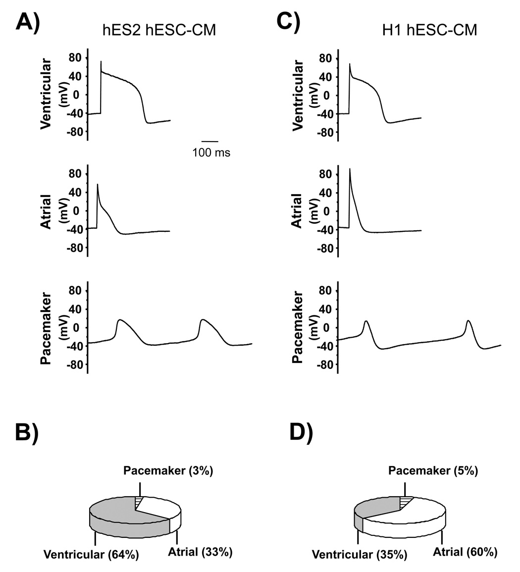
Based on the signature action potential (AP) profiles (Supplemental Table 1), chamber-specific identities were assigned. Of all the HES2-derived CMs measured (n=33; Figure 1A–B), 63.6% had the characteristic ventricular-like AP phenotype, while 33.3% were consistent with and categorized as the atrial-like phenotype. The remaining (3.1%) belonged to the pacemaker-like phenotype. By comparison, H1-derived CMs resulted in 35.0, 60.0 and 5.0% ventricular-, atrial- and pacemaker-like cells, respectively (n=20; Figure 1C–D). Thus, the H1 and HES2 cell lines undergoing cardiac differentiation via the most commonly used protocol for each cell line have distinctive preferences to become chamber-specific cells.
Figure 1.
Action potentials of ventricular, atrial and pacemaker cardiomyocytes derived from A) HES2 and C) H1 hESCs. The corresponding pie graph (B and D).
2-D DIGE of HES2 and H1
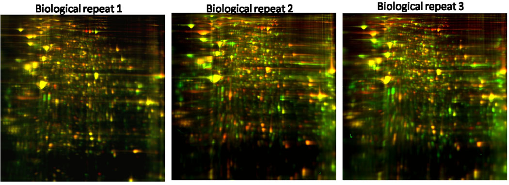
To test the hypothesis that H1 and HES2 cells are predisposed to differentiate into chamber-specific cell types with distinct preferences due to intrinsic differences in their proteomes, we next attempted to identify proteins that are differentially expressed in undifferentiated H1 and HES2 cells. ~106 undifferentiated HES2 and H1 cells were lysed and labeled with Cy3 and Cy5, respectively. As an internal standard, equal amounts of HES2 and H1 cell lysates were mixed and labeled with Cy2. Figures 2A and B show the separation of proteins present in undifferentiated HES2 and H1 cells, respectively. Superimposition of the two images enabled the visualization of the differences in protein expression: In Figure 2C, green and red protein spots corresponded to proteins that are over-expressed in HES2 and H1 cells, respectively. By contrast, yellow spots represented protein spots that were equally expressed in both hESC lines (c.f. Figure 2D). A total of 3 biological repeats were performed (Figure 3). Overall, a total of 2000 individual protein spots were separated and detected, 55 of which were significantly up-regulated in either HES2 or H1 (p<0.05; Table 1).
Figure 2.
2-D gel scanned for A) HES2 (i.e. Cy3) and B) H1 (i.e. Cy5). C) Overlay of the HES2 and H1 gels where Cy5 and Cy3 are red and green, respectively. Circled and labeled protein spots correspond to the ones isolated from the gel and identified by MALDI TOF/TOF. D). Schematic representation of the colored spots. Green and red spots represent proteins that are more highly expressed in HES2 and H1, respetively. Proteins that are equally expressed in both samples are yellow.
Figure 3.
Three biological Repeats of H1 vs HES2.
Table 1.
Summary of differential spots.
| Total Number of Proteins Identified |
Number of Up- regulated Proteins |
Number Up- regulated Proteins in HES2 |
Number of Up- regulated Proteins in H1 |
|---|---|---|---|
| >2000 | 55 | 15 | 40 |
Identification of Differentially Expressed Proteins
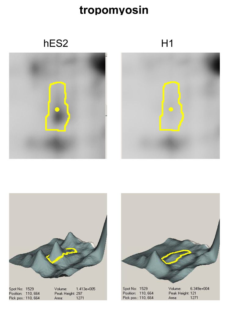
All 55 statistically significant protein spots identified were chosen for mass spectrometry. As examples, Figure 4 shows the DeCyder software analysis for one such differentially regulated protein spot - tropomyosin. Specifically, the topographical representation of the expression levels of each particular protein spot (lower panels) show that the levels of the protein in question but the neighboring proteins differed between the two samples. The corresponding original gel regions were also given (upper panels). The spots excised from the gel and in-gel digested with trypsin to obtain peptide fragments (Figure 2C), were identified by MALDI-TOF/TOF MS/MS analysis (Supplemental Table 2). Proteins found to be differentially expressed in H1 and HES2 included ones involved in cytoskeletal functions such as villin, MAPRE1, moesin, and actin. Most interestingly, tropomyosin and annexin II, two proteins known to have roles in cardiac differentiation and cardiomyocyte functions [28; 29] were up-regulated in undifferentiated HES2 cells that favored the derivation of ventricular-like CMs. These observations raise the intriguing possibility of expression of certain cardiac proteins in pluripotent hESCs may bias their cardiogenic differences.
Figure 4.
Gel images and DeCyder Software Analysis Output for tropomyosin of HES2 and H1.
Pathway Analysis
Using the Ingenuity Pathway Analysis software (www.ingenuity.com), we explored molecular functions and known pathways that are differentially expressed in pluripotent H1 and HES2 cells. Out of the 55 non-redundant proteins, 48 were successfully mapped to genes in the Ingenuity Pathways Knowledge Base (IPKB). There were 37 Network Eligible Genes which interact with other genes based on the IPKB and 34 Functions/Pathways genes that have at least one functional annotation associated with the genes in IPKB. Based on the analysis of these genes, networks that were had the highest differential expression between the H1 and HES2 lines (score >20) were: 1) Cell Death, Protein Synthesis, Gene Expression and 2) Cellular Assembly and Organization, Cell Death, Cell Morphology. From these networks, the significant molecular functions are: 1) Cellular Assembly and Organization (p = 1.43e-5), 2) RNA-Post-Transcriptional Modification (p = 9.92e-5), and 3) Carbohydrate Metabolism (p = 1.20e-4).
Discussion
Although self-renewable hESCs present a promising option for cell-based heart therapies, a number of important hurdles need to be overcome. As a first step, we functionally investigated the cardiogenic potentials of H1 and HES2 cells to become chamber-specific derivatives via electrical recordings, followed by probing the underlying proteomic differences to shed mechanistic insights into human cardiogenesis. The preferences of various murine (m) ESC lines to differentiate into the ventricular-, atrial- and pacemaker-like phenotypes have been documented [30; 31] but the same has not been systematically compared in any hESC lines. In either case, the basis for the different chamber-specific distribution is unknown [9; 10; 27]. Indeed, different ESC lines may require different methods for inducing cardiac differentiation. For instance, the methods of hanging drops and suspension have been developed for differentiating mouse and human ESCs, respectively. Specifically, H1 can be differentiated by either EB formation or END2 co-culture. By contrast, cardiac derivatives can be differentiated from HES2 only by co-culturing with END2. The different requirements may stem from the original method(s) of isolation, culturing conditions for maintaining pluripotency and/or other procedural differences. Pragmatically, however, the precise cause and nature of these phenotypic differences are perhaps not as important as the differences themselves per se; by identifying these differences, a better understanding of the basis of the resulting consequences can be obtained which in turn may subsequently lead to novel methods for directed differentiation.
Using 2-D DIGE, we have done the first quantitative studies to compare the proteomes of the H1 and HES2 cell lines. We have identified 46 unique proteins that are differentially expressed in H1 and HES2. Of particular interest are the cytoskeletal proteins and the fact that the Ingenuity Pathway Analysis suggests that H1 and HES2 cells are different in their functional pathways controlling cellular assembly and organization. Given the role that the cytoskeleton plays during differentiation [32], the initial expression levels of proteins involved in these pathways shed insights into the cardiogenic preferences of H1 and HES2. Of note, the present analysis is limited by the current information in Ingenuity Pathway Knowledge Database. 78% of the proteins identified were able to be used in the pathway analysis. The analysis results may need to be revised as the database expands.
Future experiments can involve genetic overexpression or suppression of the differentially expressed proteins identified in H1 and HES2 to obtain further insights into why HES2 cannot form EBs, why END2 co-culturing can uniquely induce cardiogenesis of HES2, and what underlie their different preferences to become chamber-specific cell types. Furthermore, it will be necessary to study the proteomes of hESC-derived CMs. Technically, however, the purification of chamber-specific derivatives for proteomic experiments remain a major challenge. During the course of this study, Huber et al reported a genetic way to identify hESC-derived ventricular derivatives by expressing GFP under the transcriptional control of the myosin light chain (MLC)-2v promoter [33]; while this promoter employed is indeed ventricular-restricted in adult, it remains unclear whether such is also the case in early CMs such as those derived from ESCs (as reviewed by Moorman et al) [34]. Successful selection methods for hESC-derived atrial and pacemaker CMs have not been reported to date. Additionally, the typically very low yield of cardiac differentiation of hESCs also makes the harvesting of enough purified proteins from hESC-CMs even more difficult. Recent protocols to substantially improve the yield of CMs have been reported [35–37] but the heterogeneity of chamber-specific CMs have not been studied.
Collectively, the present study lays an important groundwork for further understanding the basic biology of hESC, and the poorly defined human cardiogenesis. Our results suggest that the preference to become chamber-specific heart cells is predetermined in H1 and HES2, and that the same is likely to apply to other existing hESC and tissue-specific lineages.
Supplementary Material
Acknowledgements
This work was supported by the NIH (R01 HL72857 to R.A.L. and F32 HL078330 to J.C.M.), California Institute for Regenerative Medicine (to J.D.F. and R.A.L.), Hong Kong Research Grant Council (7459/04M to Drs Tse, and Li).
Footnotes
Publisher's Disclaimer: This is a PDF file of an unedited manuscript that has been accepted for publication. As a service to our customers we are providing this early version of the manuscript. The manuscript will undergo copyediting, typesetting, and review of the resulting proof before it is published in its final citable form. Please note that during the production process errors may be discovered which could affect the content, and all legal disclaimers that apply to the journal pertain.
References
- 1.Xue T, Cho HC, Akar FG, Tsang SY, Jones SP, Marban E, Tomaselli GF, Li RA. Functional integration of electrically active cardiac derivatives from genetically engineered human embryonic stem cells with quiescent recipient ventricular cardiomyocytes: insights into the development of cell-based pacemakers. Circulation. 2005;111:11–20. doi: 10.1161/01.CIR.0000151313.18547.A2. [DOI] [PubMed] [Google Scholar]
- 2.Xue T, Siu CW, Lieu DK, Lau CP, Tse HF, Li RA. Mechanistic role of I(f) revealed by induction of ventricular automaticity by somatic gene transfer of gating-engineered pacemaker (HCN) channels. Circulation. 2007;115:1839–1850. doi: 10.1161/CIRCULATIONAHA.106.659391. [DOI] [PMC free article] [PubMed] [Google Scholar]
- 3.Soonpaa MH, Field LJ. Survey of studies examining mammalian cardiomyocyte DNA synthesis. Circ Res. 1998;83:15–26. doi: 10.1161/01.res.83.1.15. [DOI] [PubMed] [Google Scholar]
- 4.Li RA, Moore JC, Tarasova YS, Boheler KR. Human embryonic stem cell-derived cardiomyocytes: therapeutic potentials and limitations. J Stem Cell. 2006;1:109–124. [Google Scholar]
- 5.Moore JC, van Laake LW, Braam SR, Xue T, Tsang SY, Ward D, Passier R, Tertoolen LL, Li RA, Mummery CL. Human embryonic stem cells: genetic manipulation on the way to cardiac cell therapies. Reprod Toxicol. 2005;20:377–391. doi: 10.1016/j.reprotox.2005.04.012. [DOI] [PubMed] [Google Scholar]
- 6.Tse HF, Xue T, Lau CP, Siu CW, Wang K, Zhang QY, Tomaselli GF, Akar FG, Li RA. Bioartificial sinus node constructed via in vivo gene transfer of an engineered pacemaker HCN Channel reduces the dependence on electronic pacemaker in a sick-sinus syndrome model. Circulation. 2006;114:1000–1011. doi: 10.1161/CIRCULATIONAHA.106.615385. [DOI] [PubMed] [Google Scholar]
- 7.Siu CW, Lieu DK, Li RA. HCN-encoded pacemaker channels: from physiology and biophysics to bioengineering. J Membr Biol. 2006;214:115–122. doi: 10.1007/s00232-006-0881-9. [DOI] [PubMed] [Google Scholar]
- 8.Kehat I, Kenyagin-Karsenti D, Snir M, Segev H, Amit M, Gepstein A, Livne E, Binah O, Itskovitz-Eldor J, Gepstein L. Human embryonic stem cells can differentiate into myocytes with structural and functional properties of cardiomyocytes. J.Clin.Invest. 2001;108:407–414. doi: 10.1172/JCI12131. [DOI] [PMC free article] [PubMed] [Google Scholar]
- 9.Mummery C, Ward-Van Oostwaard D, Doevendans P, Spijker R, Van Den BS, Hassink R, Van Der HM, Opthof T, Pera M, De La Riviere AB, Passier R, Tertoolen L. Differentiation of human embryonic stem cells to cardiomyocytes: role of coculture with visceral endoderm-like cells. Circulation. 2003;107:2733–2740. doi: 10.1161/01.CIR.0000068356.38592.68. [DOI] [PubMed] [Google Scholar]
- 10.Kehat I, Gepstein A, Spira A, Itskovitz-Eldor J, Gepstein L. High-resolution electrophysiological assessment of human embryonic stem cell-derived cardiomyocytes: a novel in vitro model for the study of conduction. Circ.Res. 2002;91:659–661. doi: 10.1161/01.res.0000039084.30342.9b. [DOI] [PubMed] [Google Scholar]
- 11.Unwin RD, Gaskell SJ, Evans CA, Whetton AD. The potential for proteomic definition of stem cell populations. Exp Hematol. 2003;31:1147–1159. doi: 10.1016/j.exphem.2003.08.012. [DOI] [PubMed] [Google Scholar]
- 12.Unlu M, Morgan ME, Minden JS. Difference gel electrophoresis: a single gel method for detecting changes in protein extracts. Electrophoresis. 1997;18:2071–2077. doi: 10.1002/elps.1150181133. [DOI] [PubMed] [Google Scholar]
- 13.Marouga R, David S, Hawkins E. The development of the DIGE system: 2D fluorescence difference gel analysis technology. Anal Bioanal Chem. 2005;382:669–678. doi: 10.1007/s00216-005-3126-3. [DOI] [PubMed] [Google Scholar]
- 14.Lee HK, Lee BH, Park SA, Kim CW. The proteomic analysis of an adipocyte differentiated from human mesenchymal stem cells using two-dimensional gel electrophoresis. Proteomics. 2006;6:1223–1229. doi: 10.1002/pmic.200500385. [DOI] [PubMed] [Google Scholar]
- 15.Oh JE, Karlmark Raja K, Shin JH, Pollak A, Hengstschlager M, Lubec G. Cytoskeleton changes following differentiation of N1E-115 neuroblastoma cell line. Amino Acids. 2006;31:289–298. doi: 10.1007/s00726-005-0256-z. [DOI] [PubMed] [Google Scholar]
- 16.Wang D, Gao L. Proteomic analysis of neural differentiation of mouse embryonic stem cells. Proteomics. 2005;5:4414–4426. doi: 10.1002/pmic.200401304. [DOI] [PubMed] [Google Scholar]
- 17.Yin X, Mayr M, Xiao Q, Wang W, Xu Q. Proteomic analysis reveals higher demand for antioxidant protection in embryonic stem cell-derived smooth muscle cells. Proteomics. 2006;6:6437–6446. doi: 10.1002/pmic.200600351. [DOI] [PubMed] [Google Scholar]
- 18.Van Hoof D, Passier R, Ward-Van Oostwaard D, Pinkse MW, Heck AJ, Mummery CL, Krijgsveld J. A quest for human and mouse embryonic stem cell-specific proteins. Mol Cell Proteomics. 2006;5:1261–1273. doi: 10.1074/mcp.M500405-MCP200. [DOI] [PubMed] [Google Scholar]
- 19.Baharvand H, Hajheidari M, Ashtiani SK, Salekdeh GH. Proteomic signature of human embryonic stem cells. Proteomics. 2006;6:3544–3549. doi: 10.1002/pmic.200500844. [DOI] [PubMed] [Google Scholar]
- 20.Thomson JA, Itskovitz-Eldor J, Shapiro SS, Waknitz MA, Swiergiel JJ, Marshall VS, Jones JM. Embryonic stem cell lines derived from human blastocysts. Science. 1998;282:1145–1147. doi: 10.1126/science.282.5391.1145. [DOI] [PubMed] [Google Scholar]
- 21.Wang K, Xue T, Tsang SY, Van Huizen R, Wong CW, Lai KW, Ye Z, Cheng L, Au KW, Zhang J, Li GR, Lau CP, Tse HF, Li RA. Electrophysiological properties of pluripotent human and mouse embryonic stem cells. Stem Cells. 2005;23:1526–1534. doi: 10.1634/stemcells.2004-0299. [DOI] [PubMed] [Google Scholar]
- 22.Reubinoff BE, Pera MF, Fong CY, Trounson A, Bongso A. Embryonic stem cell lines from human blastocysts: somatic differentiation in vitro. Nat.Biotechnol. 2000;18:399–404. doi: 10.1038/74447. [DOI] [PubMed] [Google Scholar]
- 23.Itskovitz-Eldor J, Schuldiner M, Karsenti D, Eden A, Yanuka O, Amit M, Soreq H, Benvenisty N. Differentiation of human embryonic stem cells into embryoid bodies compromising the three embryonic germ layers. Mol Med. 2000;6:88–95. [PMC free article] [PubMed] [Google Scholar]
- 24.Kehat I, Khimovich L, Caspi O, Gepstein A, Shofti R, Arbel G, Huber I, Satin J, Itskovitz-Eldor J, Gepstein L. Electromechanical integration of cardiomyocytes derived from human embryonic stem cells. Nat.Biotechnol. 2004;22:1282–1289. doi: 10.1038/nbt1014. [DOI] [PubMed] [Google Scholar]
- 25.Passier R, Ward-Van Oostwaard D, Snapper J, Kloots J, Hassink R, Kuijk E, Roelen B, Brutel de la Riviere A, Mummery C. Inreased Cardiomyocyte Differentiation from Human Embryonic Stem Cells in Serum-Free Cultures. Stem Cells. 2005 doi: 10.1634/stemcells.2004-0184. [DOI] [PubMed] [Google Scholar]
- 26.Xue T, Chan CWY, Henrikson C, Sang D, Marbán E, Li RA. Lentivirus-mediated genetic manipulations of human embryonic stem cells and their cardiac derivatives. Circulation. 2003;108:VI133–VI133. [Google Scholar]
- 27.He JQ, Ma Y, Lee Y, Thomson JA, Kamp TJ. Human embryonic stem cells develop into multiple types of cardiac myocytes: action potential characterization. Circ.Res. 2003;93:32–39. doi: 10.1161/01.RES.0000080317.92718.99. [DOI] [PubMed] [Google Scholar]
- 28.Zajdel RW, McLean MD, Denz CR, Dube S, Thurston HL, Poiesz BJ, Dube DK. Differential expression of tropomyosin during segmental heart development in Mexican axolotl. J Cell Biochem. 2006;99:952–965. doi: 10.1002/jcb.20954. [DOI] [PubMed] [Google Scholar]
- 29.Krishnan S, Deora AB, Annes JP, Osoria J, Rifkin DB, Hajjar KA. Annexin II-mediated plasmin generation activates TGF-beta3 during epithelial-mesenchymal transformation in the developing avian heart. Dev Biol. 2004;265:140–154. doi: 10.1016/j.ydbio.2003.08.026. [DOI] [PubMed] [Google Scholar]
- 30.Hescheler J, Fleischmann BK, Lentini S, Maltsev VA, Rohwedel J, Wobus AM, Addicks K. Embryonic stem cells: a model to study structural and functional properties in cardiomyogenesis. Cardiovasc Res. 1997;36:149–162. doi: 10.1016/s0008-6363(97)00193-4. [DOI] [PubMed] [Google Scholar]
- 31.Maltsev VA, Rohwedel J, Hescheler J, Wobus AM. Embryonic stem cells differentiate in vitro into cardiomyocytes representing sinusnodal, atrial and ventricular cell types. Mech Dev. 1993;44:41–50. doi: 10.1016/0925-4773(93)90015-p. [DOI] [PubMed] [Google Scholar]
- 32.Paulin D. Cytoskeleton organization in differentiating mouse teratocarcinoma cells. Biochimie. 1981;63:347–363. doi: 10.1016/s0300-9084(81)80121-6. [DOI] [PubMed] [Google Scholar]
- 33.Huber I, Itzhaki I, Caspi O, Arbel G, Tzukerman M, Gepstein A, Habib M, Yankelson L, Kehat I, Gepstein L. Identification and selection of cardiomyocytes during human embryonic stem cell differentiation. Faseb J. 2007;21:2551–2563. doi: 10.1096/fj.05-5711com. [DOI] [PubMed] [Google Scholar]
- 34.Fijnvandraat AC, Lekanne Deprez RH, Moorman AF. Development of heart muscle-cell diversity: a help or a hindrance for phenotyping embryonic stem cell-derived cardiomyocytes. Cardiovasc Res. 2003;58:303–312. doi: 10.1016/s0008-6363(03)00246-3. [DOI] [PubMed] [Google Scholar]
- 35.Kattman SJ, Huber TL, Keller GM. Multipotent flk-1+ cardiovascular progenitor cells give rise to the cardiomyocyte, endothelial, and vascular smooth muscle lineages. Dev Cell. 2006;11:723–732. doi: 10.1016/j.devcel.2006.10.002. [DOI] [PubMed] [Google Scholar]
- 36.Kouskoff V, Lacaud G, Schwantz S, Fehling HJ, Keller G. Sequential development of hematopoietic and cardiac mesoderm during embryonic stem cell differentiation. Proc Natl Acad Sci U S A. 2005;102:13170–13175. doi: 10.1073/pnas.0501672102. [DOI] [PMC free article] [PubMed] [Google Scholar]
- 37.Laflamme MA, Chen KY, Naumova AV, Muskheli V, Fugate JA, Dupras SK, Reinecke H, Xu C, Hassanipour M, Police S, O'Sullivan C, Collins L, Chen Y, Minami E, Gill EA, Ueno S, Yuan C, Gold J, Murry CE. Cardiomyocytes derived from human embryonic stem cells in pro-survival factors enhance function of infarcted rat hearts. Nat Biotechnol. 2007;25:1015–1024. doi: 10.1038/nbt1327. [DOI] [PubMed] [Google Scholar]
Associated Data
This section collects any data citations, data availability statements, or supplementary materials included in this article.


