Journal of Clinical Microbiology logoLink to Journal of Clinical Microbiology
letter
. 2009 Apr;47(4):1278–1280. doi: 10.1128/JCM.02130-08

5′ Noncoding Region Alone Does Not Unequivocally Determine Genetic Type of Human Rhinovirus Strains

Carita Savolainen-Kopra 1,*, Soile Blomqvist 1, Teemu Smura 1, Merja Roivainen 1, Tapani Hovi 1
PMCID: PMC2668324  PMID: 19339481

During the last couple of years there has been a delightful increase in interest in genetic typing of human rhinoviruses. This is to a large extent due to the discovery of a proposed novel clade, human rhinovirus C (HRV-C). As a consequence, new methods have been reported aiming at unequivocal distinction of traditional HRV prototype strains as well as the newly found uncultivable HRV-C strains. We read with interest the article by Kiang and coworkers (2) describing reverse transcription-PCR-sequencing applications for genetic typing of human rhinoviruses targeting the 5′noncoding region (5′ NCR). A similar approach with largely similar results was published earlier (6). Relatively conserved areas within this region enable broad-spectrum primer design for sensitive methods. However, there are several issues to consider when using the 5′ NCR for genetic typing of HRV.

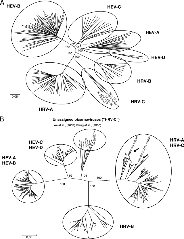
Current taxonomy and classification of picornaviruses are based on capsid region coding sequences. On the basis of this region, a group of previously characterized novel HRV strains form one distinct clade (5, 7) (Fig. 1A), a fact that has also been the basis of the proposal of the Picornavirus Study Group to form a new species, HRV-C, within the Enterovirus genus (4). However, in the article by Kiang et al., on the basis of the partial 5′ NCR sequences, the designated HRV-C strains clustered within the HRV-A clade (2). In contrast, the strains labeled HRV-C in this article formed a clade of their own. As a consequence, because 5′ NCR sequences do not segregate the designated HRV-C from HRV-A (Fig. 1B), they should not be used for typing of new strains. Nevertheless, this region is quite suitable for selected topics of molecular epidemiology, such as analysis of short-term transmission routes (1, 8) or tentative prediction of genetic type as in human enteroviruses (HEV) (9). The sequences nominated as HRV-C by Kiang et al. (2) and by Lee et al. (6) form a new clade in the 5′ NCR. The exact taxonomic position of this clade should be determined according to the clustering of these strains in the capsid region. Clearly, it is divergent from all known HRV and HEV clades in the 5′ NCR, but the decision on whether the strains represent HRV-C or some other picornavirus group cannot be made on the basis of the 5′ NCR alone.

FIG. 1.

FIG. 1.

Phylogenetic trees in the VP4/VP2 capsid coding region (A) and in the 5′ NCR (B) of different species of the enterovirus genus showing different clustering of the species in the two regions. Trees were constructed with MEGA4 using the neighbor-joining method with Tamura-Nei model and 1,000 bootstrap replicates.

The area close to the beginning of the open reading frame in the 5′ NCR is known to be a recombination hot spot in HEV. Although frequent recombination has not yet been shown for HRV, the analysis of the complete genome sequence data of all HRV prototype strains has not yet been published. Furthermore, the number of completely sequenced genomes of circulating HRV strains has remained low and is too low to conclude that the evolution in the 5′ NCR is always congruent with that of the capsid. Therefore, we would see phylogenetic analysis of the 5′ NCR of HRV as a welcome addition to HRV research, but not a surrogate of capsid coding sequence-based typing.

REFERENCES

  • 1.Jartti, T., W. M. Lee, T. Pappas, M. Evans, R. F. Lemanske, Jr., and J. E. Gern. 2008. Serial viral infections in infants with recurrent respiratory illnesses. Eur. Respir. J. 32:314-320. [DOI] [PMC free article] [PubMed] [Google Scholar]
  • 2.Kiang, D., I. Kalra, S. Yagi, J. K. Louie, H. Boushey, J. Boothby, and D. P. Schnurr. 2008. Assay for 5′ noncoding region analysis of all human rhinovirus prototype strains. J. Clin. Microbiol. 46:3736-3745. [DOI] [PMC free article] [PubMed] [Google Scholar]
  • 3.Reference deleted.
  • 4.Knowles, N. J. 2008. New species in the genus Enterovirus (Picornaviridae). http://talk.ictvonline.org/media/p/687.aspx.
  • 5.Lau, S. K., C. C. Yip, H. W. Tsoi, R. A. Lee, L. Y. So, Y. L. Lau, K. H. Chan, P. C. Woo, and K. Y. Yuen. 2007. Clinical features and complete genome characterization of a distinct human rhinovirus (HRV) genetic cluster, probably representing a previously undetected HRV species, HRV-C, associated with acute respiratory illness in children. J. Clin. Microbiol. 45:3655-3664. [DOI] [PMC free article] [PubMed] [Google Scholar]
  • 6.Lee, W. M., C. Kiesner, T. Pappas, I. Lee, K. Grindle, T. Jartti, B. Jakiela, R. F. Lemanske, P. A. Shult, and J. E. Gern. 2007. A diverse group of previously unrecognized human rhinoviruses are common causes of respiratory illnesses in infants. PLoS ONE 2:e966. [DOI] [PMC free article] [PubMed] [Google Scholar]
  • 7.McErlean, P., L. A. Shackelton, E. Andrews, D. R. Webster, S. B. Lambert, M. D. Nissen, T. P. Sloots, and I. M. Mackay. 2008. Distinguishing molecular features and clinical characteristics of a putative new rhinovirus species, human rhinovirus C (HRV C). PLoS ONE 3:e1847. [DOI] [PMC free article] [PubMed] [Google Scholar]
  • 8.Peltola, V., M. Waris, R. Osterback, P. Susi, O. Ruuskanen, and T. Hyypia. 2008. Rhinovirus transmission within families with children: incidence of symptomatic and asymptomatic infections. J. Infect. Dis. 197:382-389. [DOI] [PubMed] [Google Scholar]
  • 9.Thoelen, I., E. Moes, P. Lemey, S. Mostmans, E. Wollants, A. M. Lindberg, A. M. Vandamme, and M. Van Ranst. 2004. Analysis of the serotype and genotype correlation of VP1 and the 5′ noncoding region in an epidemiological survey of the human enterovirus B species. J. Clin. Microbiol. 42:963-971. [DOI] [PMC free article] [PubMed] [Google Scholar]
J Clin Microbiol. 2009 Apr;47(4):1278–1280.

Authors' Reply

David Kiang 1,2,3,4,*, Ishmeet Kalra 1,2,3,4, Shigeo Yagi 1,2,3,4, Janice K Louie 1,2,3,4, Homer Boushey 1,2,3,4, John Boothby 1,2,3,4, David P Schnurr 1,2,3,4

We appreciate the comments of Savolainen-Kopra et al. concerning our recent publication highlighting a novel assay for the analysis of human rhinoviruses (HRVs) based on the 5′ NCR (7). We agree that the capsid region coding sequences play a key role in the current taxonomy and classification of picornaviruses, including HRVs. The genotyping methodology based on the VP4/VP2 region reported by Savolainen et al. (16) remains useful in our laboratory as a means of typing HRVs. We developed the assay for the 5′ NCR and currently use this assay not as a surrogate of capsid coding sequence-based typing but as a supplemental tool to address the sensitivity issues regarding capsid region-based genotyping. Our motivation to develop a 5′ NCR assay was largely due to our interest in having a protocol to allow broad and sensitive detection and typing of HRVs for molecular epidemiology and surveillance. Detection and typing of HRVs can shed light on the contribution of specific serotypes of HRVs to more severe clinical outcomes associated with HRV infection, including asthma exacerbation (6, 14, 15) and lower respiratory infections (3, 5), some of which have had fatal outcomes (4, 8, 12, 17). The discovery of novel strains that coincide with the novel HRV A2/“C” strains (1, 10, 13) as well as a potential novel genogroup of HRV (7, 11), which we will refer to in this article as HRV “D” to distinguish it from the currently proposed HRV “C”, was a fortuitous and exciting finding from the evaluation of our novel assay on field isolates.

In our laboratory, genotyping results are typically, when possible, supported by data from multiple regions of the HRV genome, which can include 5′ NCR, VP4/VP2, and possibly other regions, such as VP1. Therefore, unequivocal determination of the genetic type of HRV strains based solely on the 5′ NCR of the HRV genome was not the goal of developing this assay. In fact, in the absence of whole-genome sequence analysis data of all prototype strains, it would not be prudent to conclude that any one assay can unequivocally determine the genetic type of HRV. At this stage, we cannot exclude the importance of other nonstructural and noncoding regions in the typing of existing and emerging strains of HRVs, especially if they are detected by an assay that has greater sensitivity (7, 11). The analysis of 34 fully sequenced HRVs by Kistler et al. (9) has demonstrated the importance of nonstructural genes 3C and 3D, in addition to capsid regions VP1, VP2, and VP3 as regions that contain islands of diverse sequences. The nonstructural genes, 2C and 3CD, are also indicated as species demarcation criteria for rhinoviruses in the Eighth Report of the International Committee on Taxonomy of Viruses (2). Recombination drives diversification in many genera of picornaviruses, but this has not been found for rhinoviruses (9). Analysis by Kistler et al. (9) has also revealed consistent phylogenies across the genome and only limited recombination in HRVs. Within the 5′ NCR there exist both highly conserved sequences as well as variable sequences with 45% (11) to 63% divergence (7), making this sensitive assay an ideal candidate for typing strains that have eluded capsid gene amplification. In contrast, the prediction of the genetic type of HEV cannot be performed with the 5′ NCR due to the frequent rate of recombination within the HEV genome; therefore, we would not expect the 5′ NCR to be able to segregate HEV strains. Although, the 5′ NCR assay did not segregate the newly recognized HRV-“C” strains into a clade of its own, this does not exclude its importance and utility in typing of novel strains of HRV, including the putative HRV “D” strain. There may well be instances where novel strains cannot be well segregated by the capsid coding regions, and these will require the use of noncoding regions and nonstructural genes for resolution.

REFERENCES

  • 1.Briese, T., N. Renwick, M. Venter, R. G. Jarman, D. Ghosh, S. Kondgen, S. K. Shrestha, A. M. Hoegh, I. Casas, E. V. Adjogoua, C. Akoua-Koffi, K. S. Myint, D. T. Williams, G. Chidlow, R. van den Berg, C. Calvo, O. Koch, G. Palacios, V. Kapoor, J. Villari, S. R. Dominguez, K. V. Holmes, G. Harnett, D. Smith, J. S. Mackenzie, H. Ellerbrok, B. Schweiger, K. Schonning, M. S. Chadha, F. H. Leendertz, A. C. Mishra, R. V. Gibbons, E. C. Holmes, and W. I. Lipkin. 2008. Global distribution of novel rhinovirus genotype. Emerg. Infect. Dis. 14:944-947. [DOI] [PMC free article] [PubMed] [Google Scholar]
  • 2.Fauquet, C. M., M. A. Mayo, J. Maniloff, U. Desselberger, and L. A. Ball (ed.). 2005. Virus taxonomy. Eighth report of the International Committee on Taxonomy of Viruses. Elsevier Academic Press, San Diego, CA
  • 3.Gutman, J. A., A. J. Peck, J. Kuypers, and M. Boeckh. 2007. Rhinovirus as a cause of fatal lower respiratory tract infection in adult stem cell transplantation patients: a report of two cases. Bone Marrow Transplant. 40:809-811. [DOI] [PMC free article] [PubMed] [Google Scholar]
  • 4.Hicks, L. A., C. W. Shepard, P. H. Britz, D. D. Erdman, M. Fischer, B. L. Flannery, A. J. Peck, X. Lu, W. L. Thacker, R. F. Benson, M. L. Tondella, M. E. Moll, C. G. Whitney, L. J. Anderson, and D. R. Feikin. 2006. Two outbreaks of severe respiratory disease in nursing homes associated with rhinovirus. J. Am. Geriatr. Soc. 54:284-289. [DOI] [PubMed] [Google Scholar]
  • 5.Imakita, M., K. Shiraki, C. Yutani, and H. Ishibashi-Ueda. 2000. Pneumonia caused by rhinovirus. Clin. Infect. Dis. 30:611-612. [DOI] [PubMed] [Google Scholar]
  • 6.Johnston, N. W., S. L. Johnston, J. M. Duncan, J. M. Greene, T. Kebadze, P. K. Keith, M. Roy, S. Waserman, and M. R. Sears. 2005. The September epidemic of asthma exacerbations in children: a search for etiology. J. Allergy Clin. Immunol. 115:132-138. [DOI] [PMC free article] [PubMed] [Google Scholar]
  • 7.Kiang, D., I. Kalra, S. Yagi, J. K. Louie, H. Boushey, J. Boothby, and D. P. Schnurr. 2008. Assay for 5′ noncoding region analysis of all human rhinovirus prototype strains. J. Clin. Microbiol. 46:3736-3745. [DOI] [PMC free article] [PubMed] [Google Scholar]
  • 8.Kiang, D., S. Yagi, K. A. Kantardjieff, E. J. Kim, J. K. Louie, and D. P. Schnurr. 2007. Molecular characterization of a variant rhinovirus from an outbreak associated with uncommonly high mortality. J. Clin. Virol. 38:227-237. [DOI] [PubMed] [Google Scholar]
  • 9.Kistler, A. L., D. R. Webster, S. Rouskin, V. Magrini, J. J. Credle, D. P. Schnurr, H. A. Boushey, E. R. Mardis, H. Li, and J. L. DeRisi. 2007. Genome-wide diversity and selective pressure in the human rhinovirus. Virol. J. 4:40. [DOI] [PMC free article] [PubMed] [Google Scholar]
  • 10.Lau, S. K., C. C. Yip, H. W. Tsoi, R. A. Lee, L. Y. So, Y. L. Lau, K. H. Chan, P. C. Woo, and K. Y. Yuen. 2007. Clinical features and complete genome characterization of a distinct human rhinovirus (HRV) genetic cluster, probably representing a previously undetected HRV species, HRV-C, associated with acute respiratory illness in children. J. Clin. Microbiol. 45:3655-3664. [DOI] [PMC free article] [PubMed] [Google Scholar]
  • 11.Lee, W. M., C. Kiesner, T. Pappas, I. Lee, K. Grindle, T. Jartti, B. Jakiela, R. F. Lemanske, P. A. Shult, and J. E. Gern. 2007. A diverse group of previously unrecognized human rhinoviruses are common causes of respiratory illnesses in infants. PLoS ONE 2:e966. [DOI] [PMC free article] [PubMed] [Google Scholar]
  • 12.Louie, J. K., S. Yagi, F. A. Nelson, D. Kiang, C. A. Glaser, J. Rosenberg, C. K. Cahill, and D. P. Schnurr. 2005. Rhinovirus outbreak in a long term care facility for elderly persons associated with unusually high mortality. Clin. Infect. Dis. 41:262-265. [DOI] [PMC free article] [PubMed] [Google Scholar]
  • 13.McErlean, P., L. A. Shackelton, E. Andrews, D. R. Webster, S. B. Lambert, M. D. Nissen, T. P. Sloots, and I. M. Mackay. 2008. Distinguishing molecular features and clinical characteristics of a putative new rhinovirus species, human rhinovirus C (HRV C). PLoS ONE 3:e1847. [DOI] [PMC free article] [PubMed] [Google Scholar]
  • 14.Miller, E. K., K. M. Edwards, G. A. Weinberg, M. K. Iwane, M. R. Griffin, C. B. Hall, Y. Zhu, P. G. Szilagyi, L. L. Morin, L. H. Heil, X. Lu, J. V. Williams, and New Vaccine Surveillance Network. 2009. A novel group of rhinoviruses is associated with asthma hospitalizations. J. Allergy Clin. Immunol. 123:98-104.e1. [DOI] [PMC free article] [PubMed] [Google Scholar]
  • 15.Nicholson, K. G., J. Kent, and D. C. Ireland. 1993. Respiratory viruses and exacerbations of asthma in adults. BMJ 307:982-986. [DOI] [PMC free article] [PubMed] [Google Scholar]
  • 16.Savolainen, C., S. Blomqvist, M. N. Mulders, and T. Hovi. 2002. Genetic clustering of all 102 human rhinovirus prototype strains: serotype 87 is close to human enterovirus 70. J. Gen. Virol. 83:333-340. [DOI] [PubMed] [Google Scholar]
  • 17.Wald, T. G., P. Shult, P. Krause, B. A. Miller, P. Drinka, and S. Gravenstein. 1995. A rhinovirus outbreak among residents of a long-term care facility. Ann. Intern. Med. 123:588-593. [DOI] [PubMed] [Google Scholar]

Articles from Journal of Clinical Microbiology are provided here courtesy of American Society for Microbiology (ASM)

RESOURCES