Figure 1.

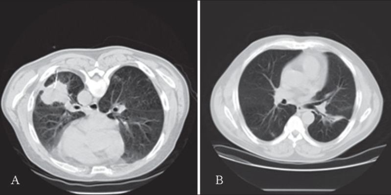
(A) Prone CT chest, placement of 22G Westcott biopsy needle in the left lower lobe mass. (B) Follow up CT chest two months after biopsy. Notice marked improvement with residual inflammation and focal bronchiectasis in the left lower lobe.

(A) Prone CT chest, placement of 22G Westcott biopsy needle in the left lower lobe mass. (B) Follow up CT chest two months after biopsy. Notice marked improvement with residual inflammation and focal bronchiectasis in the left lower lobe.