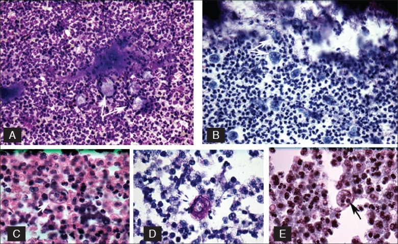
Figure 2.

Top row left (A) E. gingivalis seen as pale, irregular, macrophage-like structures (arrow), Diff-Quik, (original) ×160. Right (B) E. gingivalis (arrow) arranged along the actinomyces. Pale food vacuoles are visible, Pap Stain, (original) ×160. Second row left (C) E. gingivalis with thick border, (arrow), H/E. (original) ×400, middle (D) E. gingivalis, periodic acid schiff stain (arrow). (Original) ×630, right (E) E. gingivalis notice the distinct food vacuoles and karyosome (arrow). Wheatley stain. (Original) ×160.
