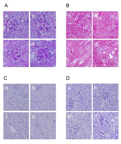
Figure 2.
Effect of deoxycorticosterone acetate (DOCA)-salt treatment on renal morphology in wild type (WT) and TRPV1-null mutant (TRPV1-/-) mice. A and B: Periodic acid-Schiff (A) or Masson’s trichrome (B) stained kidney sections showing glomerular or cortical tubulointerstitial morphological changes in control WT and TRPV1-/- mice (a and b), and WT and TRPV1-/- mice (c and d) treated for 4 weeks with DOCA-salt. Magnification ×400 (A) or ×200 (B). C and D: Immunohistochemical stained sections from the kidney cortex demonstrating proliferating cell nuclear antigen (PCNA)-positive cells (in brown) (C) or F4/80-positive cells (monocytes/macrophages in red) (D) in the renal cortex of control WT and TRPV1-/- mice (a and b), and WT and TRPV1-/- mice (c and d) treated for 4 weeks with DOCA-salt. Magnification ×400.

