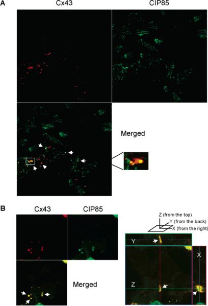
Figure 7.
Subcellular colocalization of Cx43 and CIP85. Panel A, laser-scanning confocal microscopy was performed to determine the subcellular localization of Cx43 and endogenous CIP85 in Hela cells (clone C4) utilizing TRITC-conjugated or FITC-conjugated secondary antibodies. The white arrows indicate regions of colocalization of CIP85 and Cx43. The boxed area in the merged image was magnified to show the regional colocalization of Cx43 and CIP85 in a gap junction plaque. Panel B, HEK293 cells were transiently transfected with Cx43 and Flag-CIP85. Laser-scanning confocal microscopy was performed to determine the subcellular localization of Cx43 and CIP85 with TRITC-conjugated or FITC-conjugated secondary antibodies. The white arrows indicate regions of colocalization of CIP85 and Cx43. Right panel section: an orthoimage showing the colocalization of CIP85 and Cx43 in a gap junction plaque in the X-, Y-, and Z-axes.

