Abstract
Objectives
This study was designed to evaluate the normal development of the nasal septum in Koreans using sagittal MRI for the valuable clinical information on septal procedures.
Methods
Two hundred eighty patients who had their whole nasal septum visualized in the midline sagittal view were selected among the 3,904 patients with brain MRI from January, 2004 to December, 2006 at Dankook University Hospital. The patients who had a history of nasal septal surgery or nasal trauma were excluded. Following parameters are calculated and analyzed: lengths of bony and cartilage dorsum and septal cartilage-nasal bone overlap, total septal area, septal cartilage area and, the proportion of the cartilage area to septal area and the maximal harvestable cartilage for grafting were calculated using the PAC™ program.
Results
All the parameters were increased until adolescence. Thereafter, bony dorsal length, cartilage dorsal length, total dorsal length, total septal area and maximal harvestable cartilage for grafting have not changed significantly with age, while SC-NB overlap length, septal cartilage area, and proportion of the cartilage area to the total septal area were significantly decreased with age. The SC-NB overlap length was positively correlated with the septal cartilage area and the proportion of the cartilage area to the total septal area.
Conclusion
The small septal cartilage area and its proportion to the total septal area were significantly correlated with a short overlap length of the septal cartilage under the nasal bone. Septal procedures should be carefully performed in the elderly due to the risk of incurring saddle nose.
Keywords: Septum, Cartilage, Magnetic resonance imaging, Development
INTRODUCTION
The nasal septum is placed in the middle of the nasal cavity and it divides the nasal cavity and supports the external nose. It is an important structure for maintaining the facial skeletal structure, and it consists of the septal cartilage anteriorly, the perpendicular plate of the ethmoid bone and the vomer bone posteriorly. This structure comes in contact with other structures such as nasal bone, frontal bone, cribriform plate, maxillary bone, palatine bone and sphenoidal bone (1). The septal cartilage starts to develop at the fetal stage and it continues to grow after birth. It reaches the size and structure of an adult by puberty, with some difference in the time frame between individuals (2). It is known that the cartilage becomes denatured and undergoes degenerative changes, including decreasing cartilage cells, and it becomes calcified with age (3).
Because it is important to thoroughly understand the developmental process of the nasal septum to safely carry out nasal septal procedures, many studies have been performed about the embryology and development of the nasal septum. However, most of these were studies based on autopsy or simple X-ray findings (4-7). Studies that are based on autopsies allow researchers to make realistic, concrete analysis. However, there are difficulties in generalizing the ideas from these autopsy studies as these studies did not have so many subjects. The studies conducted on the X-ray finding have limitations for providing specific information on the three-dimensional nasal septum structure. On the other hand, if the sagittal plane of magnetic resonance imaging (MRI) is used, each structure could be better identified. Therefore, a wide range of studies on the structure and development of nasal septum become possible with using MRI. However, there have been no studies using the sagittal plane of MRI for determining the structure and development of the nasal septum.
We used the sagittal planes of MRI to analyze the changes of the structures of the nasal bone and nasal septum, according to age and gender, so as to help physicians conduct safe nasal septal procedures.
MATERIALS AND METHODS
Two hundred eighty patients, who had their entire septum well visualized on the midline sagittal view, were selected among the 3,904 patients who underwent brain MRI from January, 2004 to December, 2006. Those patients who had a history of septal surgery or nasal trauma were excluded.
Two subjects were analyzed from the patients from 1 year to 70 year of age, and this was done the same with each gender. A total of 280 subjects were retrospectively analyzed by examining their saggital plane MRI.
Signa Excite (GE Medical System, Twin speed: 1.5 Tesla, Huntley, IL, USA) and Signa Excite (GE Medical System, High definition: 1.5 Tesla, Huntley, IL, USA) magnetic resonance imaging machines were used. The T1 weighted sagittal plane images were used, which are the easiest method for observing the anatomic structure of the nasal septum. The repetition time (TR) was 500-600 msec, and the echo time (TE) was 12-15 msec. The slice thickness was 5 mm, and the skip thickness was 1.5 mm. PACS (Picture Archiving and Communication System: Multivox, Seoul, Korea) was used to measure and analyze the length and area of each part in the nasal bone and the nasal septum.
The bony dorsal length was measured from the most posterior part of the bony nasofrontal angle to the rhinion (Fig. 1A). The cartilage dorsal length was measured as the length from the rhinion to the imaginary anterior septal angle. The imaginary anterior septal angle was defined as the point where the extensional line drawn from the anterior nasal spine to the caudal margin direction meets the nasal dorsum. The total dorsal length was measured from the most posterior part of the bony nasofrontal angle to the imaginary anterior septal angle (Fig. 1B). The length of the nasal septal cartilage (SC)-nasal bone (NB) overlap was measured from the rhinion to the most posterior part of the plane in which the nasal septal cartilage and the nasal bone come into contact (Fig.1C).
Fig. 1.
Measurement of the length and area of the septum. (A) The bony dorsal length (from the most posterior point in the bony nasofrontal angle to the rhinion), (B) The cartilage dorsal length (from the rhinion to the imaginary anterior septal angle), (C) The SC-NB overlap length (from the rhinion to the most posterior point of the septal cartilage contacting the nasal bone), (D) The total septal area, (E) The septal cartilage area, (F) The maximal area for grafting. SC: septal cartilage; NB: nasal bone.
The total area of the nasal septum and the area of the nasal septal cartilage were measured, and we also measured the area of the nasal septal cartilage that excluded 1 cm each of the dorsal and caudal portions of the nasal septal cartilage (Fig. 1D-F).
Statistical analysis was performed using the Statistical Package for the Social Science, version 12.0 software system (SPSS, Inc., Chicago, IL, USA), and independent T-tests were carried out for each age of the subjects. Simple regression tests were used to determine the correlation (Pearson's correlation coefficient) between the length of the overlapped part of the nasal septum and nasal bone and each parameter. Statistical significance was set at a P-value of 0.05.
RESULTS
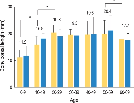
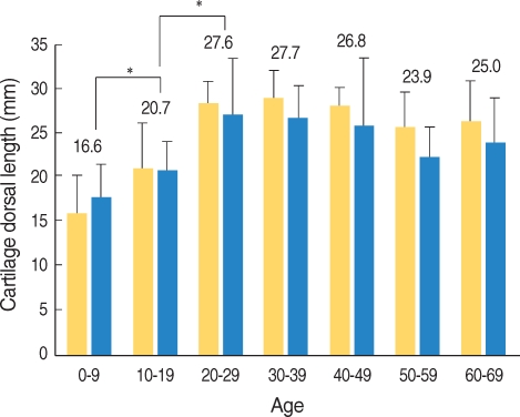
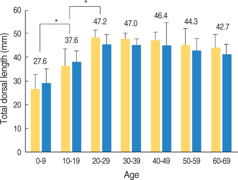
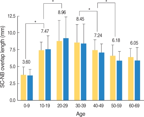
The bony dorsal length gradually increased after birth both in males and females up into their teens (16.9±2.7 mm) and twenties (19.3±2.9 mm) (Fig. 2). The cartilaginous dorsal length increased after birth until their twenties (27.6±4.6 mm) and it did not show significant changes later in adults, and no significant differences between the male and female were shown in each age group (Fig. 3). The total dorsal length was also observed to grow until the twenties; it did not show significant changes later in adults, and there were no significant gender differences (Fig. 4). The length of the overlapped part of the nasal septal cartilage and the nasal bone increased in appearance until the twenties (9.9±3.2 mm) and it later showed a significant decrease with no significant gender differences (Fig. 5).
Fig. 2.
Changes of the bony dorsal length according to age and gender. The bony dorsal length was increased until the 3rd decade and then it was not significantly changed with age. There was no significant gender difference. The diagonally stripped bars indicate males, the open bars the females. *P<0.05.
Fig. 3.
Changes of the cartilage dorsal length according to age and gender. The cartilaginous dorsal length was increased until the 3rd decade and then it was not changed significantly with age. There was no significant gender difference. The diagonally stripped bars indicate the males and the open bars the females. *P<0.05.
Fig. 4.
Changes of the total dorsal length according to age and gender. The total dorsal length was increased until the 3rd decade and then it was not changed significantly with age. There was no significant gender difference. The diagonally stripped bars indicate the males and the open bars the females. *P<0.05.
Fig. 5.
Changes of the septal cartilage-nasal bone overlap length according to age and gender. The septal cartilage-nasal bone overlap length was increased until the 3rd decade and then it was decreased significantly with age. There was no significant gender difference.
SC: septal cartilage; NB: nasal bone. The diagonally stripped bars indicate the males and the open bars the females. *P<0.05.
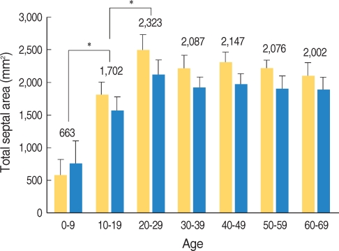
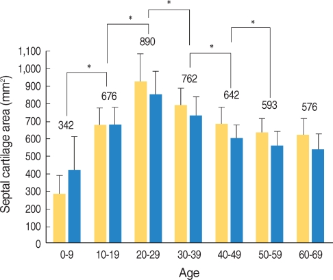
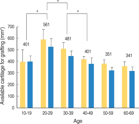
The total nasal septal area was increased up to the twenties (2,323.5±243.3 mm2) and it showed no significant change after the twenties. There were no significant differences between the males and females (Fig. 6). The area of the nasal septal cartilage was increased to the maximum by the twenties (889.4±151.6 mm2) (Fig. 7). The proportion of the area of the nasal septal cartilage to the total nasal septal area was shown to decrease as both the males and females got older (Fig. 8). The area of nasal septal cartilage, with excluding 1 cm of the dorsal and caudal portion, was measured as 426.8 mm2 on average for the adults and the range was from 253 mm2 to 750 mm2 (Fig. 9).
Fig. 6.
Changes of the total septal area according to age and gender. The total septal area was increased until the 3rd decade and then it was not changed significantly with age. There was no significant gender difference. The diagonally stripped bars indicate the males and the open bars the females. *P<0.05.
Fig. 7.
Changes of the septal cartilage area according to age and gender. The septal cartilage area was increased until the 3rd decade and then it was decreased significantly with age. There was no significant gender difference. The diagonally stripped bars indicate the males and the open bars the females. *P<0.05.
Fig. 8.
Changes of the proportion of the cartilage area according to age and gender. The proportion of the cartilage area was decreased significantly with age. Solid lines indicate males, the dotted lines the females.
Fig. 9.
Changes of the maximal available cartilage for grafting according to age and gender. The maximal available cartilage for grafting was increased until the 3rd decade and then it was decreased significantly with age. There was no significant gender difference. Diagonally stripped bars indicate the males, the open bars the females. *P<0.05.
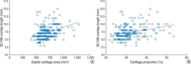
The length of the overlapped portion between the nasal septum and the nasal bone showed a significant correlation with the total dorsal length, based on the adults in or beyond their twenties (r=0.138, P=0.049), and this length also showed a significant correlation with the area of the nasal septal cartilage and the relative proportion of the nasal cartilage to the total nasal septum (r=0.386, P<0.001 and r=0.327, P<0.001, respectively) (Fig. 10).
Fig. 10.
(A) Correlation between the SC-NB overlap length and the septal cartilage area (r=0.386, P<0.001), (B) Correlation between the SCNB overlap length and the proportion of the cartilage area (r=0.327, P<0.001). SC, septal cartilage; NB, nasal bone.
DISCUSSION
Embryologically, the nasal septum consists of cartilaginous bone, and the ossification of the nasal bone and vomer is started at two ossification centers of the cartilaginous synchondroses. The ossification of the vomer occurs earlier than that of the nasal bone, and the two ossification centers are later united below the caudal part of the nasal septal cartilage (8, 9). Most of the nasal septum of a young child consists of cartilage and this size increases until puberty as the ossification of the vomer, the nasal bone and the perpendicular plate of the ethmoid bone proceeds (10, 11).
Many studies focused on the changes of the nasal septal cartilage according to age have been performed. Vetter et al. (3) reported that the anterior part of the nasal septal cartilage has a high cell density and it shows active proliferative ability, and the central part of the nasal septum shows the highest growth rate in childhood, with the cell density increasing until puberty, and the central part of the nasal septum shows a decreasing growth rate after puberty. Antoszewski et al. (12) claimed that nasal plastic surgery should be performed after the age of 18 because the growth of the nose increases until the age of 18 and this does not show a significant difference beyond that age. Rees et al. (13) announced that females before 16 years and males before 17 years should not undergo nasal plastic surgery. In this study, the total dorsal length of the nasal septum and the cartilaginous dorsal length increased until the age of twenty and there was no significant difference in these lengths beyond the age of twenty. The growth rate of nasal bone was also increased up to the twenties and there were no significant differences in the older adults, except a little decrease in the sixties.
Godley et al. (7) carried out an anatomical study on the nose by using 60 adult cadavers and they reported that the cartilaginous dorsal length was 21±5 mm on average and this length was shorter in the females by approximately 5±4 mm. They also reported that the length of the dorsal part of the nasal septum that overlaps under the nasal bone with the perpendicular plate of ethmoid bone is 4±2 mm on average. In this study, which was based on radiological measurements, the cartilage dorsal length in adult was 26±4 mm on average and there were no significant differences in the dorsal length between the males and females. The overlapped part of the nasal septal cartilage and nasal bone was measured as 7±2 mm on average in the adults, and this seemed to gradually decrease with age. We observed some individual differences for the overlapped part according to the age, and these differences ranged from 3 mm up to 15 mm.
Takahashi et al. (14) demonstrated that the cartilage component occupies about 19% of the total nasal septum, However, Miles et al. (6) reported that it is about 47.5% and the cartilage available for grafting, with excluding the L-strut, is about 420 mm2. Weiler et al. (15) reported, in the animal study using rat, that the size of the nasal septal cartilage increases after birth, it reaches a peak and it gradually decreases after, and that this was statistically significant for males. Van Loosen et al. (10), who analyzed the size of the nasal septal cartilage using 30 cadevers concluded the size of nasal septum after birth and of nasal septum throughout life. The size of the cartilage part of nasal septum gradually decreases as the bony part is increased. In this study, the total area of the nasal septum was 2,127 mm2 on average and the area did not show significant difference in the adults, whereas the area of the nasal septal cartilage was 692 mm2 on average and this area showed decreasing tendency with age. The relative proportion of the cartilage is approximately 32% of the total area of nasal septum in adults, and it was noted to decrease with age. In addition, it was noted that the maximal amount of nasal septal cartilage that can be used as graft material is about 427 mm2, which was not different from the previous reports, and that this maximal amount of nasal septal cartilage decreased with age.
The septal cartilage and upper lateral cartilage are engaged with each other below the nasal bone, and this is an important part that supports the entire cartilage structure of the nose, which attacks with the pyriform aperture dorsally, the nasal septum ventrally and the lower lateral cartilage caudally (16). The importance of this structure has been reported in several articles. Most et al. (17) introduced operative techniques that could reduce damage to these part. Rohrich et al. (18) explained that special care is needed when approaching this part during nasal plastic surgery or when performing bone dissection for removal of a hump.
In this study, the keystone area, for which the dorsal part of the nasal septal cartilage lies upon the bottom of the nasal bone, was measured and studied by using two-dimensional MR images. This study showed that the length of the overlapped part between the nasal septum and the nasal bone had positive correlation not only with the absolute numerical value of the nasal septal cartilaginous area, but also with the relative proportion of the nasal septal cartilage. We speculate that the reduction of this length with age seems to be due to the decrease in the relative proportion of the nasal septal cartilage.
This study has a limitation of excluding people with nasal septal deviation from the analysis because the subject of the study was set on the nasal septum in the median plane, as shown in two-dimensional planes. There were limitations for the analysis about the specific point when the growth of the nasal septum stops since the age range was divided into 10-year intervals. Therefore, further studies focused on these points are necessary in the future.
CONCLUSION
The cartilaginous proportion of the whole nasal septum was relatively decreased with aging. The area that the nasal bone and septal cartilage overlap also decreased, as the relative proportion and the area of the nasal septal cartilage got smaller. Clinically, if this overlapped area is damaged when performing nasal septal surgery or nasal plastic surgery, then severe complication such as saddle nose can occur. Much attention should be paid when treating older patients because of the risk of damaging their smaller overlapped nasal septal cartilage area.
References
- 1.Koo BS, Jin YD, Park CH, Park YK, Park SW, Rha KS. Anatomic changes of intranasal and surrounding bony structures in patients with nasal septal deviation: a CT analysis. Korean J Otolaryngol-Head Neck Surg. 2004 Nov;47(11):1125–1129. Korean. [Google Scholar]
- 2.Russell WH, Paul EK, Allison RM. The nasal septum. In: Cummings CW, Fredrickson JM, Jarker LA, Krause CJ, Schuller DE, editors. Otolaryngology-head and neck surgery. 4th ed. St. Louis, USA: Mosby-Year Book Inc; 2005. pp. 1001–1002. [Google Scholar]
- 3.Vetter U, Heit W, Helbing G, Heinze E, Pirsig W. Growth of the human septal cartilage: cell density and colony formation of septal chondrocytes. Laryngoscope. 1984 Sep;94(9):1226–1229. doi: 10.1288/00005537-198409000-00016. [DOI] [PubMed] [Google Scholar]
- 4.Akguner M, Barutcu A, Karaca C. Adolescent growth patterns of the bony and cartilaginous framework of the nose: a cephalometric study. Ann Plast Surg. 1998 Jul;41(1):66–69. doi: 10.1097/00000637-199807000-00012. [DOI] [PubMed] [Google Scholar]
- 5.Burke PH, Hughes-Lawson CA. Stereophotogrammetric study of growth and development of the nose. Am J Orthod Dentofacial Orthop. 1989 Aug;96(2):144–151. doi: 10.1016/0889-5406(89)90255-2. [DOI] [PubMed] [Google Scholar]
- 6.Miles BA, Petrisor D, Kao H, Finn RA, Throckmorton GS. Anatomical variation of the nasal septum: analysis of 57 cadaver specimens. Otolaryngol Head Neck Surg. 2007 Mar;136(3):362–368. doi: 10.1016/j.otohns.2006.11.047. [DOI] [PubMed] [Google Scholar]
- 7.Godley FA. Nasal septal anatomy and its importance in septal reconstruction. Ear Nose Throat J. 1997 Aug;76(8):498–501. 504–506. [PubMed] [Google Scholar]
- 8.Blustone CD. Phylogenetic aspects and embryology. In: Blustone CD, Stool SE, Scheetz MD, editors. Pediatric otolaryngology. 4th ed. Pennsylvania: Saunders; 2003. pp. 5–7. [Google Scholar]
- 9.Sandikcioglu M, Molsted K, Kjaer I. The prenatal development of the human nasal and vomeral bones. J Craniofac Genet Dev Biol. 1994 Apr–Jun;14(2):124–134. [PubMed] [Google Scholar]
- 10.van Loosen J, Verwoerd-Verhoef HL, Verwoerd CD. The nasal septal cartilage in the newborn. Rhinology. 1988 Sep;26(3):161–165. [PubMed] [Google Scholar]
- 11.Melsen B. Histological analysis of the postnatal development of the nasal septum. Angle Orthod. 1977 Apr;47(2):83–96. doi: 10.1043/0003-3219(1977)047<0083:HAOTPD>2.0.CO;2. [DOI] [PubMed] [Google Scholar]
- 12.Antoszewski B, Sitek A, Kruk-Jeromina J. [Analysis of nose growth] Otolaryngol Pol. 2005;59(6):925–931. Polish. [PubMed] [Google Scholar]
- 13.Rees TD. Surgical correction of the severely deviated nose by extramucosal excision of the osseocartilaginous septum and replacement as a free graft. Plast Reconstr Surg. 1986 Sep;78(3):320–330. doi: 10.1097/00006534-198609000-00007. [DOI] [PubMed] [Google Scholar]
- 14.Takahashi R. The formation of the nasal septum and the etiology of septal deformity. The concept of evolutionary paradox. Acta Otolaryngol Suppl. 1987;443:1–160. [PubMed] [Google Scholar]
- 15.Weiler E, Farbman AI. The septal organ of the rat during postnatal development. Chem Senses. 2003 Sep;28(7):581–593. doi: 10.1093/chemse/bjg047. [DOI] [PubMed] [Google Scholar]
- 16.Lam SM, Williams EF., 3rd Anatomic considerations in aesthetic rhinoplasty. Facial Plast Surg. 2002 Nov;18(4):209–214. doi: 10.1055/s-2002-36488. [DOI] [PubMed] [Google Scholar]
- 17.Most SP. Anterior septal reconstruction: outcomes after a modified extracorporeal septoplasty technique. Arch Facial Plast Surg. 2006 May–Jun;8(3):202–207. doi: 10.1001/archfaci.8.3.202. [DOI] [PubMed] [Google Scholar]
- 18.Rohrich RJ, Hollier LH, Jr, Janis JE, Kim J. Rhinoplasty with advancing age. Plast Reconstr Surg. 2004 Dec;114(7):1936–1944. doi: 10.1097/01.prs.0000143308.48146.0a. [DOI] [PubMed] [Google Scholar]


