Abstract
Although colorectal granular cell tumors (GCTs) are rare, their incidental finding has increased as the use of diagnostic colonoscopy has become more common. Here we describe the case of a 41-yr-old man with a GCT in the descending colon that was detected after a screening colonoscopy. Endoscopic examination revealed a yellowish submucosal tumor, 13×12 mm in diameter, in the descending colon. Endoscopic mucosal resection (EMR) followed by histological examination revealed that the tumor was composed of plump histiocyte-like cells with an abundant granular eosinophilic cytoplasm and small round nuclei. The tumor cells expressed S-100 protein and stained with periodic acid-Schiff, but were negative for desmin and cytokeratin. The resected tumor was diagnosed as a GCT. Colonoscopists should consider the possibility of GCT in the differential diagnosis of yellowish submucosal tumors of the colon. In such patients, EMR seems to be a feasible and safe approach for diagnosis and treatment.
Keywords: Colon, Neoplasms, Endoscopic Mucosal Resection, Granular Cell Tumor, Submucosal Tumor
INTRODUCTION
Granular cell tumors (GCTs) are relatively rare and thought to be both clinically and histologically benign. Although it may arise virtually anywhere in the gastrointestinal tract, the occurrence of GCTs in the colon is uncommon (1, 2). Instead, these tumors tend to be found incidentally during colonoscopic examinations performed for other reasons, as they are frequently asymptomatic. Endoscopically, a GCT usually appears as a small submucosal nodule, measuring less than 2 cm in diameter (2-5). In the present report, we describe a rare case involving a 41-yr-old man diagnosed with a GCT arising in the descending colon. The tumor was resected by endoscopic mucosal resection (EMR) for histological confirmation and treatment. In addition, we carefully reviewed the English literature for reports of other cases of colorectal GCT.
CASE REPORT
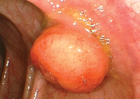
A 41-yr-old man underwent colonoscopic examination as part of a routine medical checkup. He had been healthy without specific complaints and had no past medical history. He appeared well, and physical examinations showed no abnormalities. The laboratory findings were all within normal limits. At the time of colonoscopy, however, two small polyps were detected in the sigmoid colon and rectum. These were polypectomized using a snare, with the biopsy findings showing two adenomas. In the descending colon, a yellowish polypoid lesion, 13×12 mm in diameter and consisting of a firm nodule covered by intact mucosa, was found (Fig. 1). A submucosal tumor, such as a carcinoid tumor, was suspected and the lesion was resected by EMR for histological confirmation and treatment. After injection of 4 mL of 3% hypertonic saline solution with epinephrine into the submucosa around the lesion for lifting, the tumor was cut electrically with a high-frequency snare. The patient was observed for 30 min and then discharged. No immediate or delayed complications occurred.
Fig. 1.
Colonoscopy detected an approximately 13×12 mm yellowish, submucosal tumor in the descending colon. It was hard in consistency without ulceration.
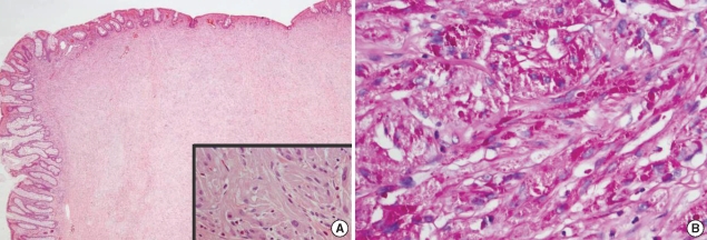
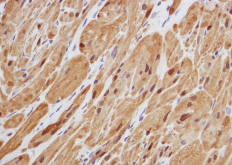
Histological examination of the resected specimen revealed a submucosal tumor composed of plump histiocyte-like cells with an abundant granular eosinophilic cytoplasm positive for acidophilic periodic acid-Schiff (PAS) staining (Fig. 2). Immunohistochemical analysis showed that the tumor cells expressed S-100 protein (Fig. 3) but were negative for desmin and cytokeratin. The resected tumor was diagnosed as a GCT of the descending colon.
Fig. 2.
Histological findings of the tumor. (A) The resected tumor was covered with normal mucosa (H&E, ×20). A nested growth of nonuniform large tumor cells with slightly pleomorphic nuclei (inlet, H&E, ×400). (B) Some granules were positive for periodic acid-Schiff (PAS, ×400).
Fig. 3.
Histological findings of the tumor showing positive immunoreaction for S-100 protein (immunohistochemical stain, ×400).
During 7 months follow-up, he has been well without disease recurrence.
DISCUSSION
Recent advances in diagnostic techniques, especially the widespread use of colonoscopy, have enabled clinicians to identify small and asymptomatic colorectal tumors. GCT is a rare tumor that usually appears as a solitary, small, nodular growth and it follows a benign course. While GCTs may occur at any site of the body, they are most frequently detected in the oral cavity, skin, and subcutaneous tissue (1). In the gastrointestinal tract, where GCTs are uncommon, the esophagus is the most frequent site, followed by the colon and stomach (1, 6). As Lack et al. (1) noted in their review, GCT was first described in 1926 by Abrikossoff as a myogenic tumor; however, subsequent studies favored a Schwann cell origin (6, 7). Recently, this hypothesis was supported by positive staining of the cytoplasm and nuclei of granular cells for S-100 protein. The additional staining of granular cells for myelin proteins and myelin-associated glycoproteins provided further evidence.
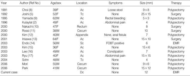
In light of our findings, we reviewed reports of other GCTs in the colon and rectum. The reports were identified by a computerized search of the PubMed database (articles in English between 1992 and 2007) and of KoreaMed (articles in Korean published between 1990 and 2007). Fifteen of the cases identified by the computerized search were reviewed (Table 1) (2, 5, 8-20). The results showed that most colorectal GCTs are asymptomatic and are found incidentally during a routine medical checkup or investigations for nonspecific gastrointestinal symptoms, as was the case in the present patient. Johnston et al. (6) also reported that 17 of 20 colorectal GCTs were found incidentally during investigations for hemorrhoids or routine medical checkup. In other reports (9, 14), rectal bleeding or a positive fecal occult blood test was described, but it is unlikely that either of these findings was caused by a GCT, which is covered by an intact colonic mucosa without ulceration and inflammation. Colorectal GCTs may be located anywhere between the rectum and the cecum, with a preferential location in the ascending colon and cecum based on the results of our literature review (Table 1). However, the most common locations for colorectal GCTs are the rectum and cecum in the literature including approximately 100 cases of colorectal GCTs (6, 11, 21). Colorectal GCTs occurred multiply in 10 to 20% of all reported cases (5, 22).
Table 1.
Granular cell tumors occurring in the colon and rectum, as reported in the English literature
Ac, ascending colon; Tc, transverse colon; Sc, sigmoid colon; Dc, descending colon; FOBT, fecal occult blood test; EMR, endoscopic mucosal resection.
Endoscopic findings of colorectal GCTs are yellow or yellow-white submucosal nodules covered by normal mucosa or sessile polyps, or less frequently, pedunculated polyps (1-3, 13). Thus, colonoscopists should generally consider the possibility of a GCT in the differential diagnosis of a yellowish submucosal tumor of the colon. Nonetheless, Palazzo et al. (23) reported that the correct diagnosis of GCT is seldom made based on macroscopic and endoscopic examinations, even if a diagnosis is made based on the results of endoscopic biopsy, and that these techniques yield a definite diagnosis in only 50% of all patients with GCTs. In some cases, endoscopic biopsy finding was misinterpreted as an other diagnosis (10). To obtain a definite diagnosis of a submucosal tumor by endoscopic biopsy, it is necessary to sample such tumors either by boring or by performing a jumbo biopsy, and/or if feasible, by carrying out a polypectomy or a mucosal resection. Actually, 12 (75%) of 16 patients with colonic GCTs in our analysis were diagnosed by polypectomy or EMR. Endoscopic ultrasonography has recently been applied for evaluating gastrointestinal submucosal tumors, but it may not sufficiently distinguish a benign submucosal tumor, such as a GCT, from malignant neoplasia, such as a carcinoid (10, 14). However, it can contribute to planning the endoscopic resection by determining the depth of tumor invasion (23).
Histologically, a GCT is composed of a nested growth comprising large tumor cells with ample granular cytoplasm and small round nuclei. The histological findings consist of plump neoplastic cells with abundant granular eosinophilic cytoplasm containing acidophilic, p-aminosalicylic-acid-positive, diastase-resistant granules; small, uniform nuclei in which mitotic figures are absent; and neural markers, including S-100 proteins or neuron-specific enolase. Granules either arise from autophagocytosis of the cell membrane or represent derivatives of the Golgi apparatus (24). The histological features of GCTs are uniformly expressed and should be adequate to establish the diagnosis (1, 6-9, 19). In our patient, the endoscopic and histological findings of the resected specimen were indeed compatible with GCT.
Few data exist, however, regarding the malignant potential and thus the appropriate management of colonic GCT. No evidence of malignant GCT in our reviewed 16 patients were noted. Johnston et al. (6) also found no evidence of malignant GCT in 78 cases of GCTs in the gastrointestinal tract and perianal lesions. Nonetheless, a malignant variety of GCT has been reported (25, 26). The malignant GCT seems to be correlated with the size of the tumor since most malignant forms are larger than 4 cm. Rapid growth and invasion of adjacent tissue as well as large size were reported to be more indicative of malignant behavior than the histological features. Other reported features associated with malignancy include cell necrosis, spindling of tumor cells, cytologic atypia and high mitotic activity, vescicular nuclei with large nucleoli, a high nuclear-to-cytoplasm ratio, and high expression of p53 and Ki-67 (25-27). In our patient, any evidence of malignancy was not seen in the resected tumor.
It is usually accepted that endoscopic resection is the best treatment for colonic GCTs, since colonic GCTs are considered to be usually benign. Actually, 12 (75%) of 16 patients in our analysis and 26 (79%) of 33 patients in other report (14) were successfully treated with polypectomy or EMR without complications. Although there are no guidelines for the endoscopic treatment of colonic GCTs at present, benign lesions <2 cm in diameter and separated from the muscularis propria can be removed by EMR (3, 9, 14). In our analysis, the remaining four patients underwent surgical resections, based on visualization of the tumors involving the muscularis propria by endoscopic ultrasound, or an incidental intraoperative finding during surgery for another indication. In addition, the treatment may become surgical resection for a benign GCT because of the misinterpretation of the biopsied lesion. In surgical cases, colorectal GCTs were misinterpreted as a possible adenocarcinoma in the biopsy findings (10) or not differentiated from a gastrointestinal stromal tumor and a carcinoid with malignant potentials (13). Although endoscopic resections of colorectal GCTs are curative in most cases, local recurrence has also been reported (1). Neither size, mitotic activity, nor the resection margins are reliable indicators of recurrence. Follow-up colonoscopic examination is necessary when the tumors are multiple or a risk of malignancy exists. In addition, endoscopic examination should include a baseline gastroscopy to exclude the presence of GCTs in the esophagus and the stomach, which are the other sites in the gastrointestinal tract where GCTs can be found.
In conclusion, colonoscopists should consider the possibility of GCTs in the differential diagnosis of submucosal tumors of the colon as these tumors are increasingly being detected as incidental findings. EMR of colonic GCT for diagnosis and treatment seems to be feasible and safe, especially when tumors are small.
References
- 1.Lack EE, Worsham GF, Callihan MD, Crawford BE, Klappenbach S, Rowden G, Chun B. Granular cell tumor: a clinicopathologic study of 110 patients. J Surg Oncol. 1980;13:301–316. doi: 10.1002/jso.2930130405. [DOI] [PubMed] [Google Scholar]
- 2.Kulaylat MN, King B. Granular cell tumor of the colon. Dis Colon Rectum. 1996;39:711. doi: 10.1007/BF02056957. [DOI] [PubMed] [Google Scholar]
- 3.Yasuda I, Tomita E, Nagura K, Nishigaki Y, Yamada O, Kachi H. Endoscopic removal of granular cell tumors. Gastrointest Endosc. 1995;41:163–167. doi: 10.1016/s0016-5107(05)80604-2. [DOI] [PubMed] [Google Scholar]
- 4.Melo CR, Melo IS, Schmitt FC, Fagundes R, Amendola D. Multicentric granular cell tumor of the colon: report of a patient with 52 tumors. Am J Gastroenterol. 1993;88:1785–1787. [PubMed] [Google Scholar]
- 5.Joshi A, Chandrasoma P, Kiyabu M. Multiple granular cell tumor of the gastrointestinal tract with subsequent development of esophageal squamous cell carcinoma. Dig Dis Sci. 1992;37:1612–1618. doi: 10.1007/BF01296510. [DOI] [PubMed] [Google Scholar]
- 6.Johnston J, Helwig EB. Granular cell tumors of the gastrointestinal tract and perianal region: a study of 74 cases. Dig Dis Sci. 1981;26:807–816. doi: 10.1007/BF01309613. [DOI] [PubMed] [Google Scholar]
- 7.Armin A, Connelly EM, Rowden G. An immunoperoxidase investigation of S-100 protein in granular cell myoblastomas: evidence for Schwann cell derivation. Am J Clin Pathol. 1983;79:37–44. doi: 10.1093/ajcp/79.1.37. [DOI] [PubMed] [Google Scholar]
- 8.Choi JK, Choi MG, Choi KY, Chung IS, Cha SB, Chung KW, Sun HS, Kim BS, Choi YJ, Lee AH. A case of colonoscopically removed granular cell tumor in the ascending colon. Korean J Gastrointest Endosc. 1991;11:383–386. [Google Scholar]
- 9.Yamada T, Fujiwara Y, Sasatomi E, Nakano S, Tokunaga O. Granular cell tumor in the ascending colon. Intern Med. 1995;34:657–660. doi: 10.2169/internalmedicine.34.657. [DOI] [PubMed] [Google Scholar]
- 10.Nakachi A, Miyazato H, Oshiro T, Shimoji H, Shiraishi M, Muto Y. Granular cell tumor of the rectum: a case report and review of the literature. J Gastroenterol. 2000;35:631–634. doi: 10.1007/s005350070064. [DOI] [PubMed] [Google Scholar]
- 11.Rossi GB, de Bellis M, Marone P, Chiara AD, Losito S, Tempesta A. Granular cell tumor of the colon: report of a case and review of the literature. J Clin Gastroenterol. 2000;30:197–199. doi: 10.1097/00004836-200003000-00014. [DOI] [PubMed] [Google Scholar]
- 12.Kim HS, Cho KA, Hwang DY, Kim KU, Kang YW, Park WK, Yoon SG, Lee KR, Lee JK, Lee JD, Kim KY. A case of granular cell tumor in the appendix. Korean J Gastroenterol. 2000;36:404–407. [Google Scholar]
- 13.Irisawa A, Hernandez LV, Bhutani MS. Endosonographic features of a granular cell tumor of the colon. J Ultrasound Med. 2001;20:1241–1243. doi: 10.7863/jum.2001.20.11.1241. [DOI] [PubMed] [Google Scholar]
- 14.Endo S, Hirasaki S, Doi T, Endo H, Nishina T, Moriwaki T, Nakauchi M, Masumoto T, Tanimizu M, Hyodo I. Granular cell tumor occurring in the sigmoid colon treated by endoscopic mucosal resection using a transparent cap (EMR-C) J Gastroenterol. 2003;38:385–389. doi: 10.1007/s005350300068. [DOI] [PubMed] [Google Scholar]
- 15.Kim DH, Kim YH, Kwon NH, Song BG, Jung JH, Kim MH, Rhee PL, Kim JJ, Rhee JC. A case of granular cell tumor in the rectum. Korean J Gastrointest Endosc. 2003;27:88–91. [Google Scholar]
- 16.Lee SH, Kim SH, Kim BR, Kim HJ, Bhandari S, Jung IS, Hong SJ, Ryu CB, Kim JO, Cho JY, Lee JS, Lee MS, Shim CS, Kim BS, Jin SY. Granular cell tumor of the ascending colon: report of a case. Intestinal Research. 2003;1:59–63. [Google Scholar]
- 17.Ryu JH, Choi MH, Kim GS, Choi CS, Suh YA, Jang HJ, Eun CS, Kae SH, Lee J. A case of granular cell tumor of the ascending colon. Korean J Gastrointest Endosc. 2003;26:439–442. [Google Scholar]
- 18.Sohn DK, Choi HS, Chang YS, Huh JM, Kim DH, Kim DY, Kim YH, Chang HJ, Jung KH, Jeong SY. Granular cell tumor of the colon: report of a case and review of literature. World J Gastroenterol. 2004;10:2452–2454. doi: 10.3748/wjg.v10.i16.2452. [DOI] [PMC free article] [PubMed] [Google Scholar]
- 19.Mori T, Orikasa H, Shigematsu T, Yamazaki K. An ultrastructural and immunohistochemical study of a combined submucosal granular cell tumor and lipoma of the colon showing a unique nodule-in-nodule structure: putative implication of CD34 or prominin-2-positive stromal cells in its histopathogenesis. Virchows Arch. 2006;449:137–139. doi: 10.1007/s00428-006-0210-9. [DOI] [PubMed] [Google Scholar]
- 20.Park NY, Kim KJ, Kim YJ, Roh JH, Im DG, Nam JH, Monn W, Park IP, Park SJ, Cheon BK. A case of granular cell tumor of the colon treated by colonoscopy. Korean J Gastrointest Endosc. 2006;32:67–70. [Google Scholar]
- 21.Parfitt JR, McLean CA, Joseph MG, Streutker CJ, Al-Haddad S, Driman DK. Granular cell tumours of the gastrointestinal tract: expression of nestin and clinicopathological evaluation of 11 patients. Histopathology. 2006;48:424–430. doi: 10.1111/j.1365-2559.2006.02352.x. [DOI] [PubMed] [Google Scholar]
- 22.Shimoyama M, Sakai Y, Takaku H, Takii Y, Okamoto H, Suda T, Hatakeyama K. A case of multiple granular cell tumors in the cecum. Gastroenterol Endosc. 1999;41:1330–1335. [Google Scholar]
- 23.Palazzo L, Landi B, Cellier C, Roseau G, Chaussade S, Couturier D, Barbier J. Endosonographic features of esophageal granular cell tumors. Endoscopy. 1997;29:850–853. doi: 10.1055/s-2007-1004320. [DOI] [PubMed] [Google Scholar]
- 24.Mittal KR, True LD. Origin of granules in granular cell tumor: intracellular myelin formation with autodigestion. Arch Pathol Lab Med. 1988;112:302–303. [PubMed] [Google Scholar]
- 25.Uzoaru I, Firfer B, Ray V, Hubbard-Shepard M, Rhee H. Malignant granular cell tumor. Arch Pathol Lab Med. 1992;116:206–208. [PubMed] [Google Scholar]
- 26.Matsumoto H, Kojima Y, Inoue T, Takegawa S, Tsuda H, Kobayashi A, Watanabe K. A malignant granular cell tumor of the stomach: report of a case. Surg Today. 1996;26:119–122. doi: 10.1007/BF00311775. [DOI] [PubMed] [Google Scholar]
- 27.Fanburg-Smith JC, Meis-Kindblom JM, Fante R, Kindblom LG. Malignant granular cell tumor of soft tissue: diagnostic criteria and clinicopathologic correlation. Am J Surg Pathol. 1998;22:779–794. doi: 10.1097/00000478-199807000-00001. [DOI] [PubMed] [Google Scholar]


