Figure 1.
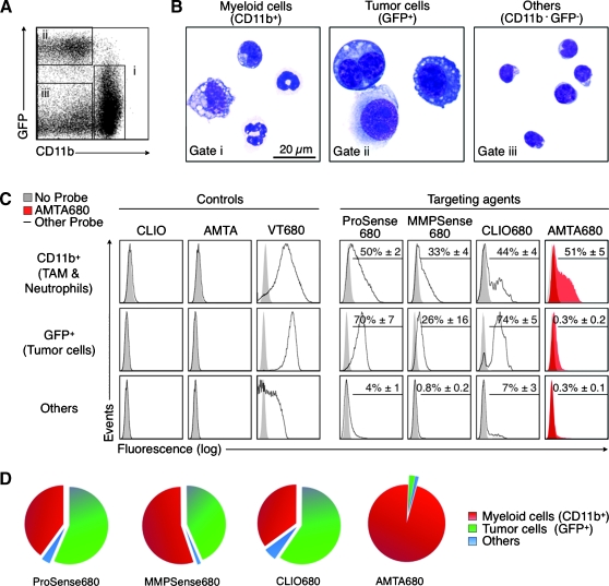
In vivo targeting of endogenous tumor-associated myeloid cells with AMTA680. (A) Flow cytometric analysis of cell suspensions obtained from excised tumors and stained with anti-CD11b mAb (tumor cells stably express EGFP) identifies myeloid cells (i), tumor cells (ii), and “other” cells (iii), n = 9. (B) Morphological analysis of sorted cells. (C) Analysis of tumor cell suspensions 24 hours after intravenous administration of “control” (CLIO, AMTA, VT680) or “targeting” agents (ProSense680, MMPSense680, CLIO680, and AMTA680). Each histogram shows a representative example of the fluorescence intensity (uptake) of each agent for each cell type. Figures in the upper right of the histograms show the percentage (mean ± SEM) of cells labeled with the agent when compared with the corresponding cells in mice that did not receive any agent. All agents but AMTA680: n = 3; AMTA680: n = 27. (D) Distribution of the targeting agents into different cell types that are present in the tumor environment. The analysis shows the specificity of AMTA680 (but not of the other agents) for tumor-associated myeloid cells.

