Abstract
Pathogenic strains of Helicobacter pylori secrete a cytotoxin, VacA, that in the presence of weak bases, causes osmotic swelling of acidic intracellular compartments enriched in markers for late endosomes and lysosomes. The molecular mechanisms by which VacA causes this vacuolation remain largely unknown. At neutral pH, VacA is predominantly a water-soluble dodecamer formed by two apposing hexamers. In this report, we show by using atomic force microscopy that below pH ≈5, VacA associates with anionic lipid bilayers to form hexameric membrane-associated complexes. We propose that water-soluble dodecameric VacA proteins disassemble at low pH and reassemble into membrane-spanning hexamers. The surface contour of the membrane-bound hexamer is strikingly similar to the outer surface of the soluble dodecamer, suggesting that the VacA surface in contact with the membrane is buried within the dodecamer before protonation. In addition, electrophysiological measurements indicate that, under the conditions determined by atomic force microscopy for membrane association, VacA forms pores across planar lipid bilayers. This low pH-triggered pore formation is likely a critical step in VacA activity.
Keywords: gastritis, ulcers, AB toxins, VacA, membrane protein
Colonization of the stomach mucosa with Helicobacter pylori is the principal cause of chronic superficial gastritis in humans and is a major risk factor for the development of peptic ulcer and gastric carcinoma (1–3). One of the primary virulence factors secreted by H. pylori is VacA (4–5), a ≈90-kDa protein that, at neutral pH, self-associates into flower-shaped, predominantly dodecameric complexes comprised of two apposing hexamers (6). In cultured cells, addition of VacA, together with weak bases, induces marked swelling, or vacuolation, of acidic compartments enriched in markers for late endosomes and lysosomes (5, 7).
The molecular mechanisms by which VacA causes cell vacuolation are largely unknown. The first step appears to involve the binding of VacA to the plasma membrane (8), followed by internalization (8–9). Expression of VacA in transfected cells results in cell vacuolation, which suggests that VacA has an intracellular site of action (10). Although endocytosis has not yet been firmly demonstrated as a requirement for vacuolation, a critical low-pH step has been proposed (6, 11–12). However, the effects of low pH on VacA association with membranes has remained largely unexplored.
In this study, we have applied atomic force microscopy (AFM) to examine the interaction of purified VacA with model lipid membranes under a variety of conditions. We show that when the pH is lowered below ≈5, VacA associates with anionic phospholipid bilayers as hexameric membrane-associated complexes. In addition, electrophysiological measurements demonstrate that under these conditions, VacA forms pores in planar lipid bilayers. These results thus demonstrate that low pH indeed causes a critical change in the structure of the VacA oligomer, resulting in its interaction with a selected lipid species. The pores formed by VacA oligomers in target membranes are likely to be directly related to the toxic effects of VacA on host cells.
METHODS
Materials.
VacA was purified from broth culture supernatant of H. pylori 60190 described in ref. 6 by using gel filtration chromatography with a Superose 6 HR 16/50 column and phosphate-buffered saline containing 1 mM EDTA and 0.1% NaN3 (PBS buffer). The zwitterionic lipids dioleoylphosphatidylcholine (DOPC), egg phosphatidylcholine (eggPC), dioleoylphosphatidylethanolamine (DOPE), and egg phosphatidylethanolamine (eggPE), the anionic lipids dioleoylphosphatidylserine (DOPS), 1-palmitoyl-2-oleoylphosphatidylserine (POPS), dioleoylphosphatidic acid (DOPA), dioleoylphosphatidylglycerol (DOPG), and cardiolipin (CDL), and the total lipid extract from bovine heart (HL, a mixture of neutral, zwitterionic, and anionic lipids) were purchased from Avanti Polar Lipids. Cholesterol was purchased from Sigma.
Preparation of Supported Lipid Bilayers and Imaging of Membrane-Bound VacA.
The supported membrane was formed by sequentially depositing two separately prepared monolayers to mica (13). After a monolayer of eggPC was first deposited using a Langmuir trough (JL Automation, Sunderland, U.K.), a monolayer of HL [1 mg/ml in chloroform/methanol (2:1 vol/vol)] was next applied to the air/buffer interface of a small Teflon well (≈30 μl). The buffer in the well was typically 1 mM citric acid (pH 2.6), although similar results were obtained with higher ionic strength (0.1 M NaCl), higher buffer concentrations (50 mM citric acid), and pH values up to ≈5, as described in the text. After evaporation of the organic solvent, the coated mica fragment was horizontally lowered onto this interface, forming the bilayer. VacA (0.5 mg/ml) was then injected into the well (final concentration of ≈30 μg/ml). For those incubations at pH <5, the pH of the protein solution was first lowered by incremental addition of 20 mM HCl prior to its injection into the well. After 1–2 hr of incubation at room temperature, the sample was washed extensively either with 1 mM citric acid (pH 6.0) or, when the influence of pH on VacA binding to membranes was measured, with the incubation buffers. To improve the mechanical stability of the sample, chemical fixation with 1% glutaraldehyde for 1–2 minutes was applied, followed by extensive washing. The samples were always under solution during transport to and imaging within the AFM. Calibration of the piezoscanner (14 μm, D scanner, Digital Instruments, Santa Barbara, CA) was performed using mica and the cholera toxin B subunit. AFM images of these (14–15) and other (16–19) samples are consistent with those structures determined by using electron microscopy or x-ray crystallography. Imaging was performed in the contact mode with a Nanoscope II AFM (Digital Instruments, Santa Barbara, CA), using oxide-sharpened “twin tip” Si3N4 cantilevers, 200 μm in length, with a spring constant of 0.06 N/m. The scan rate was typically ≈9 Hz, and the applied force was minimized to ≈0.1 nN. All images presented here were reproducible with different tips and different fast-scan directions. All lateral dimensions were determined from full-width at half-height in the unprocessed images, except for the lateral measurements of the peripheral domains, which were determined from the processed image. Images were processed by using the Integrated Crystallographic Environment (20) for lattice unbending and the mrc program package (21) for six-fold symmetrization. The lateral resolution is estimated to be 1.7 nm based on an analysis of spot intensity vs. resolution and phase residuals in the Fourier transform.
Imaging Water-Soluble VacA at Neutral pH.
VacA was directly adsorbed to freshly cleaved mica by injecting 0.5 μl of the 0.5 mg/ml stock protein solution into 10 μl of buffer covering mica. After incubation, the sample was washed extensively and imaged under the same solution. The most commonly used buffer was 1 mM Hepes/0.1 M MgCl2, pH 7.5. Small scan size images of VacA bound to mica depicted dodecamers and tetradecamers, similar to what has been reported (ref. 6, and data not shown).
Electrophysiological Recordings.
Planar lipid membranes were prepared by painting a lipid solution over an aperture separating two chambers (22). A ≈10 mg/ml solution of either eggPC/cholesterol (5:2 mol) or eggPC/DOPS/cholesterol (4:1:2 mol) in n-decane was applied to the end (inner diameter, 2.5 mm) of a polystyrene tube (≈750 μl) projecting into a small Teflon chamber (≈500 μl). VacA was added to the Teflon chamber (cis) to a final concentration of ≈0.1 μg/ml. The buffer contained 0.1 M KCl/2 mM EDTA/5 mM citric acid at either pH 4 or pH 7. The Ag/AgCl electrodes were connected to the chambers through 1 M KCl–agar bridges. Similar results were measured by using a smaller aperture (≈0.4 mm) in a polypropylene container.
RESULTS
Association of VacA with Supported Bilayers.
The binding of VacA to pure lipid membranes was assessed by adding the cytotoxin to supported bilayers, incubating for 1–2 hr at various pH values, followed by extensive washing and imaging with AFM. At neutral pH or with bilayers composed of only zwitterionic lipids, no binding of VacA to the membranes was detected by using AFM. Instead, the AFM images revealed only protein aggregates within membrane defects, directly adsorbed to mica (Fig. 1a). Similarly, when the VacA stock solution was first “preactivated” [by lowering the pH to ≈3 (12)] before its addition to the bilayer at neutral pH, no binding to the membrane was detected. However, at pH values below 5 and with bilayers containing anionic phospholipids, a high density of oligomeric VacA was found to be associated with these membranes (Fig. 1b). Binding of VacA to the membrane under these conditions was observed even with VacA that was not first preactivated. These results are summarized in Table 1. The oligomers were ≈25 nm in outer diameter and projected ≈2.5 nm from the surface of the membrane. At smaller scan sizes, the membrane-associated complexes were often displaced by the tip (likely owing to the fluidity of the membrane), preventing higher resolution imaging. Nonetheless, a central cavity, with an inner diameter of ≈6 nm, could be resolved in many oligomers (Fig. 1b).
Figure 1.
AFM images show that VacA associates only with supported bilayers containing anionic phospholipids at pH < 5. (a) An eggPC bilayer after 1 hr incubation with ≈30 μg/ml VacA in 1 mM citric acid, pH ≈ 3.5. There is no VacA association with the bilayer, but there are aggregates directly adsorbed to mica within bilayer defects (dark regions). Similar results were obtained with bilayers composed of other zwitterionic lipids (see Table 1) at all pH values examined from pH 3.5 to 7.2 or with bilayers containing negatively charged lipids at pH > 5. (b) A high density of VacA oligomers is seen associated with bilayers made of HL (a total lipid extract including anionic phospholipids) under the conditions in a but at pH ≈ 3. Individual VacA oligomers can be clearly discerned in this image. (c) After adsorption of VacA to HL bilayers, raising the pH to 7 after removal of unbound VacA promoted the formation of two-dimensional crystals of VacA oligomers. Notice that the central rings of VacA oligomers are clearly visualized both in the randomly distributed oligomers and within the crystals. [Bar = 400 nm (a) and 200 nm (b and c).]
Table 1.
Lipid and pH conditions tested for VacA binding to supported bilayers
| Lipids∗ | pH | VacA binding to bilayer |
|---|---|---|
| HL (n = 105) | <5 | Yes |
| DOPC/DOPA (1:1) (n = 4) | ≈3 | Yes |
| DOPC/DOPS (1:1) (n = 3) | ≈3 | Yes |
| EggPE/POPS (1:1) (n = 1) | ||
| DOPC/CDL (4:1) (n = 3) | ≈3 | Yes |
| DOPC/DOPG (1:1) (n = 2) | ||
| Zwitterionic†(n = 21) | <5 | No |
| HL (n = 17) | >5 | No |
| EggPC (n = 5) | ≈7 | No |
DOPC, dioleoylphosphatidylcholine; DOPA, dioleolyphosphatidic acid; DOPS, dioleoylphosphatidylserine; EggPE, egg phosphatdylethanolamine; POPS, 1-palmitoyl-2-oleoylphosphatidylserine; CDL, cardiolipin; EggPC, egg phosphatidylcholine; DOPE, dioleoylphosphatidylethanolamine; DOPG, dioleoylphosphatidylglycerol; HL, bovine heart lipidextract.
Ratios reflect mol %; n = number of samples.
EggPC, DOPC, EggPE, or DOPE.
After the oligomers were adsorbed to the membrane at a pH <5 and the sample was extensively washed, raising the pH to 6 did not induce dissociation from the membrane but rather resulted in the formation of small two-dimensional crystal patches (data not shown). Increasing the pH to 7 promoted further two-dimensional crystallization into somewhat larger patches (≈250–500 nm in breadth, Fig. 1c). At pH 8 or higher, the sample was noticeably degraded, leaving poorly resolved aggregates directly adsorbed to mica (data not shown). The crystal patches were much more stable during scanning than were the isolated oligomers (14–15) and could be imaged to a greater resolution.
Structural Characterization of Membrane-Associated VacA.
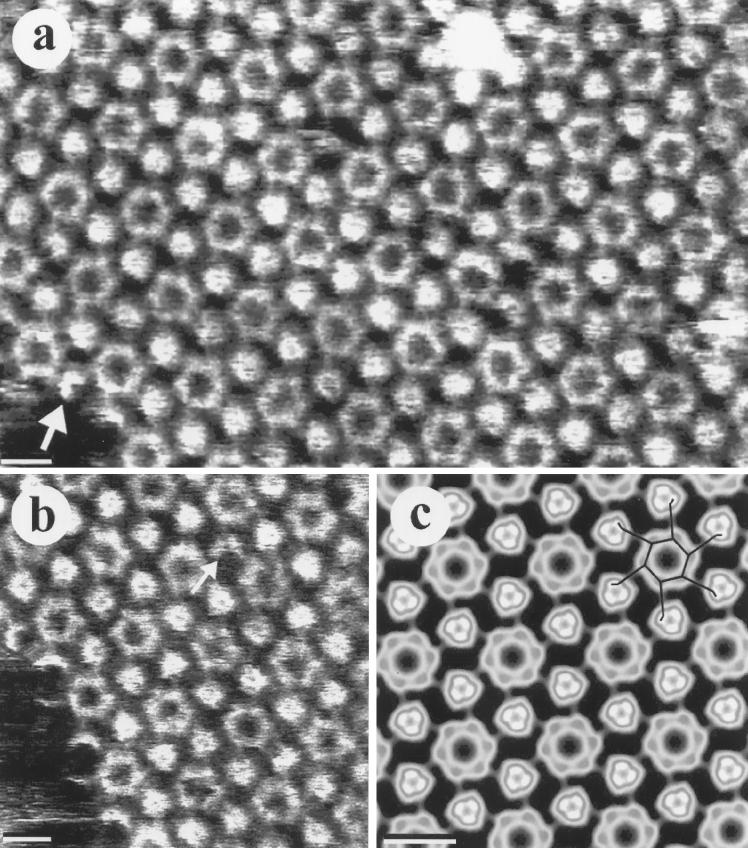
The unprocessed AFM images of the two-dimensional VacA crystals (Fig. 2 a and b) clearly reveal a hexagonal pattern of rings interspersed with smaller, triangular regions, with unit cell parameters of a = b = 22 ± 1 nm, γ = 60° ± 5° (n = 31). These topographs depict essentially flat, six-fold-symmetric oligomers (each with an overall diameter of ≈28 nm) possessing the same general characteristics as observed both with the water-soluble dodecamer (6) and with the isolated membrane-associated oligomers observed at pH <5. Although the features to be discussed below are clearly recognizable in these unprocessed images, the details of the oligomer are most easily visualized with reference to the line diagram in the processed image (Fig. 2c).
Figure 2.
Higher resolution AFM images of two-dimensional crystals of membrane-associated VacA oligomers. (a and b) Unprocessed AFM images show that the crystal consists of six-fold-symmetric VacA oligomers in a hexagonal array with lattice parameters a = b = 22 ± 1 nm, γ = 60° ± 5° (n = 31). The PDs from three nearest-neighbor oligomers are slightly higher than the centrally located hexameric ring. Notice that individual PDs can be detected at the edge of the crystal patch (arrow in a). (b) The arrow points to a region where one PD is missing, but the ring remains intact. (c) Processed AFM image after lattice unbending and six-fold symmetrization. The line diagram in the top right corner illustrates a single oligomer within the crystal with an overall diameter of ≈28 nm. Notice that the PD–connector orientation is similar to that observed at the edge of the crystal patch in a (arrow). (Bar = 15 nm.)
The central ring is a hexagon with an inner (side-to-side) diameter of 7 ± 1 nm (n = 79) and a side width and length of 3 ± 1 nm (n = 53) and 6 ± 1 nm (n = 40), respectively. The corners of the ring are slightly higher [0.4 ± 0.1 nm (n = 43)] than the adjacent sides.
The peripheral domains (PDs) from three nearest-neighbors form the triangular-shaped contact regions. Each PD is ≈5 nm long, ≈3 nm wide, and 0.8 ± 0.1 nm (n = 27) higher than the corners of the central rings. Individual PDs can be observed at the edges of the crystal patch in the unprocessed image (arrow in Fig. 2a). Notice also that in Fig. 2b, there appears to be a single PD missing from the oligomer to the left (arrow), yet the central ring seems to be intact.
The connections (connectors) between the central hexagonal ring and the PD are resolved as thin projections 3 ± 1 nm (n = 29) wide, 5 ± 1 nm (n = 50) long, and 0.8 ± 0.2 nm (n = 29) smaller in height than the corners of the central ring. Rather than a strictly radial orientation from a corner of the hexagonal ring, each connector was found to project ≈90° from one side of the ring. For comparison, with a completely radial projection of a connector, the angle between the connector and either side of the ring would be 120°. Thus, the connectors of the entire oligomer appear to have a counter-clockwise cant about the central ring. Previous deep-etch electron microscopy of the water-soluble dodecamer revealed that the interfacial surface of the hexamer has a clockwise cant of the connectors, whereas the outer surface of the dodecamer has a counterclockwise cant (6). The AFM images of the membrane-bound complex thus resemble the outer surface of the dodecamer.
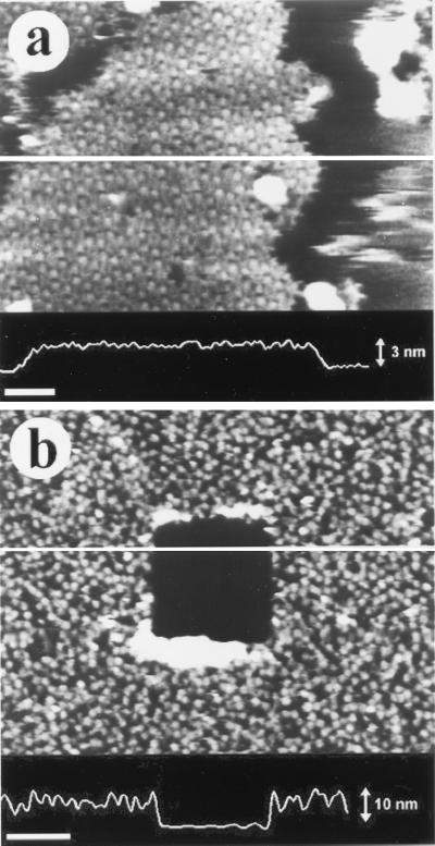
The distance by which the VacA oligomer projects from the bilayer, measured at the edge of a crystal patch, was found to be 2.9 ± 0.3 nm (n = 106) (Fig. 3a) regardless of whether or not the sample was fixed. Because the thickness of the pure lipid bilayer plus any water trapped between the bilayer and mica is ≈5 nm (15), the maximal height of the VacA oligomer associated with the membrane is 7.9 nm. This distance could be compared with the total height of the dodecamer by imaging VacA directly deposited to mica at pH 7 (Fig. 3b). In this way, the height of the water-soluble dodecamer was observed to be 9.6 ± 0.9 nm (n = 499), with or without chemical fixation, which is consistent with the range of values previously reported (6, 23–24).
Figure 3.
Height measurements of both the membrane-associated and water-soluble VacA oligomers. The cross-sectional profile along the line is shown at the bottom of each figure. (a) The height by which VacA complex protrudes from the bilayer is ≈3 nm, as determined from the edge of the crystal patch. Because the bilayer thickness is ≈5 nm (13), the maximal height of the membrane-associated oligomer is ≈8 nm. (b) Water-soluble VacA oligomers were directly adsorbed to mica, and a defect within the sample was created by the AFM tip with high force and high scan rate. The total height measured from the edge of this defect is ≈10 nm. These results were reproducible with or without chemical fixation. [Bar = 50 nm (a), 200 nm (b).]
Electrophysiological Measurements.
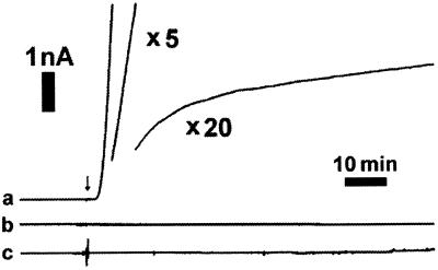
The large central ring of the membrane-associated VacA oligomer in the AFM images obviously suggests the possibility for transmembrane pores. When VacA was added to a planar lipid bilayer containing anionic lipids at pH 4, a large current was indeed observed after a delay of a few minutes (Fig. 4). This current was detected whether the potential was cis-positive or cis-negative, in contrast to the channels formed by typical A–B-type toxins, which open only for cis-positive potentials (25). No current was detected if VacA was added to bilayers at neutral pH or to bilayers without anionic lipids (Fig. 4), consistent with what was observed with AFM for membrane association. There was, however, a small, somewhat noisy current at low pH with bilayers composed only of zwitterionic lipids that began to appear ≈60 min after injection of the protein, which could indicate some weak affinity of VacA for this bilayer.
Figure 4.
VacA forms pores in planar lipid bilayers. When VacA (final concentration ≈0.1 μg/ml) was added (arrow) to bilayers composed of eggPC/DOPS/cholesterol (4:1:2 mol) in 5 mM citric acid/0.1 M KCl/2 mM EDTA, pH 4.0, a macroscopic current was detected after a few minutes (a). When VacA was added to bilayers of a similar composition as in a but at pH 7.0, no current was detected (b). When VacA was added to a zwitterionic bilayer of eggPC/cholesterol (5:2 mol) in the same pH 4.0 buffer as in a, no macroscopic current was observed (c). In all experiments, a cis-negative potential of 20 mV was applied across the bilayer. In trace a, the discontinuities were caused by a change in the gain of the recording system to avoid saturation. For the middle portion, the gain was lowered by a factor of 5 and for the final portion, by a factor of 20.
DISCUSSION
We have shown that below pH ≈ 5, VacA becomes associated with and inserts into anionic phospholipid bilayers to form oligomeric pores. The surface topography of the membrane-bound complex closely resembles the outer surface of the dodecamer observed previously, based on a comparison of both the lateral dimensions and the cant of the arms (6). However, it is unlikely that this membrane-associated oligomer is an inserted dodecamer for two reasons. First, considerable structural alterations would be necessary for the normally water-soluble exterior surface of the dodecamer to insert directly into the bilayer. Second, the height measurements clearly indicate that the maximal height of the oligomer in the membrane (7.9 nm) is markedly smaller than the total height of the soluble dodecamer (9.6 nm). We therefore conclude that the membrane-bound oligomer is not a dodecamer, but is a hexamer. Based on the resemblance to the dodecamer (6), we favor a model in which the tip-accessible surface of these membrane-bound hexamers corresponds to the outer surface of the dodecamer and the membrane-facing surface corresponds to a surface normally buried within the dodecamer. It is tempting to speculate that, by associating two hexamers into such a dodecamer and shielding a partly hydrophobic surface on each hexamer, this cytotoxin might have evolved an efficient mechanism by which two pore-forming complexes might be rapidly produced after a decrease of the pH.
Pore formation presumably requires that VacA must insert and span the entire bilayer [typical thickness ≈4.5 nm (26)]. Because the VacA hexamers protrude only 2.9 nm from the bilayer and the expected height of the hexamer in the water-soluble dodecamer is only about 4.8 nm, this suggests that the hexamers must undergo a large conformational change during the process of membrane insertion. A conformational change of this magnitude is believed to occur when another bacterial toxin, perfringolysin O, forms its pore (27).
With AFM, we have not been able to determine whether, after protonation, the dodecamer first splits into hexamers that then bind to the membrane or whether the dodecamer first disassembles into monomers in solution, followed by binding of monomers to the membrane and rapid oligomerization into hexamers, similar to several other pore-forming toxins (28). However, it should be noted that although the AFM images of water-soluble VacA revealed ≈20% tetradecamers (data not shown), no heptamers on the bilayer were ever detected either in crystal patches or with well resolved isolated oligomers, suggesting that disassembly into monomers in solution most likely precedes the formation of hexameric pores. This is also consistent with the previous studies in which the VacA dodecamer was observed to disassemble into monomers after incubation at low pH (6, 29–30). Such a dissociation step is also believed to occur with another pore-forming toxin, aerolysin, during its transition from a water-soluble dimer to a membrane-bound heptamer (31).
Because no association of preacidified VacA with the membrane at neutral pH was observed, the protonation of the monomers appears to be a critical determinant for VacA binding to anionic phospholipids. VacA also binds more avidly to the surface of HeLa cells at low pH than at neutral pH (M. S. McClain, personal communication). However, because VacA failed to bind to bilayers with negatively charged gangliosides (data not shown), the nature of VacA membrane association may be more than a simple electrostatic attraction. The role that the anionic phospholipids may play in the formation of the VacA oligomer is a very interesting subject in general (13).
Low pH-induced membrane pore formation by VacA bears some similarity to several A–B-type toxins, including diphtheria toxin and Pseudomonas exotoxin A. However, the process of VacA disassembly and reassembly to form pores accompanied by a significant reduction in subunit stoichiometry is rather different from all other A–B-type toxins. Moreover, the putative A subunit of VacA fails to cause vacuolation when expressed in the host cell (10), and neither an enzymatic activity nor the translocation of the putative A subunit of VacA has yet been demonstrated. Therefore, VacA exhibits several properties that seem to be distinct from other A–B-type toxins.
VacA pore formation at pH < 5 may be informative regarding possible cellular sites where VacA exerts its effect. In vivo, because the pH of the stomach is acidic, it is possible that VacA may be acidified and interact directly with the lipid components of the plasma membrane of the gastric epithelium, resulting in the formation of pores. In view of this possibility, it is interesting to note that another pore-forming bacterial toxin, aerolysin, can induce vacuolation of the endoplasmic reticulum, apparently without any requirement for toxin internalization (32). However, in vitro, several studies have clearly demonstrated that binding of VacA to the cell membrane at pH 7 is followed by internalization (8–9). Whether a specific receptor is directly involved in binding and internalization of VacA has yet to be firmly established (33–35). If VacA is internalized via the endocytic pathway (9), the acidic environment in the late endosome/lysosome may be sufficient to trigger the disassembly of the VacA dodecamer and subsequent formation of hexameric pores in the endosomal membrane. Pore formation in endosomal membranes would presumably be relevant to the vacuolating effects of VacA, although the details of this process remain to be elucidated. The observed inhibition of VacA-induced vacuolation by bafilomycin A1 and monensin, which raise intraendosomal pH (36–38), lends direct support for this hypothesis. In contrast, the addition of weak bases to the culture medium also increases the luminal pH of endosomes (39) but potentiates the vacuolating activity of VacA (7, 36). Therefore, further experiments are required to understand the relevance and the site of VacA-induced pore formation. Among the many possibilities, one of the most informative will be to examine a VacA mutant in which the pore-forming activity is abolished. Further studies of VacA-induced pore formation will undoubtedly provide critical information on the molecular mechanisms by which VacA exerts its cytotoxic effects in vivo.
Acknowledgments
We thank Dr. J. Heuser for the initial suggestion to work on this project and for stimulating discussions. We gratefully acknowledge Drs. G. Szabo, D. Shi, A. V. Somlyo, and A. J. Avila-Sakar for useful advice. This work was supported by grants from the National Institutes of Health (RO1-RR07720 and PO1-HL48807 to Z.S.; RO1-AI39657 and RO1-DK53623 to T.L.C.), the Medical Research Service of the Department of Veterans Affairs (T.L.C.), and the American Heart Association (Z.S.).
ABBREVIATIONS
- AFM
atomic force microscopy
- PD
peripheral domain
References
- 1.Telford J L, Covacci A, Ghiara P, Montecucco C, Rappuoli R. Trends Biotechnol. 1994;12:420–426. doi: 10.1016/0167-7799(94)90031-0. [DOI] [PubMed] [Google Scholar]
- 2.Blaser M J. Sci Am. 1996;274(2):104–107. doi: 10.1038/scientificamerican0296-104. [DOI] [PubMed] [Google Scholar]
- 3.Cover T L, Blaser M J. Adv Intern Med. 1996;41:85–117. [PubMed] [Google Scholar]
- 4.Cover T L. Mol Microbiol. 1996;20:241–246. doi: 10.1111/j.1365-2958.1996.tb02612.x. [DOI] [PubMed] [Google Scholar]
- 5.Montecucco C. Curr Opin Cell Biol. 1998;10:530–536. doi: 10.1016/s0955-0674(98)80069-0. [DOI] [PubMed] [Google Scholar]
- 6.Cover T L, Hanson P I, Heuser J E. J Cell Biol. 1997;138:759–769. doi: 10.1083/jcb.138.4.759. [DOI] [PMC free article] [PubMed] [Google Scholar]
- 7.Cover T L, Blaser M J. J Biol Chem. 1992;267:10570–10575. [PubMed] [Google Scholar]
- 8.Garner J A, Cover T L. Infect Immun. 1996;64:4197–4203. doi: 10.1128/iai.64.10.4197-4203.1996. [DOI] [PMC free article] [PubMed] [Google Scholar]
- 9.Sommi P, Ricci V, Fiocca R, Necchi V, Romano M, Telford J L, Solcia E, Ventura U. Am J Physiol. 1998;275:G681–G688. doi: 10.1152/ajpgi.1998.275.4.G681. [DOI] [PubMed] [Google Scholar]
- 10.de Bernard M, Arico B, Papini E, Rizzuto R, Grandi G, Rappuoli R, Montecucco C. Mol Microbiol. 1997;26:665–674. doi: 10.1046/j.1365-2958.1997.5881952.x. [DOI] [PubMed] [Google Scholar]
- 11.Moll G, Papini E, Colonna R, Burroni D, Telford J, Rappuoli R, Montecucco C. Eur J Biochem. 1995;234:947–952. doi: 10.1111/j.1432-1033.1995.947_a.x. [DOI] [PubMed] [Google Scholar]
- 12.de Bernard M, Papini E, de Filppis V, Gottardi E, Telford J, Manetti R, Fontana A, Rappuoli R, Montecucco C. J Biol Chem. 1995;270:23937–23940. doi: 10.1074/jbc.270.41.23937. [DOI] [PubMed] [Google Scholar]
- 13.Czajkowsky D M, Sheng S, Shao Z. J Mol Biol. 1998;276:325–330. doi: 10.1006/jmbi.1997.1535. [DOI] [PubMed] [Google Scholar]
- 14.Shao Z, Yang J. Q Rev Biophys. 1995;28:195–251. doi: 10.1017/s0033583500003061. [DOI] [PubMed] [Google Scholar]
- 15.Czajkowsky D M, Shao Z. FEBS Lett. 1998;430:51–54. doi: 10.1016/s0014-5793(98)00461-x. [DOI] [PubMed] [Google Scholar]
- 16.Hansma H G, Hoh J. Annu Rev Biophys Biomol Struct. 1994;23:115–128. doi: 10.1146/annurev.bb.23.060194.000555. [DOI] [PubMed] [Google Scholar]
- 17.Han W, Lindsay S M, Dlakic M, Harrington R E. Nature (London) 1997;386:563. doi: 10.1038/386563a0. [DOI] [PubMed] [Google Scholar]
- 18.John S A, Saner D, Pitts J D, Holzenburg A, Finbow M E, Lal R. J Struct Biol. 1997;120:22–31. doi: 10.1006/jsbi.1997.3893. [DOI] [PubMed] [Google Scholar]
- 19.Reviakine I, Bergsmaschutter W, Brisson A. J Struct Biol. 1998;121:356–362. doi: 10.1006/jsbi.1998.4003. [DOI] [PubMed] [Google Scholar]
- 20.Hardt S, Wang B, Schmid M F. J Struct Biol. 1996;116:68–70. doi: 10.1006/jsbi.1996.0012. [DOI] [PubMed] [Google Scholar]
- 21.Crowther R A, Henderson R, Smith J M. J Struct Biol. 1996;116:9–16. doi: 10.1006/jsbi.1996.0003. [DOI] [PubMed] [Google Scholar]
- 22.Hanke W, Schlue W-R. Planar Lipid Bilayers. London: Academic; 1993. [Google Scholar]
- 23.Lupetti P, Heuser J, Manetti R, Massari P, Lanzavecchia S, Bellon P L, Dallai R, Rappuoli R, Telford J L. J Cell Biol. 1996;133:801–807. doi: 10.1083/jcb.133.4.801. [DOI] [PMC free article] [PubMed] [Google Scholar]
- 24.Lanzavecchia S, Bellon P L, Lupetti P, Dallai R, Rappuoli R, Telford J L. J Struct Biol. 1998;121:9–16. doi: 10.1006/jsbi.1997.3941. [DOI] [PubMed] [Google Scholar]
- 25.Hoch D H, Mira-Romero M, Ehrich B E, Finkelstein A, DasGupta B R, Simpson L L. Proc Natl Acad Sci USA. 1985;82:1692–1696. doi: 10.1073/pnas.82.6.1692. [DOI] [PMC free article] [PubMed] [Google Scholar]
- 26.Wiener M C, White S H. Biophys J. 1992;61:437–447. doi: 10.1016/S0006-3495(92)81848-9. [DOI] [PMC free article] [PubMed] [Google Scholar]
- 27.Shepard L A, Heuck A P, Hamman B D, Rossjohn J, Parker M W, Ryan K R, Johnson A E, Tweten R K. Biochemistry. 1998;37:14563–14574. doi: 10.1021/bi981452f. [DOI] [PubMed] [Google Scholar]
- 28.Bhakdi S, Bayley H, Valeva A, Walev I, Walker B, Weller U, Kehoe M, Palmer M. Arch Microbiol. 1996;165:73–79. doi: 10.1007/s002030050300. [DOI] [PubMed] [Google Scholar]
- 29.Reyrat J-M, Charrel M, Pagliaccia C, Burroni D, Lupetti P, de Bernard M, Ji X, Norais N, Papini E, Dallai R, Rappuoli R, Telford J L. FEMS Microbiol Lett. 1998;165:79–84. doi: 10.1111/j.1574-6968.1998.tb13130.x. [DOI] [PubMed] [Google Scholar]
- 30.Molinari M, Galli C, de Bernard M, Norais N, Ruysschaert J M, Rappuoli R, Montecucco C. Biochem Biophys Res Comm. 1998;248:334–340. doi: 10.1006/bbrc.1998.8808. [DOI] [PubMed] [Google Scholar]
- 31.Wilmsen H U, Leonard K R, Tichelaar W, Buckley J T, Pattus F. EMBO J. 1992;11:2457–2463. doi: 10.1002/j.1460-2075.1992.tb05310.x. [DOI] [PMC free article] [PubMed] [Google Scholar]
- 32.Abrami L, Fivaz M, Glauser P-E, Parton R G, van der Goot F G. J Cell Biol. 1998;140:525–540. doi: 10.1083/jcb.140.3.525. [DOI] [PMC free article] [PubMed] [Google Scholar]
- 33.Yahiro K, Niidomo T, Hatakeyama T, Aoyagi H, Kurazono H, Padilla P I, Wada A, Hirayama T. Biochem Biophys Res Comm. 1997;238:629–632. doi: 10.1006/bbrc.1997.7345. [DOI] [PubMed] [Google Scholar]
- 34.Massari P, Manetti R, Burroni D, Nuti S, Norais N, Rappuoli R, Telford J L. Infect Immun. 1998;66:3981–3984. doi: 10.1128/iai.66.8.3981-3984.1998. [DOI] [PMC free article] [PubMed] [Google Scholar]
- 35.Seto K, Hayashi-Kumabara Y, Yoneta T, Suda H, Tamaki H. FEBS Lett. 1998;431:347–350. doi: 10.1016/s0014-5793(98)00788-1. [DOI] [PubMed] [Google Scholar]
- 36.Cover T L, Vaughn S G, Cao P, Blaser M J. J Infect Dis. 1992;166:1073–1078. doi: 10.1093/infdis/166.5.1073. [DOI] [PubMed] [Google Scholar]
- 37.Papini E, Bugnoli M, de Bernard M, Figura N, Rappuoli R, Montecucco C. Mol Microbiol. 1993;7:323–327. doi: 10.1111/j.1365-2958.1993.tb01123.x. [DOI] [PubMed] [Google Scholar]
- 38.Cover T L, Reddy L Y, Blaser M J. Infect Immun. 1993;61:1427–1431. doi: 10.1128/iai.61.4.1427-1431.1993. [DOI] [PMC free article] [PubMed] [Google Scholar]
- 39.Ohkuma S, Poole B. Proc Natl Acad Sci USA. 1978;75:3327–3331. doi: 10.1073/pnas.75.7.3327. [DOI] [PMC free article] [PubMed] [Google Scholar]


