Herniation of a ureter in an inguinal canal and scrotum is rare, and the general surgeon should be aware of this possibility to avoid injury to the ureter during hernial dissection. There are 2 types of inguinoscrotal herniation of a ureter: paraperitoneal (with a peritoneal hernial sac) and extraperitoneal (without a hernial sac).1 We report a case of the rarer form in which extraperitoneal herniation of the ureter in the left inguinal canal reached the upper pole of the testis and was misdiagnosed as a varicocele.
CASE REPORT
A 58-year-old man reported swelling in the left inguinoscrotal region and a dragging sensation in the left lower abdomen for 6 years. On clinical examination, the left scrotum felt like a “bag of worms.” Absent cough impulse and irreducibility ruled out an inguinal hernia. The condition was diagnosed clinically as a varicocele. However, contraindicating this diagnosis was the finding that with the patient in the supine position the swelling did not reduce. There was no other significant finding.
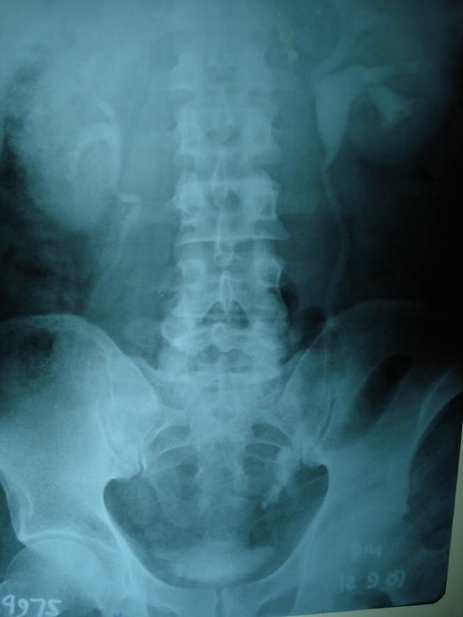
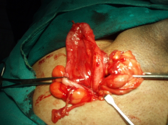
Routine investigations were within normal limits. Ultrasonography indicated that the mass was a varicocele. We performed surgery under local anesthesia. After opening the inguinal canal and the cremasteric layer, we noted a whitish tubular structure arising from the deep inguinal ring (Fig. 1). It had its own mesenteric-like blood supply. On tracking its path, we found that the structure entered the scrotum reaching the upper part of the testis; from there it reversed direction, forming a loop. During manipulation, we saw peristaltic movement in the structure, giving a clinical impression of a ureter. Aspiration with a 2-mL syringe yielded clear fluid; thus, we made an intraoperative diagnosis of ureter. We freed the ureter from adjacent tissue and reduced the hernia. Since there was no peritoneal sac, we were able to diagnose extraperitoneal herniation of a ureter. Postoperatively, a contrast-enhanced computed tomography (CT) scan showed mild dilatation of the ureter proximally and of the pelvicalyceal system of the left kidney. Intravenous urography showed a dilated, tortuous left lower ureter and mild left-sided hydronephrosis (Fig. 2).
Fig. 1. Surgical view showing the ureter as a white tubular structure in the inguinal canal. Photograph taken from the head of the patient.

Fig 2. Intravenous urogram showing a dilated and tortuous left lower ureter.
Since the patient was experiencing back pressure, we performed a second surgery: resection of the coiled tortuous extraureter with end-to-end anastomosis. The postoperative period was uncomplicated. The patient was followed up for 6 months; intravenous urography showed normal function of the left kidney with no sign of hydronephrosis.
DISCUSSION
Inguinoscrotal herniation of the ureter has been reported about 140 times in the literature. In the paraperitoneal type, a loop of herniated ureter extends alongside a peritoneal sac, whereas in the extraperitoneal type, a hernial sac is absent. Of these 2 types, about 80% are paraperitoneal and 20% are extraperitoneal.2
In paraperitoneal hernias, the ureter gets pulled into the scrotum owing to traction on underlying structures or because of adhesions connecting the ureter to the posterior peritoneum. Since paraperitoneal hernia is an indirect, sliding type of hernia, abdominal viscera may form the wall of the hernial sac. In contrast, in extraperitoneal hernias, only the ureter herniates without any peritoneal sac. Failure of ureteral differentiation during embryonic life and adhesion between the primitive ureter and the genitoinguinal ligaments causing herniation of the ureter make up the congenital basis for these hernias.3 The extraperitoneal type may be associated with other congenital renal or ureteral anomalies (such as crossed renal ectopia and nephroptosis). In our patient's case, we ruled out these anomalies with postoperative CT and intravenous urography.
CONCLUSIONS
Ureter in an inguinal canal is an uncommon presentation. Since this condition is usually misdiagnosed and there may be iatrogenically induced injury to the ureter, this entity should be kept in mind by general surgeons when they perform surgery on an atypical hernia. Intravenous urography or CT is not justifiable before surgery on every inguinal hernia and varicocele, so the surgeon should be cautious when resecting a huge lipoma and sliding fat during inguinal hernia dissection to prevent damage to the ureter.
Competing interests: None declared.
Correspondence to: Dr. R.K. Sharma Room no-16, Hostel no. 1 Bariatu Rd. Rajendra Institute of Medical Sciences Ranchi, Pin 834009 India inocent36@yahoo.com
References
- 1.Schlussel RN, Retik AB. Ectopic ureter, ureterocele, and other anomalies of the ureter. In: Walsh PC, Wein AJ, Vaughan ED, et al., editors. Campbell's urology. 8th ed. Oxford: WB Saunders; 2002. p. 2047-8.
- 2.Hwang CM, Miller FH, Dalton DP, et al. Accidental ureteral ligation during an inguinal hernia repair of patient with crossed fused renal ectopia. Clin Imaging 2002;26:306-8. [DOI] [PubMed]
- 3.Rocklin MS, Apelgren KN, Slomski CA, et al. Scrotal incarceration of the ureter with crossed renal ectopia: case report and literature review. J Urol 1989;142:366-8. [DOI] [PubMed]


