

Abstract
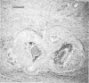
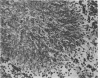
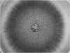
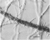
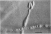
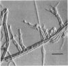
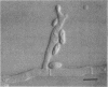
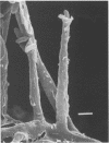
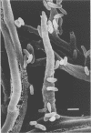
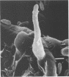
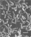
A 63-year-old Japanese man had phaeohyphomycosis that occurred as a solitary subcutaneous nodule on the dorsal aspect of his left hand. In the nodule there were foci of mixed granulomatous and suppurative infiltrations circumscribed by thick fibrous tissue reaction. The foci contained short septate hyphae and occasionally small rounded aggregates of irregularly branched septate hyphae, both of which were nonpigmented or rarely weakly pale brown. Fungal culture from the nodule was positive for a dematiaceous mold. The mycologic features of the mold were typical of Phialophora repens. The infection was successfully treated by excision of the nodule. This is the second reported case of infection due to P. repens.
Full text
PDF


Images in this article

Selected References
These references are in PubMed. This may not be the complete list of references from this article.
- Ajello L., Georg L. K., Steigbigel R. T., Wang C. J. A case of phaeohyphomycosis caused by a new species of Phialophora. Mycologia. 1974 May-Jun;66(3):490–498. [PubMed] [Google Scholar]
- Ajello L. Hyalohyphomycosis and phaeohyphomycosis: two global disease entities of public health importance. Eur J Epidemiol. 1986 Dec;2(4):243–251. doi: 10.1007/BF00419488. [DOI] [PubMed] [Google Scholar]
- Hironaga M., Mochizuki T., Watanabe S. Cutaneous phaeohyphomycosis of the sole caused by Exophiala jeanselmei and its susceptibility to amphotericin B, 5-FC and ketoconazole. Mycopathologia. 1982 Aug 20;79(2):101–104. doi: 10.1007/BF00468086. [DOI] [PubMed] [Google Scholar]
- Matsumoto T., Nishimoto K., Kimura K., Padhye A. A., Ajello L., McGinnis M. R. Phaeohyphomycosis caused by Exophiala moniliae. Sabouraudia. 1984;22(1):17–26. [PubMed] [Google Scholar]
- McGinnis M. R., Rinaldi M. G., Winn R. E. Emerging agents of phaeohyphomycosis: pathogenic species of Bipolaris and Exserohilum. J Clin Microbiol. 1986 Aug;24(2):250–259. doi: 10.1128/jcm.24.2.250-259.1986. [DOI] [PMC free article] [PubMed] [Google Scholar]
- McGinnis M. R., Sorrell D. F., Miller R. L., Kaminski G. W. Subcutaneous phaeohyphomycosis caused by Exophiala moniliae. Mycopathologia. 1981 Feb 13;73(2):69–72. doi: 10.1007/BF00562592. [DOI] [PubMed] [Google Scholar]
- Meyers W. M., Dooley J. R., Kwon-Chung K. J. Mycotic granuloma caused by Phialophora repens. Am J Clin Pathol. 1975 Oct;64(4):549–555. doi: 10.1093/ajcp/64.4.549. [DOI] [PubMed] [Google Scholar]
- Nielsen H. S., Jr, Conant N. F. A new human pathogenic Phialophora. Sabouraudia. 1968 Jun;6(3):228–231. [PubMed] [Google Scholar]
- Schwartz I. S., Emmons C. W. Subcutaneous cystic granuloma caused by a fungus of wood pulp (Phialophora richardsiae). Am J Clin Pathol. 1968 Apr;49(4):500–505. doi: 10.1093/ajcp/49.4.500. [DOI] [PubMed] [Google Scholar]
- Weitzman I., Gordon M. A., Henderson R. W., Lapa E. W. Phialophora parasitica, an emerging pathogen. Sabouraudia. 1984;22(4):331–339. doi: 10.1080/00362178485380541. [DOI] [PubMed] [Google Scholar]
- Yoshimori R. N., Moore R. A., Itabashi H. H., Fujikawa D. G. Phaeohyphomycosis of brain: granulomatous encephalitis caused by Drechslera spicifera. Am J Clin Pathol. 1982 Mar;77(3):363–370. doi: 10.1093/ajcp/77.3.363. [DOI] [PubMed] [Google Scholar]
- Zapater R. C., Albesi E. J., Garcia G. H. Mycotic keratitis by Drechslera spicifera. Sabouraudia. 1975 Nov;13(3):295–298. [PubMed] [Google Scholar]