Summary
In the mammalian liver, bile is transported to the intestine through an intricate network of bile ducts. Notch signaling is required for normal duct formation, but its mode of action has been unclear. Here, we show in mice that bile ducts arise through a novel mechanism of tubulogenesis involving sequential radial differentiation. Notch signaling is activated in a subset of liver progenitor cells fated to become ductal cells, and pathway activation is necessary for biliary fate. Notch signals are also required for bile duct morphogenesis, and activation of Notch signaling in the hepatic lobule promotes ectopic biliary differentiation and tubule formation in a dose-dependent manner. Remarkably, activation of Notch signaling in postnatal hepatocytes causes them to adopt a biliary fate through a process of reprogramming that recapitulates normal bile duct development. These results reconcile previous conflicting reports about the role of Notch during liver development and suggest that Notch acts by coordinating biliary differentiation and morphogenesis.
Keywords: Notch, Bile ducts, Liver, Mouse
INTRODUCTION
Bile plays an important role in metazoan biology by emulsifying fats and transporting the products of liver detoxification. After its synthesis by hepatocytes, bile is carried from the liver to the intestine by the bile ducts. Dysfunction of the biliary system, either through obstruction, destruction, congenital malformation or cancer, is a significant cause of morbidity and mortality. The large proximal ducts of the liver [extrahepatic bile ducts (EHBDs)] arise by branching of a primitive gut-derived diverticulum, whereas the smaller intrahepatic bile ducts (IHBDs), which constitute the largest component of the biliary tree, form in situ. During IHBD development, hepatic progenitor cells (hepatoblasts) adjacent to portal veins undergo ductal commitment, forming a structure known as the ductal plate, while progenitors located in the parenchyma, away from the portal veins, become hepatocytes (Lemaigre and Zaret, 2004). Prior to birth, tubular structures arise at discrete sites within the ductal plate, ultimately giving rise to IHBDs, while the remaining progenitor cells regress during the first few weeks of life (see Fig. 1A). It is not known how communication is established between the extra- and intrahepatic biliary systems.
Fig. 1.
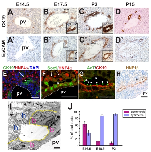
Biliary tubules arise as asymmetric structures in the ductal plate. (A-D′) Timecourse of mouse intrahepatic bile duct (IHBD) formation. Ck19+ (A-D) and Epcam+ (A′-D′) ductal plate precursor cells arise between E14.5 (A,A′) and E17.5 (B,B′). Tubules initially form as asymmetric structures (E17.5, insets), in which cells on the portal side express Ck19 (B) and Epcam (B′), whereas cells on the parenchymal side do not express these markers (arrowheads). Bile ducts achieve symmetry early in postnatal life (P2, C,C′ insets). During the first 2 weeks of life, most ductal plate cells that are not integrated into a duct regress, leaving behind mature bile ducts (D,D′). (E-H) Nascent tubules (asterisks) at E16.5 are lined by cells that express Ck19, Sox9, acetylated tubulin (AcT, arrowheads) and Hnf1β within the inner portal layer, and Hnf4α within the outer parenchymal layer (arrows). In E, note the presence of numerous Hnf4α-negative nuclei, reflecting the preponderance of hematopoietic and other `non-parenchymal' cells in the embryonic liver. (I) Transmission electron micrograph of an E17.5 asymmetric primitive ductal structure. Outer layer cells (h) and inner layer cells (b) can be distinguished by the presence of glycogen in the former (arrowheads). (J) Quantitation of ductal asymmetry during liver development (±s.e.m.). pv, portal vein; e, endothelial cell. Scale bars: 20 μm in E-H; 4 μm in I.
Notch signaling is necessary for normal bile duct development. In humans, mutations in the Notch ligand JAG1 or in the NOTCH2 receptor are responsible for Alagille syndrome (AGS), an autosomal-dominant disorder, the features of which include IHBD paucity and associated heart and skeletal abnormalities (Alagille et al., 1987; Emerick et al., 1999; Li et al., 1997; McDaniell et al., 2006; Oda et al., 1997). Importantly, inheriting a single mutant JAG1 or NOTCH2 allele is sufficient to cause human disease, suggesting that bile duct development is sensitive to small (twofold) changes in ligand or receptor levels. Several studies have examined the expression of Notch signaling components in embryonic and adult tissues (Crosnier et al., 2000; Flynn et al., 2004; Jones et al., 2000; Kodama et al., 2004; Loomes et al., 2002; Louis et al., 1999; McCright et al., 2002; Nijjar et al., 2001; Sumazaki et al., 2004; Tanimizu and Miyajima, 2004), and in vivo studies in the mouse have confirmed a requirement for Notch signaling in biliary development (Geisler et al., 2008; Kodama et al., 2004; Lozier et al., 2008; McCright et al., 2002). Nevertheless, the cellular and molecular mechanisms by which Notch regulates bile duct development are unclear.
The Notch pathway is an evolutionarily conserved signaling module. Upon ligand binding, a portion of the Notch receptor [the Notch intracellular domain (NICD)] translocates to the nucleus, where it associates with the DNA-binding protein Rbpj (also known as RBP-Jκ) and mediates changes in gene transcription. During development, Notch regulates embryonic patterning by conferring fate instructions to neighboring cells, most commonly through the Hes/Hey family of transcriptional repressors (Ehebauer et al., 2006; Kageyama et al., 2007). Notch could play a similar role in liver development by regulating a hepatocyte versus biliary epithelial cell (BEC) fate choice, and several studies have implicated Notch in the regulation of hepatoblast differentiation (Ader et al., 2006; Kodama et al., 2004; Tanimizu et al., 2003; Tanimizu et al., 2004). Arguing against such a model, mice with mutations in Notch2 or in the Notch target Hes1 exhibit abnormal duct morphology but normal biliary induction, raising the possibility that Notch signaling is dispensable for embryonic biliary specification and required only for morphogenesis (Geisler et al., 2008; Kodama et al., 2004; Lozier et al., 2008).
In the present study, we have performed a detailed in vivo analysis of Notch function during liver development by blocking or activating core components of the pathway at distinct developmental stages. We describe a novel mechanism for duct morphogenesis that relies upon sequential differentiation of adjacent layers of precursor cells. In addition, we report that Notch functions earlier than previously described in the embryonic liver, where it plays important roles in differentiation and tubule formation at distinct stages of development. Taken together, these results indicate that Notch acts in a temporal- and dose-dependent manner to coordinate biliary fate and morphogenesis.
MATERIALS AND METHODS
Mouse studies
Mice were maintained in a pathogen-free environment. All strains have been described: AFP-Cre (Kellendonk et al., 2000), Foxa3-Cre (Lee et al., 2005), Albumin-CreER (Schuler et al., 2004), RosaNICD (Murtaugh et al., 2003) and RbpjloxP/loxP (Han et al., 2002) mice were kindly provided by G. Schutz, K. Kaestner, P. Chambon, D. Melton and T. Honjo (RIKEN BioResources), respectively. A null allele of Rbpj (RbpjΔ) was made by crossing RbpjloxP/+ and Sox2-Cre mice (Hayashi et al., 2002). For activating Notch signaling in differentiated hepatocytes, 6 mg tamoxifen (TM) was administered to Albumin-CreER; RosaNICD mice on alternating days for a total of 3-5 doses, and sections were examined 5-21 days later. Serum chemistries were measured by Analytics (Gaithersburg, MD, USA). All studies were performed in accordance with policies for the humane use of animals established by the University of Pennsylvania and the NIH.
Immunostaining and immunoblotting
Tissues were fixed in zinc-buffered formalin (Polysciences), embedded in paraffin and cut at 5 μm. For antigen retrieval, slides were incubated with R-buffer A (Electron Microscopy Sciences) at 120°C using a Pickcell 2100 antigen retrieval system. Sections were blocked with 2% donkey serum or CAS Block (Invitrogen). Primary antibodies are listed in Table 1. Rabbit anti-Ck19 and anti-Hes1 antibodies were made by synthesizing peptides as described (Ito et al., 2000; Tanimizu et al., 2003), conjugating to Keyhole limpet hemocyanin (KLH), and immunizing rabbits with each peptide (Covance). For immunohistochemistry, sections were sequentially incubated with biotinylated secondary antibodies (Jackson ImmunoResearch), peroxidase-conjugated streptavidin (ABC Staining Kit, Vector Labs), DAB substrate and Hematoxylin. Alexa 488- or Alexa 594-conjugated secondary antibodies (Invitrogen) were used with DAPI counterstaining for immunofluorescence. For Hes1 and Jag1 immunostaining, tyramide signal amplification was performed using the TSA Fluorescence System (PerkinElmer). All reported results were observed in at least three animals. For western blotting, total protein was extracted from whole liver, separated by SDS-PAGE and transferred to nitrocellulose. Membranes were blocked with 5% milk powder and visualized using Chemiluminescence Reagent Plus (PerkinElmer) following incubation with an appropriate secondary antibody. Mouse anti-Gapdh (US Biological) was used as a loading control.
Table 1.
Primary antibodies used for immunostaining experiments
| Species | Source | Catalog # | Dilution | Notes | |
|---|---|---|---|---|---|
| Acetylated tubulin | Mouse | Sigma | T-6793 | 1:250 | |
| Cytokeratin 19 | Rabbit | D. Melton | – | 1:1000 | |
| Epcam | Rat | DSHB | G8.8 | 1:500 | |
| GFP | Goat | Abcam | 6673 | 1:500 | |
| Hes1 | Rabbit | Covance | – | 1:1500 | Tyramide amplification |
| Hnf1β | Goat | Santa Cruz | SC-7411 | 1:250 | |
| Hnf4α | Goat | Santa Cruz | SC-6556 | 1:250 | |
| Hnf4α | Rabbit | Santa Cruz | SC-8987 | 1:250 | |
| Jag1 | Goat | R&D | AF1277 | 1:250 | Tyramide amplification |
| Osteopontin | Goat | R&D | AF808 | 1:1000 | |
| Sox9 | Rabbit | Chemicon | AB5535 | 1:500 |
DSHB, Developmental Studies Hybridoma Bank
Duct and BEC quantification
To quantify symmetric versus asymmetric ducts during development, over 100 ductal structures (sampled from three or more animals at each time point) were scored. To quantitate Sox9+ or Ck19+ cells, four mutant animals and four controls (lacking Cre) were examined and positive cells were scored from at least ten portal tracts per animal. To measure the number of ducts per portal vein, slides at a comparable level of section from at least three mutant and three control animals were selected. Portal veins were identified by the presence of five or more biliary cells in the perivascular region, and associated bile ducts were counted. To measure proliferation, AFP-NICD and control livers (n=5 for each genotype, more than 500 cells per liver) at P2 were co-stained for Ki67 and Ck19 and the number of Ki67+ cells was calculated as a percentage of the total Ck19+ cells counted. P-values were calculated by Student's t-test.
Quantitative PCR
Total RNA was extracted from whole liver using the RNeasy Mini Kit (Qiagen) and 1 μg used to synthesize cDNA using the SuperScript Kit (Invitrogen, 11752). Quantitative PCR was performed with SYBR Green Master Mix Reagent (Applied Biosystems) using an ABI 7900 sequence detector. Transcript quantities were determined using the difference of Ct method; standard curves were constructed for each primer pair and values were normalized to Hprt. Primer sequences are listed in Table 2.
Table 2.
Primers used in this study
| Primer | Forward (5′ to 3′) | Reverse (5′ to 3′) |
|---|---|---|
| qPCR | ||
| Ck19 | CCCACGTGTTCCACAAACATC | CCATGGGAACAGTTATTTGGAGA |
| Hnf4a | TGCCTGCCTCAAAGCCAT | CACTCAGCCCCTTGGCAT |
| Oc1 | CGGAGTTCCAGCGCATGT | TCCTTCCCGTGTTCTTGCTC |
| Oc2 | CAGCATGCAAACGCAAAGA | TCTTCTGCGAGTTGTTCCTGTCT |
| Hnf1b | CCCAGCAATCTCAGAACCTC | AGGCTGCTAGCCACACTGTT |
| Hhex | GAAGACTGAAACAGGAGAATCCTCA | TCCAAACTGTCCAACGCATC |
| Sox9 | ACTCTGGGCAAGCTCTGGAG | CGAAGGGTCTCTTCTCGCTCT |
| Jag1 | CCCACGTGTTCCACAAACATC | CCATGGGAACAGTTATTTGGAGA |
| Jag2 | TCCTCCTGCTGCTTTGTGATC | TCAGGCAGGTCCCTTGCA |
| Dll1 | GATGGATCTCTGCGGCTCTTC | GCACCGGCACAGGTAAGAGT |
| Dll4 | AGCAACCCGTGTGCCAAT | TCGGCTTGGACCTCTGTTCT |
| Notch1 | CAATGTTCGAGGACCAGATGG | ACTGCAGGAGGCAATCATGAG |
| Notch2 | GACGTGCTGGACGTGAATGT | CAGGTCTGAGCTGCCTCCTC |
| Notch3 | GCACTTGCCGTGGTTACATG | CCTCACAACTGTCACCAGCATAG |
| Notch4 | CTGTGTGCCTCAGCCCAGT | TGAGCAGTTCTGTCCCTCATAGC |
| Hes1 | AAAGCCTATCATGGAGAAGAGGCG | GGAATGCCGGGAGCTATCTTTCTT |
| Hes5 | GAGATGCTCAGTCCCAAGGAGA | CGGTCCCGACGCATCTT |
| Hey1 | ACACTGCAGGAGGGAAAGGTT | CAAACTCCGATAGTCCATAGCCA |
| Hey2 | AAGCGCCCTTGTGAGGAAAC | GGTAGTTGTCGGTGAATTGGAC |
| Heyl | TCCGACGGCGAGTCTGAT | AGCGGCCTGGCCATCT |
| ChIP | ||
| Sox9 #3 | GGCTTTCTCACACGTGGGAA | TCGGAGACGGGATGTGGA |
| Sox9 #8 | GAGCATCTACAGTGCTTTTCCCA | GGAAGTGCTTGTCTACAGGGAATT |
| Sox9 #9 | GGTTCACACGGAGACCGTTC | TGCTCTCTACTCTCGGAATGTCAC |
| Sox9 #10 | GCTGCTCGGAACTGCCTG | AACTGGTAAAGTTGTCGCTCCC |
| Ctrl | TCCTGCCTTCTTTATTACTCTTAAAGC | GCAGAGGCCCTGAGACAACA |
Chromatin immunoprecipitation (ChIP) analysis
ChIP was performed using the ChIP Assay Kit (Upstate). Liver tissue (100 mg) was minced in PBS and cross-linked using 1% formaldehyde for 10 minutes. Cross-linking was quenched by the addition of glycine to a final concentration of 0.125 M. Cells were lysed with 1 ml lysis buffer supplemented with protease inhibitor (Roche). DNA was sheared into fragments of 100-500 bp by BioRuptor sonication (Diagenode), and cross-linked proteins were immunoprecipitated using Notch1 antiserum (Fang et al., 2007). After protein-A bead pull-down, cross-links were reversed and the DNA was purified using the QIAquick PCR Purification Kit (Qiagen). DNA copy number was measured by quantitative PCR, normalized to 28S ribosomal DNA sequences (Rubins et al., 2005). Enrichment of DNA was analyzed by comparing DNA copy number in ChIP samples with that of input. Primer sequences are listed in Table 2.
RESULTS
Notch signaling during IHBD development
We sought to confirm the normal sequence of events during IHBD development by examining the expression of the duct-specific cytokeratin Ck19 (Krt19 – Mouse Genome Informatics) at various stages (Fig. 1A-D). As described previously (Lemaigre, 2003), IHBD development is characterized by the appearance of ductal plate precursor cells adjacent to branches of the portal vein (∼E14-16), the appearance of dilations at discrete points along the ductal plates (∼E16-P2), and the postnatal disappearance of unincorporated biliary precursor cells (∼P2-15). In addition, we observed that nascent ducts pass through a previously undescribed intermediate stage characterized by the asymmetric expression of biliary and hepatoblast markers. Specifically, Ck19 was expressed by cells on the portal side, but not the parenchymal side, of these asymmetric tubules, whereas Hnf4α was expressed by cells on the parenchymal side, but not the portal side (Fig. 1B,E; see Fig. S1 in the supplementary material). Other markers of BECs, including Epcam, acetylated tubulin (AcT), Sox9, Hnf1β, and osteopontin (Opn; Spp1 – Mouse Genome Informatics) (Antoniou et al., 2009; Coffinier et al., 2002; Zhang et al., 2008), were also expressed exclusively by cells on the portal side of these primitive ductal structures (Fig. 1B′,F-H; see Movies 1, 2 in the supplementary material; data not shown). The asymmetry resolved after birth (P2), by which point the ducts had adopted a mature configuration and were symmetrically encircled by BECs (Fig. 1C,C′,J). These changes in biliary tubule composition were also apparent ultrastructurally (Fig. 1I; see Fig. S2 in the supplementary material). Thus, during IHBD development, lumen formation precedes the terminal differentiation of cells that will ultimately line the outer layer of the ducts.
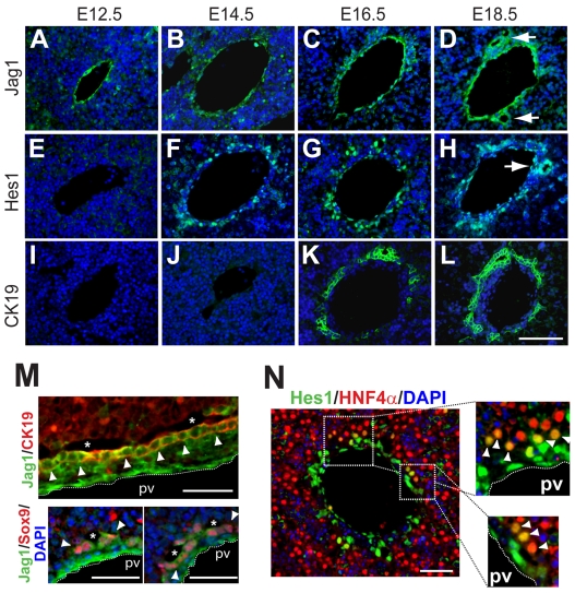
To directly examine the expression of Notch signaling components during liver development, we measured stage-specific transcript levels for all four Notch receptors, Jagged and Delta ligands, and targets from the Hes and Hey families by real-time PCR. At all stages examined, multiple signaling pathway components were expressed (see Fig. S3 in the supplementary material), indicating that functional redundancy might mitigate the loss of any single pathway component. We further characterized the expression of the Notch ligand Jag1 and Notch target Hes1 by immunostaining. Jag1 protein was detected in portal vein endothelium as early as E12.5, where it persisted throughout development (Fig. 2A-D). Jag1 was also expressed in BECs at later stages (Fig. 2D, arrows), consistent with several previous reports (Flynn et al., 2004; Loomes et al., 2002; Louis et al., 1999). Hepatoblasts surrounding the portal vein expressed Hes1 starting at ∼E14.5 (Fig. 2E,F), earlier than previously reported (Kodama et al., 2004). At later stages, Hes1 was expressed in ductal plate cells and mature ducts (Fig. 2G,H; see Fig. S4 in the supplementary material). Markers of terminal biliary differentiation such as Ck19 were expressed 1-2 days after Hes1 (Fig. 2I-L). These results suggest that activation of Notch signaling precedes the differentiation of the first (portal) layer of the ductal plate and persists in differentiated BECs.
Fig. 2.
Notch signaling is active during biliary development. (A-L) Immunofluorescence (green) for Jag1 (A-D), Hes1 (E-H) and Ck19 (I-L) demonstrates stepwise expression of Notch signaling components. Jag1 is expressed in the portal vein endothelium from E12.5 onward, whereas Hes1 is expressed in peri-portal cells (and endothelial cells) after E14.5. Expression of Ck19, a marker of terminal biliary differentiation, is observed at E16.5. Both Jag1 and Hes1 are expressed in mature ductal structures at E18.5 (arrows). All sections have been counterstained with DAPI (blue). (M) Jag1 staining is detected in portal endothelium (adjacent to dotted lines) and in cells on the portal side of primitive ductal structures at E16.5 (asterisks), where it overlaps with the expression of Sox9 and Ck19 (arrowheads). No Jag1 staining is observed in cells on the parenchymal side of asymmetric tubules. (N) Hes1 is expressed in endothelial cells and in both layers of asymmetric tubules at E16.5; in the outer layer, co-expression of Hes1 and Hnf4α is detected (arrowheads). Scale bars: 50 μm in A-L; 25 μm in M,N.
To examine the possibility that Notch activation might also precede differentiation in the second (parenchymal) layer of the ductal plate, we examined Jag1 and Hes1 expression during tubulogenesis. At E16.5, Jag1 staining was observed in portal endothelium (and possibly portal mesenchyme) as well as in cells on the portal side, but not the parenchymal side, of primitive ductal structures (Fig. 2M). Surprisingly, Hes1 was expressed by cells on both the portal and parenchymal sides of primitive ductal structures; co-staining revealed that a subset of cells comprising the second biliary layer expressed both Hes1 and Hnf4α (Fig. 2N). These results suggest that the field of Notch-responsive cells expands during biliary development in a focal manner, preceding fate acquisition in the second layer at sites of tubulogenesis.
Notch regulates embryonic biliary fate
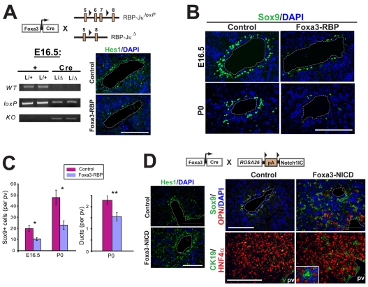
The role of Notch signaling in liver development has previously been assessed in mice bearing deletions in Jag1, Notch1, Notch2 or Hes1 (Geisler et al., 2008; Kodama et al., 2004; Loomes et al., 2007; Lozier et al., 2008; McCright et al., 2002). To circumvent possible functional redundancy, we employed mice with a conditional mutation in the Rbpj gene (Han et al., 2002). Rbpj constitutes the DNA-binding portion of the Notch transcription complex and is a necessary effector of canonical Notch signaling (Bolos et al., 2007; Oka et al., 1995). To examine the consequences of Rbpj loss on liver development, we obtained Foxa3-Cre mice (Lee et al., 2005), which permit early and efficient recombination in hepatoblasts (see Fig. S5A in the supplementary material). We then generated Foxa3-Cre; RbpjloxP/Δ (Foxa3-RBP) embryos, in which the DNA-binding domain of Rbpj was deleted on one allele and flanked by loxP recombination sequences on the other allele. Rbpj deletion was confirmed by genomic PCR, and loss of Notch signaling was documented by a reduction of Hes1 staining in the ductal plate region (Fig. 3A). Compared with controls, Foxa3-RBP mutants exhibited a reduced number of ductal plate cells at E16.5 and P0 (Fig. 3B,C; see Fig. S6 in the supplementary material) and a significant decrease in the number of bile ducts at P0 (Fig. 3C). These results indicate that Rbpj is necessary for normal ductal plate development in the embryonic liver.
Fig. 3.
Notch signaling controls embryonic biliary fate. (A) Deletion of Rbpj was achieved by creating Foxa3-Cre; RbpjloxP/Δ (Foxa3-RBP) mice, in which one allele of Rbpj has been deleted and the other allele contains loxP sites flanking crucial coding sequences. PCR for the wild-type, mutant and deleted alleles, using liver DNA as template, shows deletion of the Rbpj gene in Foxa3-RBP animals at E16.5. Since as many as half of the cells in the E16.5 liver are hematopoietic in origin (see Fig. 1E), the observed reduction in RbpjloxP PCR product reflects efficient deletion in hepatoblasts at this stage. A reduction in the number of Hes1+ cells in the peri-portal region of Foxa3-RBP mice confirms the loss of ductal plate Notch signaling at this stage. (B,C) Foxa3-RBP mutants exhibit a reduced number of Sox9+ BECs at E16.5 and P0 and a reduced number of bile ducts at P0. Bar charts show the mean (±s.e.m.) of Sox9+ cells or ducts per portal vein (pv). Each bar represents measurements from four independent animals. *P<0.05, **P<0.01. (D) Activation of Notch signaling in Foxa3-Cre; RosaNICD (Foxa3-NICD) mice results in an expansion of the Hes1 expression domain and ectopic biliary differentiation at E16.5; structures resembling mature bile ducts are found in Foxa3-NICD lobules (inset; nuclei are counterstained with DAPI). pv, portal vein. Scale bars: 100 μm.
Previous in vitro studies have suggested that Notch signaling induces biliary differentiation in hepatic cells (Kodama et al., 2004; Tanimizu et al., 2003; Tanimizu et al., 2004). To determine whether Notch plays an instructive role in biliary differentiation in vivo, we employed Rosa26Notch1IC mice (Murtaugh et al., 2003) (henceforth referred to as RosaNICD), which harbor a constitutively active form of Notch1 downstream of loxP-flanked transcriptional stop sequences (Fig. 3D) (Murtaugh et al., 2003). This strain has been used to activate the Notch signaling cascade in a variety of tissues (Cheng et al., 2007; Jadhav et al., 2006; Niranjan et al., 2008; Stanger et al., 2005). We obtained bigenic Foxa3-Cre; RosaNICD/+ (Foxa3-NICD) embryos at the expected Mendelian ratio. Widespread Hes1 staining was observed in Foxa3-NICD livers prior to E16.5, confirming that Notch signaling was activated in hepatic precursor cells (Fig. 3D). Ectopic BECs expressing a full repertoire of ductal markers were observed in the parenchyma of Foxa3-NICD livers as early as E16.5 (Fig. 3D; see Fig. S6B in the supplementary material; data not shown). Remarkably, some of these cells had assembled into bile ducts of mature appearance (Fig. 3D, inset; see Fig. S6B in the supplementary material). Taken together, these results suggest that Notch signaling (acting via Rbpj) regulates the differentiation of embryonic biliary precursors.
Notch regulates formation of the second biliary layer
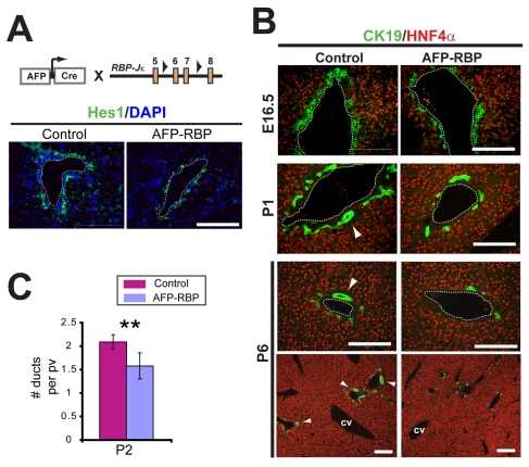
We next sought to determine whether Notch plays a role in the development of primitive ductal structures. We employed AFP-Cre mice, in which Cre recombinase is expressed under the regulatory control of the α-fetoprotein (Afp) enhancer and albumin promoter (Kellendonk et al., 2000), and confirmed by RosaYFP reporter analysis that recombination occurs later with AFP-Cre mice than with Foxa3-Cre mice. At E15.5, 36% of Hnf4α+ cells were labeled in AFP-Cre; RosaYFP mice, significantly less than the 81% of Hnf4α+ cells labeled in Foxa3-Cre; RosaYFP mice at this stage. By contrast, 88% of Hnf4α+ cells were labeled in AFP-Cre; RosaYFP mice at E16.5 (see Fig. S5 in the supplementary material). By P2, 95% of Hnf4α+ cells (hepatocytes) and 98% of Ck19+ cells (BECs) were labeled in AFP-Cre; RosaYFP mice. Thus, AFP-Cre exhibits peak activity (as measured by this assay) during the formation of the second ductal layer (∼E16.5). AFP-Cre; RbpjloxP/loxP (AFP-RBP) livers exhibited a less severe reduction in peri-portal Hes1+ cells at E16.5 than that observed with Foxa3-Cre (e.g. compare Fig. 3A with Fig. 4A). Consistent with less efficient deletion at this stage, mutant animals had ductal plates of normal appearance at E16.5 (Fig. 4B, top panels). At P1 and P2, however, AFP-RBP livers exhibited a significant reduction in the number of bile ducts (Fig. 4B,C). This defect was also apparent at P6 (Fig. 4B, bottom panels), indicating that the phenotype was not due to delayed bile duct maturation. These results suggest that following induction of the first ductal plate layer, Rbpj is required for the subsequent formation of mature ducts.
Fig. 4.
Late deletion of Rbpj preserves ductal plate formation but results in abnormal tubulogenesis. (A) Deletion of the transcriptional regulator Rbpj in AFP-Cre; RbpjloxP/loxP results in a more modest reduction in peri-portal Hes1+ cells at E16.5 than is seen in Foxa3-RBP mice (see Fig. 3). (B) AFP-RBP mutants have normal ductal plate development (E16.5) but have fewer mature bile ducts postnatally (arrowheads). cv, central vein. (C) Bar chart showing the mean number (±s.e.m.) of bile ducts per portal vein (pv); each bar represents the scoring of at least 50 portal regions (n=3 animals for each genotype). **P<0.01. Scale bars: 100 μm.
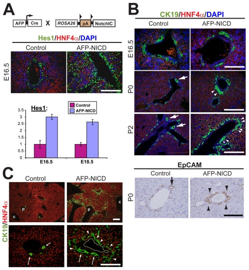
To determine whether Notch directly regulates tubulogenesis, we crossed RosaNICD mice to AFP-Cre mice, yielding bigenic AFP-Cre; RosaNICD/+ (AFP-NICD) embryos. AFP-NICD livers exhibited an increase in Hes1 transcript levels and protein, confirming that Notch signaling was activated throughout the hepatic lobule (Fig. 5A). Whereas the ductal plates appeared normal at E16.5, AFP-NICD mice exhibited an increase in portal vein-associated BECs at P0 and P2 (Fig. 5B). This change was associated with an increase in the size and number of bile ducts at P2 from a mean of 2.3 (control) to 3.5 ducts per portal vein (AFP-NICD) (n=3 for each genotype, P<0.001). Although most BECs were confined to the portal region, ectopic Ck19+ cells were also detected in the lobules starting at P2 (Fig. 5B, Fig. 6). These Ck19+ cells failed to undergo regression, leading to the persistence of BECs at P15 in a portal-to-lobular gradient (Fig. 5C). Ck19+ cells showed higher proliferation in AFP-NICD livers compared with control (1.44% versus 1.06%, respectively; P=0.042), indicating that enhanced proliferation might contribute to the phenotype.
Fig. 5.
Notch signaling promotes ectopic biliary differentiation in the periportal region. (A) Real-time PCR or staining for Hes1 (n=3 animals of each genotype per time point) shows increased expression throughout the hepatic lobule in AFP-NICD mouse embryos. (B) Ductal plate precursor cells appear at E16.5 in the correct peri-portal location in AFP-NICD embryos. An increase in peri-portal BECs at P0 and P2 is associated with more elongated and numerous ducts in AFP-NICD livers (arrowheads) compared with controls (arrows). (C) Ectopic biliary cells persist in AFP-NICD livers. (Left) By P15, control portal tracts exhibit near-complete regression of the ductal plate; most remaining Ck19+ cells are incorporated into bile ducts (arrows). (Right) Age-matched AFP-NICD livers exhibit abundant Ck19+ cells arranged in a ductal plate conformation (arrowheads). The peri-portal concentration of these cells, resulting in a portal-to-central gradient, can be appreciated at low magnification (top panels). Note the presence of enlarged bile duct lumens in AFP-NICD livers (arrows). p, portal vein; c, central vein. Scale bars: 200 μm in B (P0 and P2); 100 μm for all others.
Fig. 6.
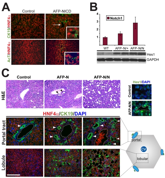
Notch promotes dose-dependent tubulogenesis at postnatal day 2. (A) Ectopic tubules exhibit expression of Ck19 and AcT. (Top) Lobular tubules in AFP-NICD mice (arrow) are lined by cells expressing either Hnf4α+ or Ck19+ (inset shows high magnification). (Bottom) AcT, a cilia marker normally confined to ductal plate progenitor cells, is ectopically expressed in the lobular tubules of AFP-NICD animals (inset shows high magnification). (B) Mice having one (AFP-NICD) or two (AFP-NICD/NICD) copies of RosaNICD exhibit graded levels of Notch1 transcripts by real-time PCR (top; n=3 animals for each genotype) and Hes1 protein by western blot analysis (bottom). (C) NICD gene dosage affects tubulogenesis at P2. Hematoxylin and Eosin staining reveals an increase in the number of lobular tubules with increasing RosaNICD gene dosage (H&E, top). A parallel increase in the size and number of peri-portal bile ducts (arrowheads) and lobular asymmetric tubules (bottom) is evident by Ck19 staining. Most cells surrounding the tubules express Hes1. Since over 90% of hepatic cells exhibit recombination at the Rosa26 locus prior to birth (see Fig. S5 in the supplementary material), these changes are likely to reflect increased `per-cell' activity of NICD rather than an increase in the number of cells expressing NICD. Scale bar: 100 μm.
Notch regulates tubulogenesis in a dose-dependent manner
At P2, AFP-NICD mice exhibited ectopic tubule formation in the lobules, an area normally occupied by hepatocyte-lined sinusoids (Fig. 6). The tubules were lined by Ck19+ cells and Hnf4α+ cells in a manner reminiscent of the asymmetric tubules present during normal biliary tubulogenesis (Fig. 6A, top right inset). Acetylated tubulin (AcT), a cilia marker that is confined to ductal plate BECs in control livers, was expressed in these ectopic structures (Fig. 6A, bottom panels). The tubules disappeared over the first 2 weeks of life and were replaced by duct-like structures (Fig. 5C; see Fig. S7E in the supplementary material), indicating that Notch-induced tubulogenesis is transient in nature.
Human bile duct development is sensitive to changes in JAG1 and NOTCH2 gene dosage. Therefore, we hypothesized that biliary tubulogenesis might be influenced by increasing the dose of Notch signaling. To test this possibility, we bred two copies of the RosaNICD allele into the AFP-Cre background (AFP-N/N), resulting in graded levels of Notch signaling in relation to RosaNICD copy number (Fig. 6B). AFP-N/N mice exhibited a profound tubulogenesis phenotype with dilated tubules in the ductal plate region as early as E16.5 (see Fig. S7C in the supplementary material). At P2, AFP-N/N livers exhibited dilated bile ducts and ectopic tubules throughout the lobule that completely disrupted normal hepatic architecture (Fig. 6C). Cells lining the tubules resembled their counterparts in AFP-NICD mice (i.e. cells expressed either Hnf4α or Ck19). A majority of the cells lining the tubules also expressed Hes1 (Fig. 6C, inset), again evoking the primitive ductal structures normally present at E16-17. In contrast to AFP-NICD mice, AFP-N/N mice exhibited bile ducts of mature appearance in the lobules at P15 (see Fig. S7F in the supplementary material). Surprisingly, these animals exhibited preserved liver chemistries (see Fig. S8 in the supplementary material). Notably, serum bilirubin was undetectable in AFP-N/N animals, raising the possibility that the ectopic ducts in these animals were functional. Notch signaling can therefore exert a direct effect on morphogenesis during liver development, promoting tubule formation and bile duct maturation in a dose-dependent manner.
Sox9 is a Notch target
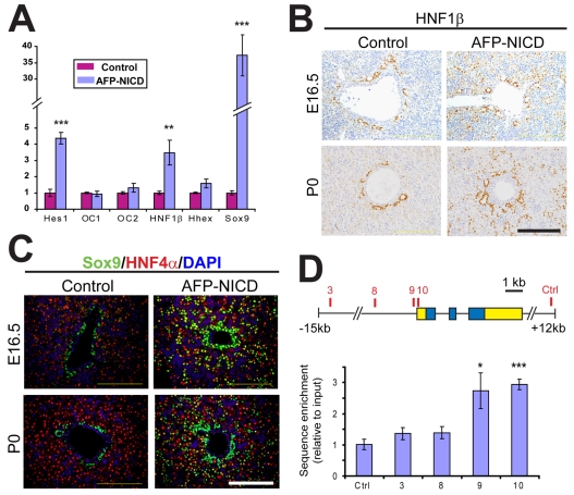
To better understand the mechanism underlying Notch-induced biliary differentiation and tubulogenesis, we examined the expression of known regulators of biliary development – Oc1 (Onecut1 – Mouse Genome Informatics), Oc2, Hnf1b, Hhex and Sox9 – by real-time PCR. Transcripts for Hnf1b and Sox9, but not Oc1, Oc2 or Hhex, were significantly increased in AFP-NICD livers compared with controls at P0 (Fig. 7A). Immunostaining confirmed that Sox9 and Hnf1β were ectopically expressed throughout the lobules of AFP-NICD livers at E16.5 and P0 (Fig. 7B,C). Conversely, no increase in Oc1 or Hhex staining was detected (data not shown). To determine whether Sox9 is direct target of Notch, we scanned upstream sequences of the Sox9 gene and identified ten consensus Rbpj binding sites. We chose four elements – three sites close to the Sox9 promoter region and one conserved element 14 kb upstream – for chromatin immunoprecipitation (ChIP) studies in AFP-NICD livers. When compared with a control sequence, the two sequences closest to the Sox9 transcriptional start site were significantly enriched following ChIP with an anti-Notch1 antibody, whereas the other sequences showed no enrichment (Fig. 7D). This result suggests that Notch1 is capable of binding directly to the Sox9 promoter in vivo. Because these two sites (#9 and #10) are within ∼400 bp of each other, this result could represent binding of NICD to both or a single site.
Fig. 7.
Notch induces Hnf1β and Sox9 expression. (A) Real-time PCR measuring transcript levels (mean±s.e.m.) for several transcription factors involved in biliary development, comparing control and AFP-NICD mice at P0 (n=3 for each group). Hnf1b and Sox9 are the only two (other than Hes1) that exhibit a significant increase. Data are representative of two independent experiments. (B,C) Immunostaining for Hnf1β (B) and Sox9 (C) confirms widespread expression of both transcription factors throughout the mutant lobule as early as E16.5. (D) Schematic view of the Sox9 gene (5′ and 3′ untranslated regions in yellow, coding sequence in blue) showing the location of putative Rbpj binding sites and the control site. Chromatin prepared from AFP-NICD livers (P10) was subjected to immunoprecipitation with a Notch1 antibody followed by real-time PCR amplification using primers specific for each candidate binding site (see Materials and methods). The data represent the mean (±s.e.m.) of five independent experiments. *P<0.05, **P<0.01, ***P<0.001; all other differences were not significant at the P=0.05 level. Scale bars: 100 μm.
Notch signaling reprograms postnatal albumin+ cells
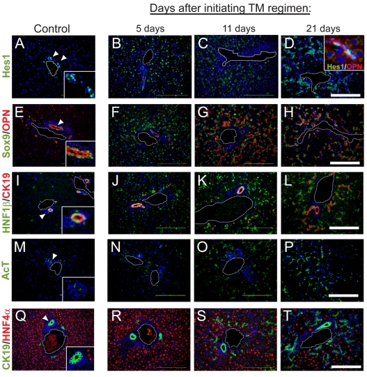
Because BECs first appear in the lobules of AFP-NICD mice postnatally, several days after the onset of ectopic Hes1, Sox9 and Hnf1β expression, we hypothesized that terminally differentiated hepatocytes might retain competence to respond to Notch signals. To test this possibility, we employed the Albumin-CreER strain (Schuler et al., 2004), which mediates loxP recombination in hepatocytes following tamoxifen (TM) administration (see Fig. S5 in the supplementary material). We induced recombination by giving TM to 6-day-old Albumin-CreER; RosaNICD/+ (AlbuminCreER-NICD) mice and examined liver sections 5, 11 or 21 days following the first dose (Fig. 8).
Fig. 8.
Notch activation in differentiated hepatocytes promotes biliary differentiation. (A-T) Albumin-CreER; RosaNICD mice were given five doses of tamoxifen (TM) (6 mg) starting at P6 and examined by immunofluorescence for the indicated markers 5, 11 or 21 days after the first dose. Expression of Hes1, Sox9, Opn, Hnf1β, AcT and Ck19 was initially restricted to the portal tract (arrowheads, left column; insets show high magnification). TM treatment leads to rapid induction of Hes1, Sox9 and Hnf1β, but a more gradual induction of AcT, Opn and Ck19. Note the rearrangement of ectopic biliary cells from a diffuse lobular distribution to a more organized ductal configuration. Clones of ectopic biliary cells generated by administering a low dose of TM are found in the lobule and show a tight correspondence between Hes1 and Opn expression (inset, upper right panel). All sections are counterstained with DAPI (blue). Scale bars: 100 μm.
Within 5 days after receiving TM, Hes1 expression was observed in a pan-lobular distribution, indicating broad activation of Notch signaling in hepatocytes. Widespread expression of Sox9, and to a lesser extent Hnf1β and AcT, was also observed at this stage. Eleven days after TM administration, lobular expression of Hes1, Sox9, Hnf1β and AcT remained high, and expression of Opn (and, to a lesser extent, Ck19) was also detected in the lobules. All markers exhibited robust staining 21 days after TM injection, resulting in an extensive lobular Ck19+ ductal network. Notably, the morphology of the BEC-like cells also changed between 5 and 21 days after TM treatment, transitioning from a scattered distribution and assembling into duct-like structures (e.g. compare Sox9 staining in Fig. 8F and 8H). To determine whether these neo-biliary cells resulted from cell-autonomous or non-cell-autonomous effects of Notch, we gave a low dose of TM to AlbuminCreER-NICD mice to induce clones with activated Notch signaling. In these clones, identified as small clusters of ectopic Opn+ cells within the hepatic lobule, Hes1 staining co-localized completely with the biliary marker Opn (Fig. 8D, inset). These results suggest that Notch acts in a cell-autonomous manner to induce a biliary program in hepatocytes.
DISCUSSION
Notch regulates biliary fate in vivo
Among the factors that could regulate biliary differentiation, the Notch pathway has long stood out as a compelling candidate. Our results provide strong in vivo evidence in support of this hypothesis: (1) spatial and temporal expression of Jag1 and Hes1 in the developing liver is consistent with a role in biliary induction; (2) early deletion of Rbpj, an essential mediator of Notch signaling, results in a reduced number of BEC precursors and bile ducts; and (3) activation of the pathway with an NICD transgene results in ectopic biliary differentiation. Remarkably, activation of Notch signaling in the postnatal liver resulted in widespread biliary differentiation. Although we cannot exclude the possibility that Notch mediates this effect by acting in a rare subpopulation of albumin+ progenitor cells, our data are most consistent with the conclusion that Notch signaling converts differentiated hepatocytes into BECs.
Transdifferentiation of hepatocytes to BEC-like cells has been observed following biliary injury (Michalopoulos et al., 2005) and in hepatocytic spheroids in vitro, where the fate change was accompanied by an increase in the expression of Notch pathway components (Nishikawa et al., 2005). The molecular mechanisms underlying biliary reprogramming by Notch are unclear. We found that the process recapitulated features of normal biliary development, including induction of Sox9 and Hnf1β. Since components of the Notch signaling pathway are upregulated in a number of adult liver diseases (Flynn et al., 2004; Nijjar et al., 2001; Nijjar et al., 2002), our finding that hepatocytes retain biliary competence in response to Notch signals raises the possibility that Notch regulates hepatobiliary remodeling following injury.
How does Notch regulate the biliary program? During development, progenitor cells near the portal vein appear to be more sensitive to the effects of ectopic Notch signaling than those within the lobules, indicating that Notch might act in concert with other factors located near the portal vein (e.g. see Fig. 5 and Fig. S7 in the supplementary material). One candidate for such a cooperating signal is the TGFβ/activin pathway, an important regulator of embryonic biliary differentiation (Clotman et al., 2005). TGFβ/Notch cross-talk occurs in several settings, including myogenesis, where Hes1 is synergistically regulated by both pathways (Blokzijl et al., 2003; Dahlqvist et al., 2003). Since TGFβ/activin is active in a portal-to-central gradient during liver development (Clotman et al., 2005), cooperation between the Notch and TGFβ/activin pathways could confer additional spatial cues during bile duct development, as has been proposed (Ader et al., 2006; Clotman and Lemaigre, 2006). Our results also suggest a role for Sox9, a transcription factor that modulates TGFβ signaling during biliary development (Antoniou et al., 2009) and the expression of which was increased in AFP-NICD mutants and decreased in Foxa3-RBP mutants. Cross-talk between Notch and Sox9 has been reported in the pancreas (Seymour et al., 2007) and central nervous system (Taylor et al., 2007), and our ChIP results suggest that Sox9 is a direct target of Notch signaling. A connection between Notch signaling and Sox9 is also consistent with recent observations that Sox9 controls the timing of maturation of primitive ductal structures (Antoniou et al., 2009).
Although our findings are consistent with the in vitro observation that Notch can induce a biliary fate, they are at odds with in vivo studies suggesting that Notch is dispensable for biliary specification (Geisler et al., 2008; Kodama et al., 2004; Lozier et al., 2008; Tanimizu et al., 2003; Tanimizu et al., 2004). This discrepancy might in part be due to functional redundancy. We and others have observed the expression of multiple Notch ligands, receptors and Hes/Hey family members in embryonic liver (Crosnier et al., 2000; Flynn et al., 2004; Jones et al., 2000; Kodama et al., 2004; Loomes et al., 2002; Louis et al., 1999; McCright et al., 2002; Nijjar et al., 2001; Sumazaki et al., 2004; Tanimizu and Miyajima, 2004). Therefore, our studies relied on deletion of Rbpj, an essential mediator of canonical Notch signaling, to achieve complete pathway inactivation. It is also possible that differences in timing might account for the earlier phenotypes we observed. Ductal plate phenotypes appeared only when the early acting Foxa3-Cre strain was used to delete Rbpj (Figs 3, 4, 5; see Fig. 6A in the supplementary material). Although deletion of Rbpj with AFP-Cre had no effect on ductal plate development, it did result in a reduced number of bile ducts at birth, similar to the phenotypes resulting from Albumin-Cre-mediated deletion of Notch2 (Geisler et al., 2008; Lozier et al., 2008). In our hands, the Albumin-Cre strain mediates recombination late in embryonic development, exhibiting kinetics similar to those of AFP-Cre (data not shown). Therefore, the absence of an embryonic phenotype in previous studies might have resulted from Notch2 loss after ductal plate specification. As discussed below, we propose that duct morphogenesis, which is a late event in liver development, occurs through Notch-dependent regulation of differentiation in the second biliary layer.
Biliary morphogenesis and Notch
We have shown that bile ducts form through a process of sequential differentiation of two adjacent cellular layers, a mechanism that is distinct from other types of tube formation in the body (Hogan and Kolodziej, 2002; Lubarsky and Krasnow, 2003). During biliary tubulogenesis, lumens form at discrete sites along the first layer of the ductal plate, giving rise to asymmetric, primitive ductal structures at E16-17. Cells lining the two sides of the lumen are similar ultrastructurally but cells comprising the inner (first) layer express a set of distinctive BEC markers: Ck19, Sox9, Hnf1β and AcT. This asymmetric intermediate has been independently observed (Antoniou et al., 2009), and our results extend their findings. We do not know why bile ducts form through this process, as opposed to the `budding' or `wrapping' mechanisms used in many other tissues (Lubarsky and Krasnow, 2003). One possibility stems from the fact that, unlike many other tubes, bile ducts must retain connectivity in two planes – hepatocyte canaliculi (x axis) and the ductal tree (z axis) – and thus lack a `terminal' branch. Sequential differentiation might facilitate the development of this interconnected arrangement by ensuring contact between hepatocytes and BECs throughout development.
Several lines of evidence suggest that Notch functions during the formation and/or maturation of primitive ductal structures. First, the expression of Notch signaling components is consistent with a role in the differentiation of the second layer. Within asymmetric tubules, Jag1 expression is detected in BECs of the first layer, whereas Hes1 expression is detected in second-layer cells that still express Hnf4α. This suggests that Hes1 expression precedes biliary differentiation in the second layer of the ductal plate. Second, late deletion of Rbpj (with AFP-Cre) permits differentiation of the first layer but results in a significant reduction in the number of bile ducts, consistent with a role in the formation of the second layer and associated tubulogenesis. Third, Notch activation results in an increase in the number of bile ducts at birth. Finally, ectopic Notch activation promotes the formation of tubules that resemble primitive ductal structures; these ectopic tubules arise in a dose-dependent manner and gradually acquire a ductal morphology (Fig. 6 and see Fig. S7 in the supplementary material).
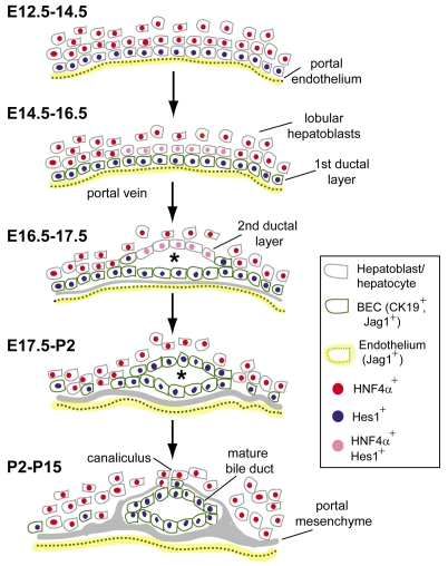
Taken together, these findings are consistent with a model in which Notch controls bile duct development by regulating biliary fate at successive stages of development (Fig. 9). Initially, endothelial Jag1 activates Notch signaling in peri-portal hepatoblasts, resulting in biliary differentiation and the appearance of the first (portal) layer of the ductal plate (E14.5-16.5). The nascent BECs also express Jag1, prompting activation of Notch signaling in the adjacent second layer, lumen formation, and the emergence of primitive ductal structures (E16.5-17.5). Subsequently, cells in the second layer complete the biliary program, giving rise to mature symmetrical ducts (P2). This model accommodates our results and reconciles conflicting reports from the literature regarding whether Notch acts as a regulator of differentiation or morphogenesis (Geisler et al., 2008; Kodama et al., 2004; Lozier et al., 2008; Tanimizu et al., 2003; Tanimizu et al., 2004). The observation that Notch functions in the differentiation of the second biliary layer, an event that is intimately associated with tubulogenesis, suggests that Notch serves both tasks, linking its activity as a regulator of cell fate with a role in morphogenesis.
Fig. 9.
Model of bile duct development. Early in liver development (E12.5-14.5), endothelial-derived Jag1 (yellow) activates Notch signaling and Hes1 expression in adjacent hepatoblasts (blue nuclei), resulting in formation of the first ductal plate layer at E14.6-16.5 (cells outlined in green). Between E16.5 and E17.5, tubulogenesis occurs at discrete sites of active Notch signaling in adjacent hepatocytes (pink nuclei), giving rise to a primitive ductal structure (asterisk). Cells comprising the second (outer) layer of this asymmetric tubule undergo biliary differentiation between E17.5 and P2. Subsequent growth of the portal mesenchyme and loss of unincorporated BECs leads to the formation of a mature portal tract by P15. Note that BECs in both the first and second layers of the ductal plate express Jag1. See text for details.
This model leaves several questions unanswered. First, how is biliary development controlled spatially? Although Notch signaling is activated throughout the first layer of the ductal plate, lumens arise at discrete locations, and it is unclear what governs the focal formation of these primitive structures. A related question concerns how expression of the ligand (Jag1) spreads to BECs. One possibility is `lateral induction', a process by which Notch signaling in a cell results in the activation, rather than repression, of ligand expression in that cell (Eddison et al., 2000; Timmerman et al., 2004). In addition, other mechanisms are likely to restrict Notch signaling. For example, Jag1 is highly expressed in newborn BECs, where it results in the induction of Hes1, but other cells adjacent to these ligand-producing cells do not express Hes1 (see Fig. 2D,H). Furthermore, although our study demonstrates that biliary tubulogenesis is responsive to increasing Notch dosage, consistent with the known sensitivity of human bile duct development to JAG1 or NOTCH2 haploinsufficiency (Li et al., 1997; McDaniell et al., 2006; Oda et al., 1997), the mechanism underlying this dose responsiveness remains unclear.
It is worth pointing out two caveats. First, despite the fact that Notch2 is the major Notch receptor involved in bile duct development (Geisler et al., 2008; McDaniell et al., 2006), our experiments used the intracellular domain of Notch1 for gain-of-function. Despite this mismatch, we believe that the Notch1 ICD serves as a reasonable surrogate for Notch activity in the liver. Domain-swapping experiments have shown that the C-terminal portion of the Notch1 and Notch2 ICDs are functionally interchangeable in vivo (Kraman and McCright, 2005). In addition, the RosaNICD strain we used is capable of rescuing a renal fate specification phenotype caused by Notch2 deficiency (Cheng et al., 2007). This indicates that Notch2 targets are appropriately activated by this transgene. Furthermore, our observations with the Notch1 ICD are in agreement with the loss-of-function phenotype resulting from Rbpj deletion. Nevertheless, confirmation that Notch2 promotes biliary differentiation will ultimately be needed. Second, our model proposes a role for Jag1 in the induction of the second layer in primitive ducts. However, conditional deletion of Jag1 in the hepatic epithelium is not associated with bile duct abnormalities during development (Loomes et al., 2007). Timing of deletion or functional redundancy with other hepatic ligands (Jag2, Dll1 or Dll4) could account for the lack of an embryonic Jag1 mutant phenotype, issues that can be addressed by earlier Jag1 deletion and with compound mutants.
Supplementary Material
Supplementary material
Supplementary material available online at http://dev.biologists.org/cgi/content/full/136/10/1727/DC1
We thank G. Schutz, K. Kaestner, P. Chambon, T. Honjo and D. Melton for sharing mice; T. Sudo, A. Miyajima and C. Bogue for sharing aliquots of Hes1, Ck19 and Hhex antisera, respectively; J. Lelay and Y. Ohtani for help with ChIP; K. Kaestner, W. Pear, K. Loomes, M. Ryan and M. Pack for helpful discussions; J. Friedman for reading the manuscript; D. Ludwig and the AFCRI Histology Core for assistance with sample preparation; and Y. Sofer and A. Stout for help with confocal imaging. Monoclonal antibody G8.8 was provided by the Developmental Studies Hybridoma Bank. B.Z.S. was supported by grant DK076583 from NIDDK and support from the Penn Center for Molecular Studies in Digestive and Liver Disease. F.L. was supported by the Interuniversity Attraction Poles Program (Belgian Science Policy), the DG Higher Education and Scientific Research of the French Community of Belgium, the Alphonse and Jean Forton Fund, and the Fund for Scientific Medical Research. A.A. and P.R. hold fellowships from the Université Catholique de Louvain. Deposited in PMC for release after 12 months.
References
- Ader, T., Norel, R., Levoci, L. and Rogler, L. E. (2006). Transcriptional profiling implicates TGFbeta/BMP and Notch signaling pathways in ductular differentiation of fetal murine hepatoblasts. Mech. Dev. 123, 177-194. [DOI] [PubMed] [Google Scholar]
- Alagille, D., Estrada, A., Hadchouel, M., Gautier, M., Odievre, M. and Dommergues, J. P. (1987). Syndromic paucity of interlobular bile ducts (Alagille syndrome or arteriohepatic dysplasia): review of 80 cases. J. Pediatr. 110, 195-200. [DOI] [PubMed] [Google Scholar]
- Antoniou, A., Raynaud, P., Cordi, S., Zong, Y., Tronche, F., Stanger, B. Z., Jacquemin, P., Pierreux, C. E., Clotman, F. and Lemaigre, F. P. (2009). Intrahepatic bile ducts develop according to a new mode of tubulogenesis regulated by the transcription factor SOX9. Gastroenterology (in press). [DOI] [PMC free article] [PubMed]
- Blokzijl, A., Dahlqvist, C., Reissmann, E., Falk, A., Moliner, A., Lendahl, U. and Ibanez, C. F. (2003). Cross-talk between the Notch and TGF-beta signaling pathways mediated by interaction of the Notch intracellular domain with Smad3. J. Cell Biol. 163, 723-728. [DOI] [PMC free article] [PubMed] [Google Scholar]
- Bolos, V., Grego-Bessa, J. and de la Pompa, J. L. (2007). Notch signaling in development and cancer. Endocr. Rev. 28, 339-363. [DOI] [PubMed] [Google Scholar]
- Cheng, H. T., Kim, M., Valerius, M. T., Surendran, K., Schuster-Gossler, K., Gossler, A., McMahon, A. P. and Kopan, R. (2007). Notch2, but not Notch1, is required for proximal fate acquisition in the mammalian nephron. Development 134, 801-811. [DOI] [PMC free article] [PubMed] [Google Scholar]
- Clotman, F. and Lemaigre, F. P. (2006). Control of hepatic differentiation by activin/TGFbeta signaling. Cell Cycle 5, 168-171. [DOI] [PubMed] [Google Scholar]
- Clotman, F., Jacquemin, P., Plumb-Rudewiez, N., Pierreux, C. E., Van der Smissen, P., Dietz, H. C., Courtoy, P. J., Rousseau, G. G. and Lemaigre, F. P. (2005). Control of liver cell fate decision by a gradient of TGF beta signaling modulated by Onecut transcription factors. Genes Dev. 19, 1849-1854. [DOI] [PMC free article] [PubMed] [Google Scholar]
- Coffinier, C., Gresh, L., Fiette, L., Tronche, F., Schutz, G., Babinet, C., Pontoglio, M., Yaniv, M. and Barra, J. (2002). Bile system morphogenesis defects and liver dysfunction upon targeted deletion of HNF1beta. Development 129, 1829-1838. [DOI] [PubMed] [Google Scholar]
- Crosnier, C., Attie-Bitach, T., Encha-Razavi, F., Audollent, S., Soudy, F., Hadchouel, M., Meunier-Rotival, M. and Vekemans, M. (2000). JAGGED1 gene expression during human embryogenesis elucidates the wide phenotypic spectrum of Alagille syndrome. Hepatology 32, 574-581. [DOI] [PubMed] [Google Scholar]
- Dahlqvist, C., Blokzijl, A., Chapman, G., Falk, A., Dannaeus, K., Ibanez, C. F. and Lendahl, U. (2003). Functional Notch signaling is required for BMP4-induced inhibition of myogenic differentiation. Development 130, 6089-6099. [DOI] [PubMed] [Google Scholar]
- Eddison, M., Le Roux, I. and Lewis, J. (2000). Notch signaling in the development of the inner ear: lessons from Drosophila. Proc. Natl. Acad. Sci. USA 97, 11692-11699. [DOI] [PMC free article] [PubMed] [Google Scholar]
- Ehebauer, M., Hayward, P. and Arias, A. M. (2006). Notch, a universal arbiter of cell fate decisions. Science 314, 1414-1415. [DOI] [PubMed] [Google Scholar]
- Emerick, K. M., Rand, E. B., Goldmuntz, E., Krantz, I. D., Spinner, N. B. and Piccoli, D. A. (1999). Features of Alagille syndrome in 92 patients: frequency and relation to prognosis. Hepatology 29, 822-829. [DOI] [PubMed] [Google Scholar]
- Fang, T. C., Yashiro-Ohtani, Y., Del Bianco, C., Knoblock, D. M., Blacklow, S. C. and Pear, W. S. (2007). Notch directly regulates Gata3 expression during T helper 2 cell differentiation. Immunity 27, 100-110. [DOI] [PMC free article] [PubMed] [Google Scholar]
- Flynn, D. M., Nijjar, S., Hubscher, S. G., de Goyet Jde, V., Kelly, D. A., Strain, A. J. and Crosby, H. A. (2004). The role of Notch receptor expression in bile duct development and disease. J. Pathol. 204, 55-64. [DOI] [PubMed] [Google Scholar]
- Geisler, F., Nagl, F., Mazur, P. K., Lee, M., Zimber-Strobl, U., Strobl, L. J., Radtke, F., Schmid, R. M. and Siveke, J. T. (2008). Liver-specific inactivation of Notch2, but not Notch1, compromises intrahepatic bile duct development in mice. Hepatology 48, 607-616. [DOI] [PubMed] [Google Scholar]
- Han, H., Tanigaki, K., Yamamoto, N., Kuroda, K., Yoshimoto, M., Nakahata, T., Ikuta, K. and Honjo, T. (2002). Inducible gene knockout of transcription factor recombination signal binding protein-J reveals its essential role in T versus B lineage decision. Int. Immunol. 14, 637-645. [DOI] [PubMed] [Google Scholar]
- Hayashi, S., Lewis, P., Pevny, L. and McMahon, A. P. (2002). Efficient gene modulation in mouse epiblast using a Sox2Cre transgenic mouse strain. Mech. Dev. 119 Suppl. 1, S97-S101. [DOI] [PubMed] [Google Scholar]
- Hogan, B. L. and Kolodziej, P. A. (2002). Organogenesis: molecular mechanisms of tubulogenesis. Nat. Rev. Genet. 3, 513-523. [DOI] [PubMed] [Google Scholar]
- Ito, T., Udaka, N., Yazawa, T., Okudela, K., Hayashi, H., Sudo, T., Guillemot, F., Kageyama, R. and Kitamura, H. (2000). Basic helix-loop-helix transcription factors regulate the neuroendocrine differentiation of fetal mouse pulmonary epithelium. Development 127, 3913-3921. [DOI] [PubMed] [Google Scholar]
- Jadhav, A. P., Cho, S. H. and Cepko, C. L. (2006). Notch activity permits retinal cells to progress through multiple progenitor states and acquire a stem cell property. Proc. Natl. Acad. Sci. USA 103, 18998-19003. [DOI] [PMC free article] [PubMed] [Google Scholar]
- Jones, E. A., Clement-Jones, M. and Wilson, D. I. (2000). JAGGED1 expression in human embryos: correlation with the Alagille syndrome phenotype. J. Med. Genet. 37, 658-662. [DOI] [PMC free article] [PubMed] [Google Scholar]
- Kageyama, R., Ohtsuka, T. and Kobayashi, T. (2007). The Hes gene family: repressors and oscillators that orchestrate embryogenesis. Development 134, 1243-1251. [DOI] [PubMed] [Google Scholar]
- Kellendonk, C., Opherk, C., Anlag, K., Schutz, G. and Tronche, F. (2000). Hepatocyte-specific expression of Cre recombinase. Genesis 26, 151-153. [DOI] [PubMed] [Google Scholar]
- Kodama, Y., Hijikata, M., Kageyama, R., Shimotohno, K. and Chiba, T. (2004). The role of notch signaling in the development of intrahepatic bile ducts. Gastroenterology 127, 1775-1786. [DOI] [PubMed] [Google Scholar]
- Kraman, M. and McCright, B. (2005). Functional conservation of Notch1 and Notch2 intracellular domains. FASEB J. 19, 1311-1313. [DOI] [PubMed] [Google Scholar]
- Lee, C. S., Sund, N. J., Behr, R., Herrera, P. L. and Kaestner, K. H. (2005). Foxa2 is required for the differentiation of pancreatic alpha-cells. Dev. Biol. 278, 484-495. [DOI] [PubMed] [Google Scholar]
- Lemaigre, F. P. (2003). Development of the biliary tract. Mech. Dev. 120, 81-87. [DOI] [PubMed] [Google Scholar]
- Lemaigre, F. and Zaret, K. S. (2004). Liver development update: new embryo models, cell lineage control, and morphogenesis. Curr. Opin. Genet. Dev. 14, 582-590. [DOI] [PubMed] [Google Scholar]
- Li, L., Krantz, I. D., Deng, Y., Genin, A., Banta, A. B., Collins, C. C., Qi, M., Trask, B. J., Kuo, W. L., Cochran, J. et al. (1997). Alagille syndrome is caused by mutations in human Jagged1, which encodes a ligand for Notch1. Nat. Genet. 16, 243-251. [DOI] [PubMed] [Google Scholar]
- Loomes, K. M., Taichman, D. B., Glover, C. L., Williams, P. T., Markowitz, J. E., Piccoli, D. A., Baldwin, H. S. and Oakey, R. J. (2002). Characterization of Notch receptor expression in the developing mammalian heart and liver. Am. J. Med. Genet. 112, 181-189. [DOI] [PubMed] [Google Scholar]
- Loomes, K. M., Russo, P., Ryan, M., Nelson, A., Underkoffler, L., Glover, C., Fu, H., Gridley, T., Kaestner, K. H. and Oakey, R. J. (2007). Bile duct proliferation in liver-specific Jag1 conditional knockout mice: effects of gene dosage. Hepatology 45, 323-330. [DOI] [PubMed] [Google Scholar]
- Louis, A. A., Van Eyken, P., Haber, B. A., Hicks, C., Weinmaster, G., Taub, R. and Rand, E. B. (1999). Hepatic jagged1 expression studies. Hepatology 30, 1269-1275. [DOI] [PubMed] [Google Scholar]
- Lozier, J., McCright, B. and Gridley, T. (2008). Notch signaling regulates bile duct morphogenesis in mice. PLoS ONE 3, e1851. [DOI] [PMC free article] [PubMed] [Google Scholar]
- Lubarsky, B. and Krasnow, M. A. (2003). Tube morphogenesis: making and shaping biological tubes. Cell 112, 19-28. [DOI] [PubMed] [Google Scholar]
- McCright, B., Lozier, J. and Gridley, T. (2002). A mouse model of Alagille syndrome: Notch2 as a genetic modifier of Jag1 haploinsufficiency. Development 129, 1075-1082. [DOI] [PubMed] [Google Scholar]
- McDaniell, R., Warthen, D. M., Sanchez-Lara, P. A., Pai, A., Krantz, I. D., Piccoli, D. A. and Spinner, N. B. (2006). NOTCH2 mutations cause Alagille syndrome, a heterogeneous disorder of the notch signaling pathway. Am. J. Hum. Genet. 79, 169-173. [DOI] [PMC free article] [PubMed] [Google Scholar]
- Michalopoulos, G. K., Barua, L. and Bowen, W. C. (2005). Transdifferentiation of rat hepatocytes into biliary cells after bile duct ligation and toxic biliary injury. Hepatology 41, 535-544. [DOI] [PMC free article] [PubMed] [Google Scholar]
- Murtaugh, L. C., Stanger, B. Z., Kwan, K. M. and Melton, D. A. (2003). Notch signaling controls multiple steps of pancreatic differentiation. Proc. Natl. Acad. Sci. USA 100, 14920-14925. [DOI] [PMC free article] [PubMed] [Google Scholar]
- Nijjar, S. S., Crosby, H. A., Wallace, L., Hubscher, S. G. and Strain, A. J. (2001). Notch receptor expression in adult human liver: a possible role in bile duct formation and hepatic neovascularization. Hepatology 34, 1184-1192. [DOI] [PubMed] [Google Scholar]
- Nijjar, S. S., Wallace, L., Crosby, H. A., Hubscher, S. G. and Strain, A. J. (2002). Altered Notch ligand expression in human liver disease: further evidence for a role of the Notch signaling pathway in hepatic neovascularization and biliary ductular defects. Am. J. Pathol. 160, 1695-1703. [DOI] [PMC free article] [PubMed] [Google Scholar]
- Niranjan, T., Bielesz, B., Gruenwald, A., Ponda, M. P., Kopp, J. B., Thomas, D. B. and Susztak, K. (2008). The Notch pathway in podocytes plays a role in the development of glomerular disease. Nat. Med. 14, 290-298. [DOI] [PubMed] [Google Scholar]
- Nishikawa, Y., Doi, Y., Watanabe, H., Tokairin, T., Omori, Y., Su, M., Yoshioka, T. and Enomoto, K. (2005). Transdifferentiation of mature rat hepatocytes into bile duct-like cells in vitro. Am. J. Pathol. 166, 1077-1088. [DOI] [PMC free article] [PubMed] [Google Scholar]
- Oda, T., Elkahloun, A. G., Pike, B. L., Okajima, K., Krantz, I. D., Genin, A., Piccoli, D. A., Meltzer, P. S., Spinner, N. B., Collins, F. S. et al. (1997). Mutations in the human Jagged1 gene are responsible for Alagille syndrome. Nat. Genet. 16, 235-242. [DOI] [PubMed] [Google Scholar]
- Oka, C., Nakano, T., Wakeham, A., de la Pompa, J. L., Mori, C., Sakai, T., Okazaki, S., Kawaichi, M., Shiota, K., Mak, T. W. et al. (1995). Disruption of the mouse RBP-J kappa gene results in early embryonic death. Development 121, 3291-3301. [DOI] [PubMed] [Google Scholar]
- Rubins, N. E., Friedman, J. R., Le, P. P., Zhang, L., Brestelli, J. and Kaestner, K. H. (2005). Transcriptional networks in the liver: hepatocyte nuclear factor 6 function is largely independent of Foxa2. Mol. Cell. Biol. 25, 7069-7077. [DOI] [PMC free article] [PubMed] [Google Scholar]
- Schuler, M., Dierich, A., Chambon, P. and Metzger, D. (2004). Efficient temporally controlled targeted somatic mutagenesis in hepatocytes of the mouse. Genesis 39, 167-172. [DOI] [PubMed] [Google Scholar]
- Seymour, P. A., Freude, K. K., Tran, M. N., Mayes, E. E., Jensen, J., Kist, R., Scherer, G. and Sander, M. (2007). SOX9 is required for maintenance of the pancreatic progenitor cell pool. Proc. Natl. Acad. Sci. USA 104, 1865-1870. [DOI] [PMC free article] [PubMed] [Google Scholar]
- Stanger, B. Z., Datar, R., Murtaugh, L. C. and Melton, D. A. (2005). Direct regulation of intestinal fate by Notch. Proc. Natl. Acad. Sci. USA 102, 12443-12448. [DOI] [PMC free article] [PubMed] [Google Scholar]
- Sumazaki, R., Shiojiri, N., Isoyama, S., Masu, M., Keino-Masu, K., Osawa, M., Nakauchi, H., Kageyama, R. and Matsui, A. (2004). Conversion of biliary system to pancreatic tissue in Hes1-deficient mice. Nat. Genet. 36, 83-87. [DOI] [PubMed] [Google Scholar]
- Tanimizu, N. and Miyajima, A. (2004). Notch signaling controls hepatoblast differentiation by altering the expression of liver-enriched transcription factors. J. Cell Sci. 117, 3165-3174. [DOI] [PubMed] [Google Scholar]
- Tanimizu, N., Nishikawa, M., Saito, H., Tsujimura, T. and Miyajima, A. (2003). Isolation of hepatoblasts based on the expression of Dlk/Pref-1. J. Cell Sci. 116, 1775-1786. [DOI] [PubMed] [Google Scholar]
- Tanimizu, N., Saito, H., Mostov, K. and Miyajima, A. (2004). Long-term culture of hepatic progenitors derived from mouse Dlk+ hepatoblasts. J. Cell Sci. 117, 6425-6434. [DOI] [PubMed] [Google Scholar]
- Taylor, M. K., Yeager, K. and Morrison, S. J. (2007). Physiological Notch signaling promotes gliogenesis in the developing peripheral and central nervous systems. Development 134, 2435-2447. [DOI] [PMC free article] [PubMed] [Google Scholar]
- Timmerman, L. A., Grego-Bessa, J., Raya, A., Bertran, E., Perez-Pomares, J. M., Diez, J., Aranda, S., Palomo, S., McCormick, F., Izpisua-Belmonte, J. C. et al. (2004). Notch promotes epithelial-mesenchymal transition during cardiac development and oncogenic transformation. Genes Dev. 18, 99-115. [DOI] [PMC free article] [PubMed] [Google Scholar]
- Zhang, L., Theise, N., Chua, M. and Reid, L. M. (2008). The stem cell niche of human livers: symmetry between development and regeneration. Hepatology 48, 1598-1607. [DOI] [PubMed] [Google Scholar]
Associated Data
This section collects any data citations, data availability statements, or supplementary materials included in this article.


