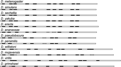
Figure 6.—
AT-hook organization of the D1 proteins of 12 Drosophila species. The D1 protein sequences are drawn to relative scale as rectangles. The AT-hook motifs predicted by Pfam (http://pfam.janelia.org/) are illustrated as shaded boxes, with light shading indicating matches of lower confidence. The proteins are ordered to reflect the evolutionary relatedness of the species (http://insects.eugenes.org/species/).

