

Abstract
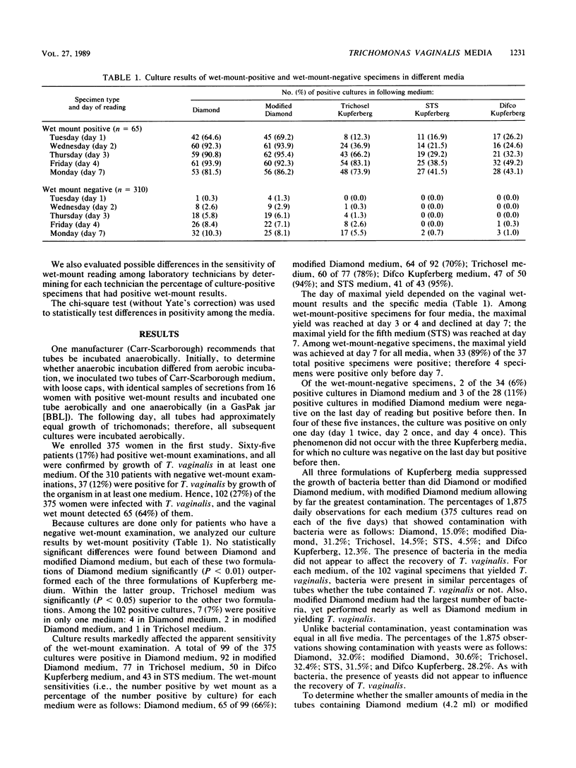
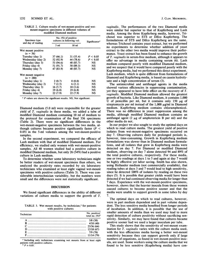
Many media have been formulated for the growth of Trichomonas vaginalis, but the relative sensitivities of these media have not been determined. We evaluated the ability of six media, including all five media commercially available in the United States, to grow Trichomonas vaginalis from vaginal secretions. In a first experiment, we evaluated the ability of five media to grow T. vaginalis from vaginal secretions of 375 women and determined the optimal days on which to read culture tubes, by inoculating aliquots of secretions into each medium and reading the tubes 1, 2, 3, 4, and 7 days later. Sixty-five patients (17%) had a positive wet-mount examination for T. vaginalis, and all the positive results were confirmed by growth in at least one medium. Of 310 wet-mount-negative specimens, 37 (12%) grew T. vaginalis; overall, 102 women (27%) had a positive culture. Diamond and modified Diamond media (the latter being the only medium not commercially available) detected 99 (97%) and 92 (90%) isolates, respectively, compared with three formulations of Kupferberg medium, which detected 77 (75%), 50 (49%), and 43 (42%) isolates. The optimal single day to read wet-mount-negative cultures was day 7, but 4 (11%) of the 37 positive specimens were positive only before day 7. In a second study, we compared the ability of modified Diamond medium with that of a sixth medium, Lash medium, to grow T. vaginalis from 48 wet-mount-positive specimens; modified Diamond medium supported growth in all cases, whereas Lash medium supported growth in only 26 (54%) cases. We conclude that formulations of Diamond medium are superior to formulations of Kupferberg or Lash medium for growth of t. vaginalis.
Full text
PDF
Selected References
These references are in PubMed. This may not be the complete list of references from this article.
- Fouts A. C., Kraus S. J. Trichomonas vaginalis: reevaluation of its clinical presentation and laboratory diagnosis. J Infect Dis. 1980 Feb;141(2):137–143. doi: 10.1093/infdis/141.2.137. [DOI] [PubMed] [Google Scholar]
- Krieger J. N., Tam M. R., Stevens C. E., Nielsen I. O., Hale J., Kiviat N. B., Holmes K. K. Diagnosis of trichomoniasis. Comparison of conventional wet-mount examination with cytologic studies, cultures, and monoclonal antibody staining of direct specimens. JAMA. 1988 Feb 26;259(8):1223–1227. doi: 10.1001/jama.259.8.1223. [DOI] [PubMed] [Google Scholar]
- LASH J. J. A simplified casein hydrolysate-serum medium for the cultivation of Trichomonas vaginalis. Am J Trop Med Hyg. 1950 Sep 5;30(5):641–642. doi: 10.4269/ajtmh.1950.s1-30.641. [DOI] [PubMed] [Google Scholar]
- Lossick J. G. The diagnosis of vaginal trichomoniasis. JAMA. 1988 Feb 26;259(8):1230–1230. [PubMed] [Google Scholar]
- Smith R. F. Incubation time, second blind passage, and cost considerations in the isolation of Trichomonas vaginalis. J Clin Microbiol. 1986 Jul;24(1):139–140. doi: 10.1128/jcm.24.1.139-140.1986. [DOI] [PMC free article] [PubMed] [Google Scholar]
- Thomason J. L., Gelbart S. M., Sobun J. F., Schulien M. B., Hamilton P. R. Comparison of four methods to detect Trichomonas vaginalis. J Clin Microbiol. 1988 Sep;26(9):1869–1870. doi: 10.1128/jcm.26.9.1869-1870.1988. [DOI] [PMC free article] [PubMed] [Google Scholar]