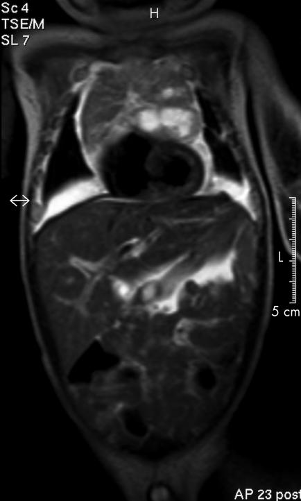
Thymic enlargement is a common physiological and incidental finding on chest x rays in healthy neonates and infants. There are usually no clinical symptoms or complications, and pathological lesions in infant thymi are rare. We report on a 5‐week‐old boy with recurrent respiratory distress since birth, pursed‐lips breathing with prolonged duration of expiration and tachypnoea. MRI showed massive congenital thymic hyperplasia (fig l) with huge life‐threatening mediastinal bleeding from ruptured thymic cysts into the pleural spaces. Thoracotomy and complete thymectomy were carried out, and the baby was discharged from the hospital on the fourteenth postoperative day without further complications.
Figure 1 T2‐weighted MRI of the thorax showing a large anterior mediastinal mass extending to the thoracic aperture and partially compressing the heart. There are some hyperdense area within the mass, suspicious for bleeding. Pleural effusion is also present.
The differential diagnosis of anterior mediastinal masses in neonates includes lymphangioma, teratoma, lipoma, lymphofollicular hyperplasia in myasthenia gravis, thyroid enlargement, physiological reaction to breast feeding and thymic disorders.1 Of these, thymic hyperplasia is the most common. Congenital thymic cysts are remnants of the thymopharyngeal duct.2 However, haemorrhage from ruptured thymic cysts in a neonate is exceedingly rare; it may occur in haemorrhagic disease of newborns.3
Footnotes
Competing interests: None.
References
- 1.Sauter E R, Arensman R M, Falterman K W. Thymic enlargement in children. Am Surg 19915721–23. [PubMed] [Google Scholar]
- 2.Khariwala S S, Nicollas R, Triglia J M.et al Cervical presentations of thymic anomalies in children. Int J Pediatr Otorhinolaryngol 200468909–914. [DOI] [PubMed] [Google Scholar]
- 3.Bees N R, Richards S W, Fearne C.et al Neonatal thymic haemorrhage. Br J Radiol 199770210–212. [DOI] [PubMed] [Google Scholar]


