Abstract
Few experimental and complementary theoretical studies have investigated high-frequency (>20 MHz) nonlinear responses from polymer-shelled ultrasound contrast agents. Three polymer agents with different shell properties were examined for their single-bubble backscatter when excited with a 40 MHz tone burst. Higher-order harmonic responses were observed for the three agents; however, their occurrence was at least partly due to nonlinear propagation. Only one of the agents (1.1 μm mean diameter) showed a subharmonic response for longer excitations (≈10-15 cycles) and midlevel pressure excitations (≈2.5 MPa). Theoretical calculations of the backscattered spectrum revealed behavior similar to the experimental results in specific parameter regimes.
1. Introduction
Applications involving microcirculation, such as ophthalmic disease diagnosis and small-animal imaging, particularly the normal and abnormal vascular development of genetically engineered mouse embryos and neonates, are ideal for high-frequency ultrasound (HFU, >20 MHz) contrast agents. The acoustic contrast agents in current use were designed to have an optimal response in the 1-10 MHz range of standard clinical ultrasound probes. These agents usually have a mean diameter of 2-4 μm where a larger bubble typically has a lower resonance frequency. Agents specifically designed for use with HFU have yet to become available, but many existing and experimental agents have been studied for HFU responses while utilizing a broadband pulse.1-3 The results typically showed backscatter centered at the transducer center frequency but no harmonic content was reported.
Goertz et al.4 recently reported the generation of harmonic and subharmonic backscatter components for the lipid-shelled agent Definity (Bristol-Myers Squibb, New York, NY). The contrast agents were excited with a tone burst consisting of 4-10 cycles at either 20 or 30 MHz. The observation of harmonics is surprising because few agents are typically present in the submicron size range in a given dose and, from the theory of free bubbles, the expectation would be that relatively high pressures would be required to excite a bubble nonlinearly with HFU. Previous theoretical work by Allen et al.5 suggests that shell waves may contribute to an enhanced high-frequency dipole response. Deng et al.2 reported experimental results that suggest a high-frequency response from conventional-size protein-shelled agents may be due to elastic properties of the shell. A new generation of polymer agents is becoming available, but most studies with these agents have been done with low MHz ultrasound6-8 and not much is known about their high-frequency response.
In this paper we report experimental and theoretical backscatter results for nitrogen-filled, polycaprolactone-shelled agents (POINT Biomedical, San Carlos, CA) with mean diameters of 3.4 (Point 1668, P1), 1.1 (Point 1466, P2) and 0.56 (Point 1470, P3) μm. These agents were excited with tone bursts from 1-15 cycles using a 40 MHz, focused transducer. The backscatter signals from single agents were examined for harmonic content. Experimental results were compared to theoretical calculations for the case of the polymer shell remaining intact during excitation.
2. Methods
2.1 Theory
For polymer-shelled agents, experimental evidence indicates the elasticity and thickness of the shell are important components to the overall dynamics.9 The model of Church,10 developed for protein-shelled agents, and a more-recent model based on finite elasticity,11 are the most appropriate models for these considerations. Both models were examined for this study but here, for brevity, we report initial results only from the Church model.
The model was modified to include compressibility corrections for the surrounding liquid as has been done for free bubble equations. Adiabatic behavior of the interior gas was assumed using a polytropic exponent relationship (γ =1.4) and van der Waals hard core terms for air. Shell thickness was estimated to be 5 nm and surface tension values of 50 and 5 dyn/cm were used for the shell/liquid and shell/gas interfaces, respectively. The density of the shell was assumed to be 1.1 gm/cm3 and the shell shear viscosity was approximated as 5.0 Poise. We estimated a shear modulus of 8.0 MPa for the shell under the assumption that the polymer behaved as a rubbery material. The radius-time, R(t), curve of the bubble response was calculated and the radiated pressure was computed from p(t)∝R2R̈+2RṘ2.12 The theoretical calculations did not account for the bandpass-filtering effect of the transducer or acoustic attenuation in water.
2.2 Experimental system
Experiments were performed with a dilute solution of contrast agent passing through a flow phantom. The flow phantom had a 3 mm channel with fluid ports on each side. The rate of flow was controlled by a planetary gear-driven digital pump (Cole Parmer, Vernon Hills, IL). The focal region of a 40 MHz probe attached to a Vevo 770 (Visual Sonics, Toronto, Canada) ultrasound backscatter microscope was placed within the flow phantom, and the passage of the contrast agents was visualized in real time. A pc containing a PCI-based digitizer (Acqiris DP105, Monroe, NY) was linked to the Vevo 770, and a custom LABVIEW program (National Instruments, Austin, TX) permitted acquisition of radio-frequency (rf) data corresponding to the B-mode display of the Vevo 770.
The agents were reconstituted from a dry state, according to manufacturer's instructions, using milli-Q water at a final concentration of 50 mg/ml. The samples were pumped from a reservoir into the flow phantom at a flow rate of 1 μl/s and diluted until single bubble flow could be observed on the Vevo 770 B-mode display. While an agent passed through the transducer focal zone, rf data frames were digitized at rates of up to 2 frames/s. The rf frames were acquired at a sampling rate of 500 MHz and consisted of 384 scan lines with 5000 points/line. The pulse repetition frequency was 8 kHz and the data frames spanned 8 mm, which corresponded to ≈21 μm separation between scan lines. Data were acquired for several Vevo 770 settings between 1% and 100% power and excitation pulses consisting of 1, 5, 10, and 15 cycles. Acoustic pressure levels for the power settings were measured with a calibrated hydrophone (Precision Acoustics Ltd., Dorset, UK) and negative peak pressures will be utilized to characterize the exposure settings. In addition, pulse/echo reference spectra were acquired using a 25-μm-diam wire situated at the transducer geometric focus.
2.3 Data analysis
The rf data were analyzed manually to examine the backscatter from single bubbles passing through the transducer focal zone. A Hamming window was placed over an instance of a single contrast agent, and the windowed rf data were padded to 1024 points. The spectrum of the signal was then calculated. The spectra we report were not normalized to remove the system frequency response. We chose the rf line in the “middle” of the bubble for our analysis. Because the rf data were acquired with a real-time imaging system, each bubble received multiple exposures to the ultrasound burst. In fact, bubbles could be observed moving through the image frame over multiple rf-frame acquisitions. These exposure conditions represent a more realistic examination of how a contrast agent withstands a long-term exposure during a HFU imaging procedure. In these studies, we generally did not observe obvious contrast agent destruction, which indicates the agents are fairly robust at high frequency on a time scale of several seconds.
In addition to calculating the spectrum of single-bubble backscatter, we performed a time-frequency analysis of the data using spectrograms. For our studies, spectrograms are an ideal tool to evaluate how bubbles respond to short-duration (e.g., 1 or 5 cycles) or long-duration excitation signals (e.g., 10 or 15 cycles) and to evaluate the onset and nature of nonlinear behavior (e.g., subharmonic or harmonic components). To obtain the spectrogram, the short-time Fourier transform was computed over 128 samples (i.e., =0.26 μs) and then zero padded to 8192 samples. To achieve adequate temporal resolution, consecutive Fourier transforms overlapped by 120 points.
3. Results
3.1 Experiment
The experimental results for the three agents along with the wire phantom reference spectra are summarized in Fig. 1. Figure 1(a) shows reference spectra obtained with the wire reflector for three 40 MHz tone-burst excitation conditions. The case with 1 cycle and 4.9 MPa essentially shows the system impulse response with a broadband spectrum centered at 33 MHz and additional harmonic activity. When the tone burst was increased to 15 cycles, the center frequency was seen to shift to 40 MHz (the quoted transducer frequency) and harmonics from nonlinear propagation appeared at 80 and 120 MHz. The asymmetric sidelobes seen in the spectra relate to the convolution of the frequency response of the transducer (centered at 33 MHz) and the 40 MHz tone burst.
Fig. 1.
(Color online)(a) Wire reflector spectra for 1 cycle and 15 cycle tone bursts. (b) Spectra for agents P1 and P3 reveal harmonic components while only (c) P2 reveals any subharmonic activity.
The backscatter spectra for representative cases of P1 and P3 are shown in Fig. 1(b). The case for P1 with 1 cycle and 4.9 MPa shows a response similar to that seen in Fig. 1(a), except that the center frequency shifted to a lower value (20 MHz). The remaining spectra for P1 and P3 are for excitations with 5-15 cycles. No subharmonic responses were observed at any of the Vevo 770 excitation conditions but higher-order harmonics appeared with longer tone bursts and higher excitation pressures. These observations are consistent with what has been reported in the literature for lipid-shelled agents.4 However, based on the reference spectra, a large portion of the higher-order harmonics appear to result from nonlinear propagation rather than a nonlinear bubble response.
Figure 1(c) shows spectra for P2 using a 15 cycle excitation at three different drive pressures. The 1.5 MPa setting is interesting because a clear subharmonic was present, but there were no significant higher harmonics. When the pressure was raised to 2.5 MPa, the sub-harmonic was maintained and the second and third harmonic also appeared. Finally, when the pressure was raised to 5.9 MPa, the subharmonic was suppressed but the higher harmonics were still visible. This behavior is consistent with what is generally observed for a subharmonic response.13 However, our observations are somewhat surprising considering the rigid polymer shell of the agent and the suboptimal driving conditions for a resonance response. One explanation may be that the POINT agent ruptured and that we were actually seeing the backscatter from a free bubble or an unconstrained bubble fragment still linked to the fractured shell.
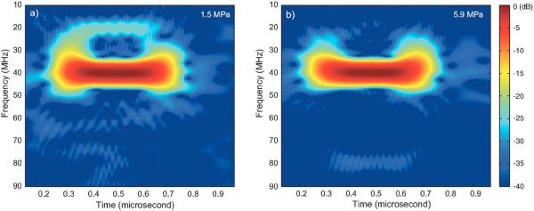
Figure 2 displays spectrograms of the P2 agent excited with a 40 MHz, 15 cycle pulse at two exposure levels: 1.5 and 5.9 MPa [black and blue curves in Fig. 1(c)]. Figure 2(a), obtained at the lower pressure, displays a significant spectral component near the 40 MHz drive frequency throughout the bubble response. However, the spectrogram also reveals a significant nonlinear spectral component near 20 MHz, 25 dB below the 40 MHz component. Figure 2(b) obtained at the higher exposure pressure, also shows energy at 40 MHz, but with no visible subharmonic component. However, there was some energy near 80 MHz at 35 dB below fundamental. As noted above, a portion of the higher harmonic content is probably due to nonlinear propagation. However, we believe that a nonlinear response from the agents also contributes.
Fig. 2.
(Color online) Selected spectrograms for the P2 agent, 15 cycle excitation at 40 MHz with (a) 1.5 MPa and (b) 5.9 MPa negative peak pressure excitations.
3.2 Theoretical
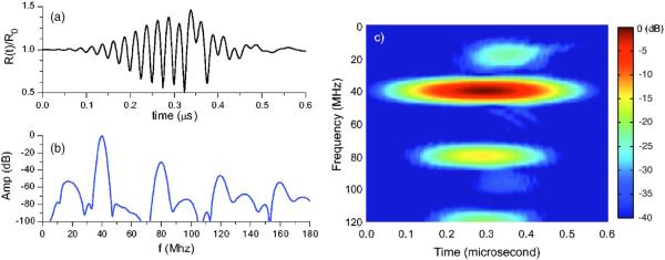
Figure 3(a) shows the R(t) curve of a 1.1-μm-diam agent forced with a Gaussian weighted, 40 MHz, 15 cycle, cosine pulse with a peak pressure of 5.9 MPa. An initial delay time of 0.1 μs was used for the pulse. At first, the agent, which was forced well above its resonance frequency, appears to oscillate at the forcing frequency of the transducer. As the pulse begins to decay, the onset of the subharmonic becomes visible in the R(t) curve [Fig. 3(a)]. Due to the density difference between the shell and fluid, additional nonlinearities influence the dominating inertial response of the agent, especially as it is forced above its resonance. The spectrum pressure curve is shown in Fig. 3(b). The subharmonic is visible at a similar level to the experimental results and higher harmonic components appear, although at higher levels than observed experimentally. The spectrogram of the p(t) curve [Fig. 3(c)] shows the time evolution of the harmonic responses. The subharmonic initiates towards the middle of excitation at a later time than the higher-order harmonics, an observation that provides information on the level of dissipation in the system.
Fig. 3.
(Color online) Theoretical (a) R(t)/R0 curve, (b) spectrum of the pressure calculated from the R(t) curve, and (c) spectrogram of pressure curve for P2 excited by a 40 MHz, 15 cycle, 5.9 MPa excitation.
We investigated a range of parameter space for the shell properties by varying the thickness and elasticity to estimate the values used in this study. Future experimental measurements of the shell properties of polymer agents will facilitate a more direct comparison of experiments to theoretical calculations. We note that the theoretical subharmonic response occurs at somewhat higher pressure amplitude than observed experimentally; however, the simulations reveal this response is quite sensitive to small variations in wave form, pressure amplitude, shell thickness, and elastic modulus. Also, an earlier response in time of the subharmonic in the experimental data compared with the theory suggests that a transient process such as the nonspherical buckling of the shell or escape of gas might contribute to its onset.
4. Discussion and conclusions
Three polymer-shelled agents from POINT Biomedical were examined to determine their response to a 40 MHz, tone-burst excitation. The three agents showed indications of higher harmonics, but a large portion of the response could be attributed to nonlinear propagation rather than a total nonlinear agent response. Only one agent, P2, was observed to have a subharmonic response for the tested exposure conditions. We should point out that the results reported here were repeatable, but did show variability. The bubbles in each batch of agents had a size distribution and they do not all respond in an identical fashion.
A theoretical analysis of the P2 agent's radial response revealed some similarity to the experimental results. These preliminary results suggest that polymer-shelled contrast agents are viable candidates for high-frequency imaging using the fundamental frequency. However, for an optimal nonlinear response, the polymer shell material properties need to be carefully selected. More comprehensive experiments, measurement of material properties, and a complementary theory are needed to fully understand and optimize polymer-shelled contrast agents for HFU applications.
From the theory of free gas bubbles, we might expect smaller agents to be more attractive for high-frequency nonlinear imaging. However, our results for the three polymer-shelled agents indicate that this is not necessarily the case. One hypothesis for a larger agent giving a better nonlinear response is that the agent must first rupture for it to become acoustically active. If this were true, smaller agents would be more resistant to acoustic rupture because of their stiffer shell and, therefore, more likely to scatter only the fundamental. Our experimental results also indicate that even if the agents were to have ruptured, the fragments or gas bubbles remain acoustically active for several seconds.
Acknowledgments
The authors wish to thank POINT Biomedical and Bob Short for making available the polymer-shelled agents used in these studies and Visual Sonics for providing access to the engineering mode of the Vevo 770. This work was supported in part by NIH Grant No. HL078665 (D.H.T.).
References and links
- 1.Morgan K, Dayton P, Klibanov A, Brandenburger G, Kaul S, Wei K, Ferrara K. Properties of contrast agents insonified at frequencies above 10 MHz. IEEE Ultrason. Symp. 1996:1127–1130. [Google Scholar]
- 2.Deng CX, Lizzi FL, Silverman RH, Ursea R, Coleman DJ. Imaging and spectrum analysis of contrast agents in the in vivo rabbit eye using very-high-frequency ultrasound. Ultrasound Med. Biol. 1998;24:383–394. doi: 10.1016/s0301-5629(97)00288-3. [DOI] [PubMed] [Google Scholar]
- 3.Moran CM, Watson RJ, Fox KA, McDicken WN. In vitro acoustic characterization of four intravenous ultrasonic contrast agents at 30 MHz. Ultrasound Med. Biol. 2002;28:785–791. doi: 10.1016/s0301-5629(02)00520-3. [DOI] [PubMed] [Google Scholar]
- 4.Goertz DE, Cherin E, Needles A, Karshafian R, Brown AS, Burns PN, Foster FS. High frequency nonlinear b-scan imaging of microbubble contrast agents. IEEE Trans. Ultrason. Ferroelectr. Freq. Control. 2005;52:65–79. doi: 10.1109/tuffc.2005.1397351. [DOI] [PubMed] [Google Scholar]
- 5.Allen JS, Kruse DE, Ferrara KW. Shell waves and acoustic scattering from ultrasound contrast agents. IEEE Trans. Ultrason. Ferroelectr. Freq. Control. 2001;48:409–418. doi: 10.1109/58.911723. [DOI] [PubMed] [Google Scholar]
- 6.Bouakaz A, Versluis M, de Jong N. High-speed optical observations of contrast agent destruction. Ultrasound Med. Biol. 2005;31:391–399. doi: 10.1016/j.ultrasmedbio.2004.12.004. [DOI] [PubMed] [Google Scholar]
- 7.Bloch SH, Short RE, Ferrara KW, Wisner ER. The effect of size on the acoustic response of polymer-shelled contrast agents. Ultrasound Med. Biol. 2005;31:439–444. doi: 10.1016/j.ultrasmedbio.2004.12.016. [DOI] [PubMed] [Google Scholar]
- 8.Chen WS, Matula TJ, Brayman AA, Crum LA. A comparison of the fragmentation thresholds and inertial cavitation doses of different ultrasound contrast agents. J. Acoust. Soc. Am. 2003;113:643–651. doi: 10.1121/1.1529667. [DOI] [PubMed] [Google Scholar]
- 9.Leong-Poi H, Song J, Rim SJ, Christiansen J, Kaul S, Lindner JR. Influence of microbubble shell properties on ultrasound signal: Implications for low-power perfusion imaging. J. Am. Soc. Echocardiogr. 2002;15:1269–1276. doi: 10.1067/mje.2002.124516. [DOI] [PubMed] [Google Scholar]
- 10.Church CC. The effects of an elastic solid surface layer on the radial pulsations of gas bubbles. J. Acoust. Soc. Am. 1995;97:1510–1521. [Google Scholar]
- 11.Allen JS, Rashid MM. Dynamics of a hyperelastic gas-filled spherical shell in a viscous fluid. J. Appl. Mech. 2004;71:195–200. [Google Scholar]
- 12.Leighton TG. The Acoustic Bubble. Academic; London: 1994. [Google Scholar]
- 13.Shi WT, Forsberg F, Raichlen JS, Needleman L, Goldberg BB. Pressure dependence of subharmonic signals from contrast microbubbles. Ultrasound Med. Biol. 1999;25:275–283. doi: 10.1016/s0301-5629(98)00163-x. [DOI] [PubMed] [Google Scholar]


