The fetal acetylcholine receptor (AChR) is present until 33 weeks gestation, when the fetal (γ) subunit is replaced by the adult (ɛ) subunit. Most infants of myasthenic mothers are asymptomatic despite intrauterine exposure to AChR antibodies (AChR Ab). A higher fetal to adult AChR Ab ratio can lead to transient neonatal myasthenia gravis (TNMG) in 10–15% of infants or rarely to arthrogryposis multiplex congenita (AMC). Here we report three brothers with facial diplegia, highly arched palate, velopharyngeal incompetence, conductive hearing loss, and cryptorchidism. The maternal fetal AChR Ab were elevated. We propose the term fetal acetylcholine receptor inactivation syndrome and suggest this phenotype results from inactivation of the fetal subunit during a critical period of fetal muscle development.
Case report.
A 33-year-old woman developed ptosis, facial weakness, generalized fatigue, and elevated AChR Ab (>1,000, normal <0.4). Thymectomy, pyridostigmine, prednisone, and plasmapheresis improved her symptoms. Subsequently, she had three successive pregnancies with polyhydramnios and normal fetal movements while continuing prednisone. Each son was born at term by C-section with no respiratory distress.
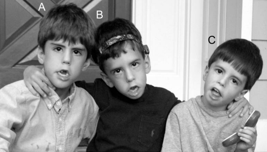
During her first pregnancy, the mother received no plasmapheresis. At birth, the infant (figure, A) had hypotonia, poor suck, and inability to swallow. He remained in the neonatal intensive care unit (NICU) with TNMG for 5 weeks and improved with IV immunoglobulin (IVIg) and nasogastric (NG) tube feedings. At 5 years he has microcephaly, facial diplegia with inability to close mouth or eyes completely, excessive drooling, and hypernasal poorly intelligible speech. Brain MRI, serum CK, and repetitive median and accessory nerve stimulation at 3 Hz are normal.
Figure First child at age 6 years 10 months (A), second child at age 5 years (B), and third child at age 3 years 10 months (C)
The three brothers share the following characteristics, with decreasing severity from the eldest to the youngest: facial diplegia with incomplete mouth closure, highly arched narrow palate, excessive drooling, dysarthria, lingual weakness, bilateral middle ear effusion managed with myringotomy tubes, intact extraocular movements, and surgically corrected cryptorchidism and inguinal hernias; the second child has a bone conduction hearing aid.
During the second pregnancy, the mother received plasmapheresis biweekly after the first trimester. At birth, the infant (figure, B) had hypotonia and poor suck and swallow. He remained in the NICU with TNMG for 5 weeks and improved with NG tube feedings and IVIg. At 3 years he has facial diplegia, incomplete mouth and eye closure, excessive drooling, and hypernasal speech.
The third child (figure, C) is the least severely affected. The mother received plasmapheresis biweekly throughout pregnancy. At birth, he was hypotonic and sucked poorly but swallowed. He remained in the NICU with TNMG for 3 weeks, improving with NG tube feedings. At 2 years he has mild facial diplegia, full mouth and eye closure, and mild speech difficulties.
All three siblings have normal neurobehavioral milestones, intact axial and limb muscle strength, pes cavus deformities, areflexia, no joint contractures, surgically corrected cryptorchidism and inguinal hernias, and middle ear effusions with conductive hearing loss managed with myringotomy tubes. The second child required bone conduction hearing aid. Serum, obtained from the mother when asymptomatic after her third pregnancy and assayed by radioimmunoassay,1 was positive for AChR antibodies. The titer was 1,890 nM against fetal AChR and 157 nM against adult AChR (normal values <0.5 nM).
Discussion.
We found nine other reported cases with persisting bulbar and facial weakness following TNMG.2–6 In one report6 maternal antibodies selectively inhibited the fetal AChR subunit. His mother had six further pregnancies all affected by lethal AMC while she remained clinically asymptomatic.
The maternal fetal/adult AChR Ab ratio is a stronger predictor of severity in offspring than the total AChR Ab. In our case, the maternal fetal AChR Ab titer was 10 times higher than the adult titer. More aggressive plasmapheresis treatment in the second and third pregnancies correlated with decreasing phenotypic severity. Another case report supports plasmapheresis therapy.7 A myasthenic mother had two successive pregnancies culminating in AMC and neonatal death. Prednisone and plasmapheresis during her third pregnancy resulted in a newborn with TNMG only.
The persistent weakness of selective muscle groups in these children suggests a differential vulnerability during fetal development. Bidirectional signaling between the muscle and nerve modulates the maturation of the postsynaptic membrane. Fast and delayed synapsing muscle groups have heterogeneous synaptic maturation rates and distinct response patterns to disturbances in the agrin-MuSK pathway. Fetal AChR inactivation impairs fetal movements and neuromuscular development as seen with gamma mutant mice and humans with the lethal and Escobar variants of multiple pterygium syndrome. A fetal akinesia sequence model, developed by injecting plasma from a myasthenic mother with affected babies into pregnant mice, also has been reported.1
Mothers with a previously affected child have a risk for future pregnancies approaching 100% irrespective of their clinical symptomatology. These findings should be considered during prenatal counseling of myasthenic mothers. Long-term follow-up of infants with TNMG may identify previously unrecognized speech impairment and hearing loss.
ACKNOWLEDGMENT
The authors thank the parents of the children presented in this report for their helpful comments and their permission to show the facial features of their three children to increase awareness of this clinical phenotype.
Supported by the SMA Foundation, New York, NY (M.O., W.K.C., D.C.D.); Helene Pelletier Foundation (M.O.), Colleen Giblin Foundation (D.C.D.); NIH CTSA Award 1 UL1 RR024156-01 (P.K.).
Disclosure: M.O., W.K.C., P.K., and D.C.D. have received grants from the SMA Foundation and D.C.D. has received grants from the Colleen Giblin Foundation for other research activities not reported in this article. A.V. has received royalties for antibodies used in the study of neurological diseases. The remaining authors report no conflicts of interest.
Received April 2, 2008. Accepted in final form July 2, 2008.
Address correspondence and reprint requests to Dr. Darryl C. De Vivo, 710 168th Street, 2nd floor, New York, NY 10032; dcd1@columbia.edu
&NA;
- 1.Jacobson L, Polizzi A, Morriss-Kay G, Vincent A. Plasma from human mothers of fetuses with severe arthrogryposis multiplex congenita causes deformities in mice. J Clin Invest 1999;103:1031–1038. [DOI] [PMC free article] [PubMed] [Google Scholar]
- 2.Rieder AA, Conley SF, Rowe L. Pediatric myasthenia gravis and velopharyngeal incompetence. Int J Pediatr Otorhinolaryngol 2004;68:747–752. [DOI] [PubMed] [Google Scholar]
- 3.Morel E, Eymard B, Vernet-der Garabedian B, Pannier C, Dulac O, Bach JF. Neonatal myasthenia gravis: a new clinical and immunologic appraisal on 30 cases. Neurology 1988;38:138–142. [DOI] [PubMed] [Google Scholar]
- 4.Ahlsten G, Lefvert AK, Osterman PO, Stalberg E, Safwenberg J. Follow-up study of muscle function in children of mothers with myasthenia gravis during pregnancy. J Child Neurol 1992;7:264–269. [DOI] [PubMed] [Google Scholar]
- 5.Jeannet PY, Marcoz JP, Kuntzer T, Roulet-Perez E. Isolated facial and bulbar paresis: a persistent manifestation of neonatal myasthenia gravis. Neurology 2008;70:237–238. [DOI] [PubMed] [Google Scholar]
- 6.Brueton LA, Huson SM, Cox PM, et al. Asymptomatic maternal myasthenia as a cause of the Pena-Shokeir phenotype. Am J Med Genet 2000;92:1–6. [DOI] [PubMed] [Google Scholar]
- 7.Carr SR, Gilchrist JM, Abuelo DN, Clark D. Treatment of antenatal myasthenia gravis. Obstet Gynecol 1991;78:485–489. [PubMed] [Google Scholar]


