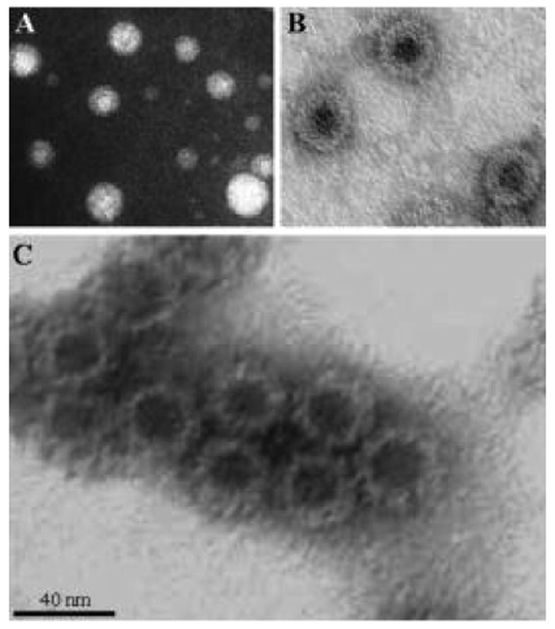
Fig. 2.

Plant produced VLPs visualized by negative staining and electron microscopy. (A) HBsAg particles from transgenic tobacco, (B) HBcAg particles from N. benthamiana infected with the MagnICON viral vector, (C) NVCP particles from transgenic tobacco.
