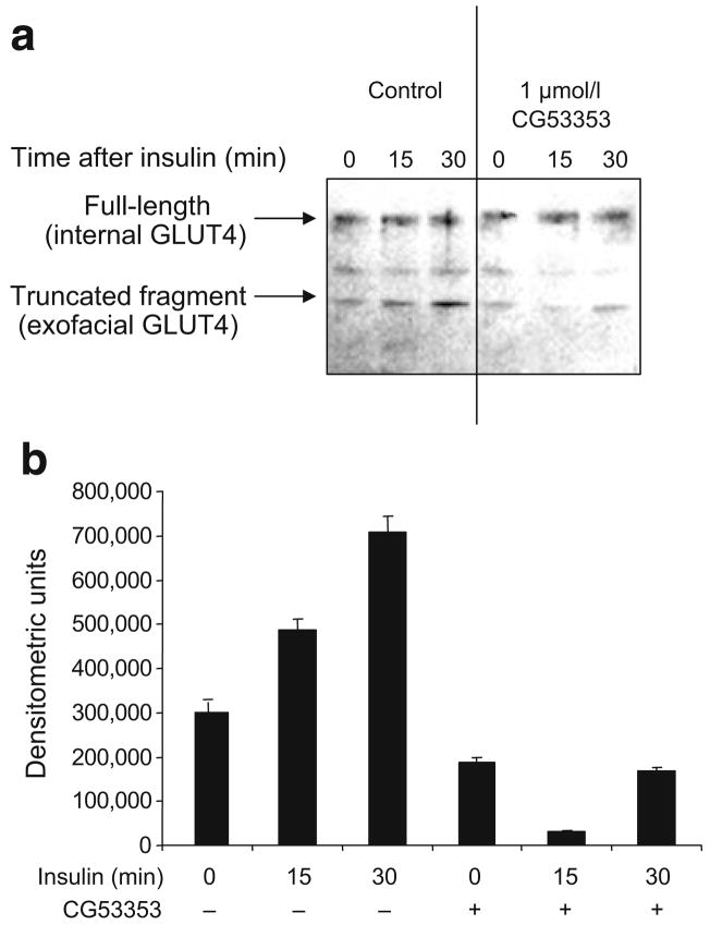
Fig. 7.
a GLUT4 in the plasma membrane was reduced by inhibiting PKCβII activity. L6 cells were grown as described in the Methods. Prior to insulin treatment (10 μIU/ml for 30 min), cells were treated for 30 min with or without 1 μmol/l CG53353 or DMSO. Cells were then treated with KCN to halt metabolic activity and trypsin to cleave exofacial proteins. GLUT4 transporters inserted into the plasma membrane would be susceptible to proteolysis of the exofacial loops by trypsin. Cells were then lysed, membranes isolated and proteins were separated by SDS-PAGE, transferred to nitrocellulose and probed using anti-GLUT4 antibodies recognising the intracellular region of the protein. This detects the full-length (near membrane) and the cleaved (inserted in plasma membrane) protein. The experiment was performed on three occasions with similar results. b The change in the lower molecular mass, fully cleaved fragment determined by scanning as described in the legend of Fig. 1

