

Abstract
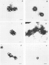
A new solid-phase immune electron microscopy double-antibody colloidal-gold technique (SPIEMDAGT) was developed and compared with direct electron microscopy, direct immune electron microscopy, and enzyme immunoassay for detecting rotavirus. Guinea pig and rabbit antirotavirus antisera were used as capture and detector antibodies, respectively, and goat anti-rabbit immunoglobulin G-gold complexes were employed as a label. Animal rotavirus in cell culture media and human virus in stool specimens were detected by this method. On average, SPIEMDAGT detected 800 times more virus particles than direct electron microscopy and 45 times more particles than direct immune electron microscopy and yielded 20% more positives than enzyme immunoassay. SPIEMDAGT could detect not only viral antigen associated with morphologically recognizable particles but also antigen present when whole virus particles were not visible.
Full text
PDF
Images in this article
Selected References
These references are in PubMed. This may not be the complete list of references from this article.
- Chernesky M., Castriciano S., Mahony J., Spiewak M., Schaefer L. Ability of TESTPACK ROTAVIRUS enzyme immunoassay to diagnose rotavirus gastroenteritis. J Clin Microbiol. 1988 Nov;26(11):2459–2461. doi: 10.1128/jcm.26.11.2459-2461.1988. [DOI] [PMC free article] [PubMed] [Google Scholar]
- Derrick K. S. Quantitative assay for plant viruses using serologically specific electron microscopy. Virology. 1973 Dec;56(2):652–653. doi: 10.1016/0042-6822(73)90068-8. [DOI] [PubMed] [Google Scholar]
- Faulk W. P., Taylor G. M. An immunocolloid method for the electron microscope. Immunochemistry. 1971 Nov;8(11):1081–1083. doi: 10.1016/0019-2791(71)90496-4. [DOI] [PubMed] [Google Scholar]
- Giraldo G., Beth E., Lee J., de Harven E., Chernesky M. Solid-phase immune electron microscopy-double-antibody technique for rapid detection of papovaviruses. J Clin Microbiol. 1982 Mar;15(3):517–521. doi: 10.1128/jcm.15.3.517-521.1982. [DOI] [PMC free article] [PubMed] [Google Scholar]
- Hopley J. F., Doane F. W. Development of a sensitive protein A-gold immunoelectron microscopy method for detecting viral antigens in fluid specimens. J Virol Methods. 1985 Oct;12(1-2):135–147. doi: 10.1016/0166-0934(85)90014-x. [DOI] [PubMed] [Google Scholar]
- Kjeldsberg E. Specific labelling of human rotaviruses and adenoviruses with gold-IgG complexes. J Virol Methods. 1985 Oct;12(1-2):47–57. doi: 10.1016/0166-0934(85)90007-2. [DOI] [PubMed] [Google Scholar]
- Nicolaieff A., Obert G., van Regenmortel M. H. Detection of rotavirus by serological trapping on antibody-coated electron microscope grids. J Clin Microbiol. 1980 Jul;12(1):101–104. doi: 10.1128/jcm.12.1.101-104.1980. [DOI] [PMC free article] [PubMed] [Google Scholar]
- Wu B. G., Mahony J., Chernesky M. Comparison of three protein A-gold immune electron microscopy methods for detecting rotaviruses. J Virol Methods. 1989 Jul;25(1):109–118. doi: 10.1016/0166-0934(89)90105-5. [DOI] [PubMed] [Google Scholar]