CELL BIOLOGY Correction for “Identification of small-molecule inducers of pancreatic β-cell expansion,” by Weidong Wang, John R. Walker, Xia Wang, Matthew S. Tremblay, Jae Wook Lee, Xu Wu, and Peter G. Schultz, which appeared in issue 5, February 3, 2009, of Proc Natl Acad Sci USA (106:1427–1432; first published January 22, 2009; 10.1073/pnas.0811848106).
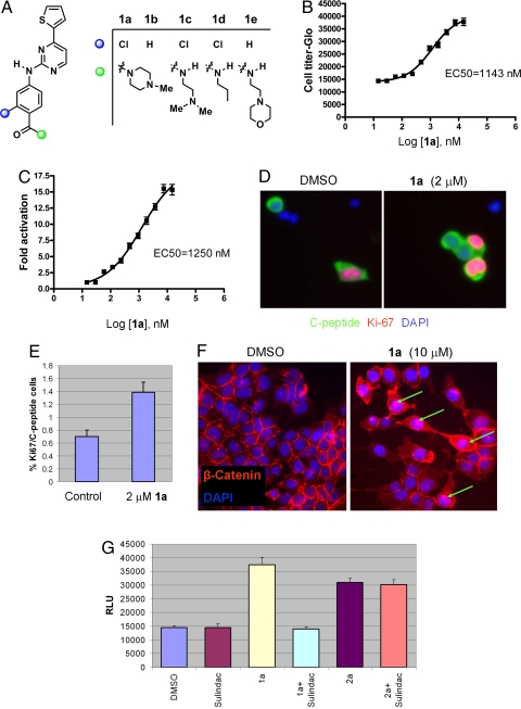
The authors note that due to a printer's error, on page 1428, Fig. 1 appeared incorrectly. In B, the notation “EC50=1143 nm” was missing. Additionally, in F, the text was missing from the box on the lower left, and in G the label “Sulindac” appeared incorrectly. The corrected figure and its legend appear below.
Fig. 1.
Novel Wnt agonists are inducers of β-cell proliferation. (A) Chemical structures of 5-thiophene pyrimidines. (B) Dose-dependent effects of 1a on β-cell proliferation. Relative luminescence unit (RLU) was measured by CellTiter-Glo assay after 7-day incubation of growth-arrested R7T1 β cells with 1a, which was added at day 0 and refreshed at day 4; experiments were performed in quadruplicate. All data in this paper are presented as mean ± SD, unless otherwise specified. (C) Dose-dependent effects of 1a on the activation of Super (8X) TOPFlash reporter. HEK293 cells transfected with Super (8X) TOPFlash reporter were treated with 1a at the indicated concentrations 24 h after transfection. Luciferase activity was measured 48 h after compound treatment. (D and E) The proliferative effect of 1a on rat primary β cells. (D) After incubation for 72 h with 2 μM 1a, replicating β cells were identified by double-immunofluorescence staining by using anti-C-peptide antibody to mark β cells (green) and antibody against Ki-67, a proliferation marker (red). Nuclear DNA was stained with DAPI (blue). (Scale bar, 10 μm.) (E) The percentage of Ki-67-positive cells of all C-peptide-positive cells, which corresponds to the fraction of replicating β cells. The number of proliferating primary β cells increased after incubation for 72 h in the presence of 2 μM 1a compared with DMSO control: 1a at 2 μM increases the replicating β cells ≈2-fold. (F) Compound 1a stimulates the translocation of β-catenin into the nucleus. Nuclear β-catenin accumulation (green arrows) was increased in 10 μM 1a-treated HEK293 cells; in DMSO-treated control cells, β-catenin staining is weak and appears to concentrate at the cell surface. (G) 1a-stimulated β-cell proliferation was abolished by the Wnt signaling antagonist Sulindac. R7T1 cells were treated with 10 μM 1a, 60 μM Sulindac, or 1 μM 2a as indicated in the figure. CellTiter-Glo activity was measured 7 days after compound treatment.


