Abstract
Purpose
Kidney volume is regarded as the most precise indicator of kidney size. However, it is not widely used clinically, because its measurement is difficult due to the complex kidney shape. We attempted to evaluate the normal kidney volume in young Korean men by using multi-detector computed tomography (MDCT).
Materials and Methods
We retrospectively reviewed MDCT data of young Korean men (113 patients). After data processing, we measured the volume and length of the kidneys. Body parameters (height, body weight, body-surface area, and total body water) and laboratory data were collected. Glomerular filtration rate (GFR) was calculated using Cockcroft-Gault (CG) equation.
Results
The mean kidney volume was 205.29 ± 36.81 cm3; and mean kidney length was 10.80 ± 0.69 cm. The former correlated significantly with height, body weight, body-surface area, and total body water (p < 0.05, correlation coefficient : γ = 0.328, 0.649, 0.640, and 0.638, respectively). The latter also correlated significantly with all body indexes, however the correlation was weaker, except with height (p < 0.05, correlation coefficient : γ = 0.457, 0.473, 0.505, and 0.503, respectively). Only kidney volume significantly predicted estimated GFR (adjusted R2 = 0.431, F = 85.90 and p < 0.05).
Conclusion
The kidney volume measured with MDCT is correlated well with body parameters, and is useful to predict renal function.
Keywords: Kidney, volume measurement, computed tomography
INTRODUCTION
The determination of kidney size is important in clinical field, because it can facilitate to make decisions on the management strategy.1 Of several indexes of kidney size, kidney length was traditionally used because it can conveniently be measured using ultrasonography (US). However, considering the complexity of the kidney shape, kidney length can not appropriately represent kidney mass. Additionally, kidney length, as measured by US, is prone to interobserver variability and poor repeatability.2
Kidney volume is a more sensitive index of kidney size than kidney length for the detection of renal abnormalities.3 It is also excellent predictor of renal function and correlates very well with body indexes.4 Furthermore, pre-transplant kidney volume is an independent determinant of the prognosis of the graft kidney.5 Kidney volume can be measured by US, but it needs calculation using ellipsoid formulae of three-dimensional value of the kidney.3 Moreover, the calculation using ellipsoid formulae tends to underestimate kidney volume.6
With development of more elaborate imaging methods such as magnetic resonance imaging (MRI) and multi-detector computed tomography (MDCT), measurement of the mass or organ volume has become feasible. The MDCT which generates 3-D reconstruction images is particularly accurate in measurement of organ volume.7,8
In this study, we estimated the normal kidney volume of healthy young Korean men by using MDCT and evaluated its predictability of renal function and relationship with body indexes.
MATERIALS AND METHODS
Patient population
We retrospectively reviewed the abdominal CT scans of 113 young Korean male patients who had visited the Armed Forces Yang-Ju Hospital between February 2005 and December 2007. The patient population comprised outpatients and inpatients that required CT examination due to common clinical indications such as weight loss, unexplained abdominal pain, unresolved nausea, and abdominal trauma. We also reviewed the medical records and laboratory findings of the patients and the radiologist's report for each CT examination. The exclusion criteria were as follows: 1) underlying diseases such as diabetes mellitus or hypertension, 2) any abnormal findings on CT examination that could affect kidney volume, including intrarenal diseases such as renal cysts, hydronephrosis, and single kidney, and 3) abnormal laboratory findings. Based on these selection criteria, we recruited 113 subjects. This study was approved by the ethics board of our institution.
CT and assessment of kidney size
This study was performed using a 4-slice helical CT scanner (GE Medical Systems, Milwaukee, WI, USA). Images were obtained prior to and after the administration of 150 mL of iodinated contrast media during the parenchymal phases of enhancement. The kidney size was measured using GE Advantage Windows Workstation (version 4.2; General Electric Medical Systems, Milwaukee, WI, USA), and kidney length was measured using coronal sections. We calculated the maximum kidney length from all coronal sections. The kidney volume was measured from contiguous slices. In coronal section images with parenchymal enhancement, the region of interest was drawn around the kidney, and the slices were reconstructed at 1-mm intervals to obtain a 3-D volume-rendered image of the kidney. The volume was calculated by multiplying the sum of areas from each slice by the reconstruction interval at the workstation.
Body index and renal function assessment
We used data on the patients' heights and weights, as registered in medical records, to deduce the body parameters. Height and weight were measured by automatic height/weight measurement system (DONG SAN JENIX Inc, Seoul, Korea) during regular physical check-up. Body-surface area (BSA) and total body water (TBW) were calculated using the following formulae.9,10

TBW (liters) of males = 2.447 + 0.3362 × weight (kg) + 0.1074 × height (cm) - 0.09516 × age (year)
As the surrogate of renal function of the subjects, we estimated the glomerular filtration rate (GFR) by using the Cockcroft-Gault (CG) equation.11 The equation used was as follows:
Cockcroft-Gault (CG): CrCl × BSA/1.73 m2
CrCl = (140-age) × (weight (kg)) × [0.85 if female]/(72 × Serum creatinine (mg/dL))
CG-GFR estimate12 : GFR = 0.84 × CrCl by CG equation
Statistical analysis
The results are presented as mean ± SD. We used the independent-samples t-test to compare the means between Lt. and Rt. kidneys and used Pearson's correlation coefficient to evaluate the relationship between the parameters. To predict renal function based on the renal length and volume, multiple linear regression analysis was performed. The results were considered significant when the p value was below 0.05. Statistical analysis was performed using the Statistical Package for the Social Sciences (SPSS) version 11.5 (SPSS Inc., Chicago, IL, USA).
RESULTS
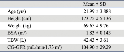
All 113 patients in this study were young Korean men. The mean age of the patients was 21.99 years (19-38 years). Their mean height was 173.75 cm (163-186 cm), mean body weight, 69.65 kg (53-106 kg), mean BSA, 1.83 m2 (1.58-2.29 m2), and TBW, 42.32 L (36.42-55.2 L) (Table 1).
Table 1.
Charateristics of Subjects
BSA, body surface area; TBW, total body water; CG-GFR, Cockcroft-Gault-glomerular filtration rate.
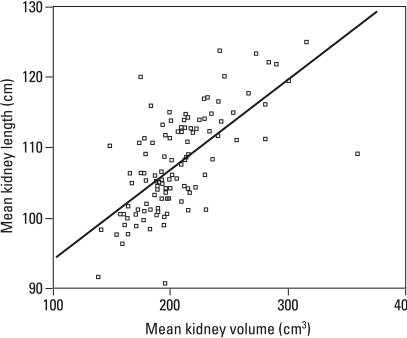
The mean kidney volume was 205.15 cm2 (138.53-359.6 cm2). The left kidney was significantly (p < 0.05) larger than the right kidney, and they were highly correlated (correlation coefficient : r = 0.874, p < 0.05). The mean kidney length was 108.02 cm (9.09-12.49 cm). The left kidney was also significantly (p < 0.05) longer than the right kidney. The kidney length and kidney volume were highly correlated reciprocally (r = 0.671, p < 0.05) (Fig. 1). The mean value of CG-GFR was 104.90 mL/min (60.99-216.38) (Table 2).
Fig. 1.
Relationship of mean kidney length and mean kidney volume.
Table 2.
Mean Value of Kidney Length and Volume According to Position
*p < 0.05 vs. Rt kidney length, †p < 0.05 vs. Rt kidney volume (± Data presentedare Mean ± SD).
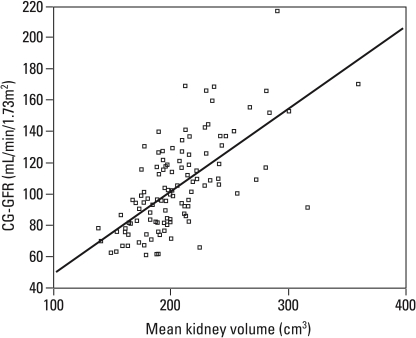
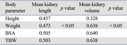
The mean length and volume of both kidneys were significantly correlated with height, weight, BSA, and TBW. The correlation coefficients (r) of the mean kidney volume with the height, weight, BSA, and TBW were 0.328, 0.649, 0.640, and 0.683, respectively (p < 0.05), and those of the mean kidney length were 0.457, 0.473, 0.505, and 0.503, respectively (p < 0.05). The correlation of kidney volume with weight, BSA and TBW was relatively higher, while the correlation coefficient of the kidney length with height was higher. Using linear regression, we found kidney volume to be a good predictor of CG-GFR estimate (adjusted R2 = 0.431, F = 85.90 and p < 0.05) (Fig. 2). However, kidney length was not a significant predictor of CG-GFR. However CG-GFR was best predicted by the following equation : CG-GFR = -2.979 + 0.525 × kidney volume.
Fig. 2.
Distribution of CG-GFR according to kidney volume. CG-GFR, Cockcroft-Gault-glomerular filtration rate.
DISCUSSION
Our present results present the normal kidney volume measured with MDCT in young Korean men. We also demonstrated that kidney volume is a better indicator of body parameters and predictor of renal function than kidney length, thus suggesting that kidney volume is more useful than kidney length in clinical field.
In this study, the mean kidney volume was 205.29 ± 36.81 cm3 in young Korean men. A few studies measured normal kidney volume by various imaging tools including MDCT.13,14 However, all of these studies have been performed in the Western population. Normal body parameters, including organ size, show racial variations. Hence, it is not rational to extrapolate the normal kidney volume data of Western adult population to Korean population.
It has been conclusively proven that age and gender are independent determining factors of renal size. Previous studies showed that the kidney becomes shorter and thicker with age,15 and that no significant age-related changes occurs in the kidney length until the 60s, but its size significantly decreases after the 70s.16 Thus, it is not reasonable to include various age groups and both genders when evaluating the normal kidney size.
In South Korea, military service is compulsory for all healthy young men, therefore the army population reflects the general population. Consequently, the results of this study might represent the kidney volume of young Korean men. Furthermore, the study population comprised subjects of only one gender with relatively less age variation. This enabled the ageand gender-independent evaluation of normal kidney volume and its relationship to body parameters and renal function.
In this study, mean kidney length measured with MDCT was 10.8 ± 0.69 cm in young Korean men. It was reported that the mean value of actual length of the left kidney in Korean men is 11.15 ± 1.02 cm,17 which is slightly longer than that observed in this study. However, that of the right and left kidneys, as measured on coronal CT sections, was 10.3 ± 0.7 cm and 10.7 ± 0.9 cm, respectively,17 which is slightly shorter than that observed in this study. According to an another study, the mean kidney length in Korean men measured using US was 10.71 ± 0.74 cm.16 It is very difficult in practice to calculate the longest axis of the kidney by using imaging tools such as US or CT. Therefore, we cannot exclude the possibility that the kidney length was underestimated in our study.
Our results on the relationship between the kidney volume and body parameters are consistent with those of previous reports.15 Both kidney volume and kidney length were significantly correlated to all body indexes, but the former showed a relatively greater correlation. We also evaluated the predictability of kidney volume and kidney length to renal function, by using the CG equation which is regarded as accurate and less biased equation to estimate GFR in healthy adults.18,19 The result showed that the kidney volume predicted the renal function significantly, whereas the kidney length did not.
Limitation of this study is that we couldn't compare the kidney volume with real size of kidney. However, it is already proved that contiguous CT slice examination is an objective and reliable method to measure the kidney volume.20,21 In addition, compared to conventional spiral CT, MDCT produces more fine slices, which can minimize disparity between real size and measured size. Recently, a new software and method of processing 3-D reconstruction images have been developed by using MDCT. This is expected to make measurement of organ volume by using CT more convenient and easier.22,23
In conclusion, kidney volume is more reliable index of kidney size than kidney length, and measurement of kidney volume with MDCT is useful method. Hence, MDCT facilitates measurement of kidney volume and enables us to use it in clinical field.
Table 3.
Correlation Coefficients (r) between Body Parameters, GFR and Kidney Size
GFR, glomerular filtration rate; BSA, body surface area; TBW, total body water
References
- 1.Lalli AF. Renal enlargement. Radiology. 1965;84:688–691. doi: 10.1148/84.4.688. [DOI] [PubMed] [Google Scholar]
- 2.Ablett MJ, Coulthard A, Lee RE, Richardson DL, Bellas T, Owen JP, et al. How reliable are ultrasound measurements of renal length in adults? Br J Radiol. 1995;68:1087–1089. doi: 10.1259/0007-1285-68-814-1087. [DOI] [PubMed] [Google Scholar]
- 3.Jones TB, Riddick LR, Harpen MD, Dubuisson RL, Samuels D. Ultrasonographic determination of renal mass and renal volume. J Ultrasound Med. 1983;2:151–154. doi: 10.7863/jum.1983.2.4.151. [DOI] [PubMed] [Google Scholar]
- 4.Widjaja E, Oxtoby JW, Hale TL, Jones PW, Harden PN, McCall IW. Ultrasound measured renal length versus low dose CT volume in predicting single kidney glomerular filtration rate. Br J Radiol. 2004;77:759–764. doi: 10.1259/bjr/24988054. [DOI] [PubMed] [Google Scholar]
- 5.Poggio ED, Hila S, Stephany B, Fatica R, Krishnamurthi V, del Bosque C, et al. Donor kidney volume and outcomes following live donor kidney transplantation. Am J Transplant. 2006;6:616–624. doi: 10.1111/j.1600-6143.2005.01225.x. [DOI] [PubMed] [Google Scholar]
- 6.Bakker J, Olree M, Kaatee R, de Lange EE, Moons KG, Beutler JJ, et al. Renal volume measurements: accuracy and repeatability of US compared with that of MR imaging. Radiology. 1999;211:623–628. doi: 10.1148/radiology.211.3.r99jn19623. [DOI] [PubMed] [Google Scholar]
- 7.Sommer G, Bouley D, Frisoli J, Pierce L, Sandner-Porkristl D, Fahrig R. Determination of 3-dimensional zonal renal volumes using contrast-enhanced computed tomography. J Comput Assist Tomogr. 2007;31:209–213. doi: 10.1097/01.rct.0000236423.12890.d6. [DOI] [PubMed] [Google Scholar]
- 8.Schlosser T, Mohrs OK, Magedanz A, Voigtländer T, Schmermund A, Barkhausen J. Assessment of left ventricular function and mass in patients undergoing computed tomography (CT) coronary angiography using 64-detector-row CT: comparison to magnetic resonance imaging. Acta Radiol. 2007;48:30–35. doi: 10.1080/02841850601067611. [DOI] [PubMed] [Google Scholar]
- 9.Mosteller RD. Simplified calculation of body-surface area. N Engl J Med. 1987;317:1098. doi: 10.1056/NEJM198710223171717. [DOI] [PubMed] [Google Scholar]
- 10.Watson PE, Watson ID, Batt RD. Total body water volumes for adult males and females estimated from simple anthropometric measurements. Am J Clin Nutr. 1980;33:27–39. doi: 10.1093/ajcn/33.1.27. [DOI] [PubMed] [Google Scholar]
- 11.Cockcroft DW, Gault MH. Prediction of creatinine clearance from serum creatinine. Nephron. 1976;16:31–41. doi: 10.1159/000180580. [DOI] [PubMed] [Google Scholar]
- 12.National Kidney Foundation. K/DOQI clinical practice guidelines for chronic kidney disease: evaluation, classification, and stratification. Am J Kidney Dis. 2002;39(2) Suppl 1:S1–S266. [PubMed] [Google Scholar]
- 13.Geraghty EM, Boone JM, McGahan JP, Jain K. Normal organ volume assessment from abdominal CT. Abdom Imaging. 2004;29:482–490. doi: 10.1007/s00261-003-0139-2. [DOI] [PubMed] [Google Scholar]
- 14.Cheong B, Muthupillai R, Rubin MF, Flamm SD. Normal values for renal length and volume as measured by magnetic resonance imaging. Clin J Am Soc Nephrol. 2007;2:38–45. doi: 10.2215/CJN.00930306. [DOI] [PubMed] [Google Scholar]
- 15.Emamian SA, Nielsen MB, Pedersen JF, Ytte L. Kidney dimensions at sonography: correlation with age, sex, and habitus in 665 adult volunteers. AJR Am J Roentgenol. 1993;160:83–86. doi: 10.2214/ajr.160.1.8416654. [DOI] [PubMed] [Google Scholar]
- 16.Lee BH, Ahn HJ, Kang WH, Seo GH, Kim B, Lee SG, et al. Estimation of kidney size by ultrasonography in normal Korean adults. Korean J Nephrol. 1999;18:46–51. [Google Scholar]
- 17.Kang KY, Lee YJ, Park SC, Yang CW, Kim YS, Moon IS, et al. A comparative study of methods of estimating kidney length in kidney transplantation donors. Nephrol Dial Transplant. 2007;22:2322–2327. doi: 10.1093/ndt/gfm192. [DOI] [PubMed] [Google Scholar]
- 18.Mahajan S, Mukhiya GK, Singh R, Tiwari SC, Kalra V, Bhowmik DM, et al. Assessing glomerular filtration rate in healthy Indian adults: a comparison of various prediction equations. J Nephrol. 2005;18:257–261. [PubMed] [Google Scholar]
- 19.Al-Khader AA, Tamim H, Sulaiman MH, Jondeby MS, Taher S, Hejaili FF, et al. What is the most appropriate formula to use in estimating glomerular filtration rate in adult Arabs without kidney disease? Ren Fail. 2008;30:205–208. doi: 10.1080/08860220701810554. [DOI] [PubMed] [Google Scholar]
- 20.Lerman LO, Bentley MD, Bell MR, Rumberger JA, Romero JC. Quantitation of the in vivo kidney volume with cine computed tomography. Invest Radiol. 1990;25:1206–1211. doi: 10.1097/00004424-199011000-00009. [DOI] [PubMed] [Google Scholar]
- 21.Kotre CJ, Owen JP. Method for the evaluation of renal parenchymal volume by X-ray computed tomography. Med Biol Eng Comput. 1994;32:338–341. doi: 10.1007/BF02512534. [DOI] [PubMed] [Google Scholar]
- 22.Rao M, Stough J, Chi YY, Muller K, Tracton G, Pizer SM, et al. Comparison of human and automatic segmentations of kidneys from CT images. Int Radiat Oncol Biol Phys. 2005;61:954–960. doi: 10.1016/j.ijrobp.2004.11.014. [DOI] [PubMed] [Google Scholar]
- 23.Cai W, Holalkere NS, Harris G, Sahani D, Yoshida H. Dynamic-threshold level set method for volumetry of porcine kidney in CT images in vivo and ex vivo assessment of the accuracy of volume measurement. Acad Radiol. 2007;14:890–896. doi: 10.1016/j.acra.2007.03.005. [DOI] [PubMed] [Google Scholar]


