Abstract
Recent studies have reported that the "cholinergic anti-inflammatory pathway" regulates peripheral inflammatory responses via α 7 nicotinic acetylcholine receptors (α 7 nAChRs) and that acetylcholine and nicotine regulate the expression of proinflammatory mediators such as TNF-α and prostaglandin E2 in microglial cultures. In a previous study we showed that ATP released by β-amyloid-stimulated microglia induced reactive oxygen species (ROS) production, in a process involving the P2X7 receptor (P2X7R), in an autocrine fashion. These observations led us to investigate whether stimulation by nicotine could regulate fibrillar β amyloid peptide (1-42) (fAβ1-42)-induced ROS production by modulating ATP efflux-mediated Ca2+ influx through P2X7R. Nicotine inhibited ROS generation in fAβ1-42-stimulated microglial cells, and this inhibition was blocked by mecamylamine, a non-selective nAChR antagonist, and α-bungarotoxin, a selective α7 nAChR antagonist. Nicotine inhibited NADPH oxidase activation and completely blocked Ca2+ influx in fAβ1-42-stimulated microglia. Moreover, ATP release from fAβ1-42-stimulated microglia was significantly suppressed by nicotine treatment. In contrast, nicotine did not inhibit 2',3'-O-(4-benzoyl)-benzoyl ATP (BzATP)-induced Ca2+ influx, but inhibited ROS generation in BzATP-stimulated microglia, indicating an inhibitory effect of nicotine on a signaling process downstream of P2X7R. Taken together, these results suggest that the inhibitory effect of nicotine on ROS production in fAβ1-42-stimulated microglia is mediated by indirect blockage of ATP release and by directly altering the signaling process downstream from P2X7R.
Keywords: acetylcholine; adenosine triphosphate; amyloid β-protein; microglia; NADPH oxidase; nicotine; reactive oxygen species; receptors, nicotinic; purinoceptor P2Z
Introduction
The neuropathological hallmarks of Alzheimer's disease (AD) include extracellular deposition of the beta amyloid peptide (Aβ) in the form of senile plaques and the appearance of intracellular neurofibrillary tangles composed of hyperphosphorylated tau (Akiyama et al., 2000). Another neuropathological feature of AD is the loss of both cholinergic neurons (Davies and Maloney, 1976; McGeer et al., 1984; Muir, 1997) and nicotinic acetylcholine receptors (nAChRs) (Burghaus et al., 2000; Mousavi et al., 2003) in the basal forebrain, which contributes to cognitive dysfunction. The nAChR is a ligand-gated ion channel consisting of five subunits with eight different α (α2-α9) subunits and three different β (β2-β4) components (Gotti and Clementi, 2004). Of these nAChRs, α7 and α4β are the most abundant subunits in the brain (Buisson and Bertrand, 2002). The administration of nAChR agonists in aging animals and humans induced cognitive improvement (Newhouse et al., 1997; Terry and Buccafuso, 2003) and prevented neuronal death induced by Aβ(O'Neill et al., 2002).
A recent study suggests that α7 nAChRs, expressed in peripheral macrophages, are essential for operation of the so-called "cholinergic anti-inflammatory pathway" that regulates systemic inflammatory responses in the peripheral nervous system (Wang et al., 2003). Recent studies have revealed that the α7 nAChRs are expressed in murine microglia in addition to neurons and peripheral macrophages, and are involved in the suppression of neuroinflammation (Shytle et al., 2004; De Simone et al., 2005). Such unique receptors should play an important role in neuroprotection, because of the activation of α7 nAChRs in capable of modulating the activity of microglia, changing the microglial cell from an overactive inflammatory cell to a protective cell type (Suzuki et al., 2006).
One of the mechanisms explaining Aβ neurotoxicity is that microglia-mediated oxidative stress, via NADPH oxidase activation, plays a critical role in the pathogenesis of AD. Fibrillar Aβ (fAβ) stimulates reactive oxygen species (ROS) production from cultured microglial cells via activation of NADPH oxidase (Bianca et al., 1999). In addition, fAβ mediated neurotoxicity in mixed neuron-microglia cultures by causing the production of ROS (Qin et al., 2002). Furthermore, NADPH oxidase activation has been identified in AD brains (Shimohama et al., 2000). Our previous study demonstrated that one of the mechanisms of ROS production in Aβ-stimulated microglia involved fAβ1-42-induced ATP release, which in turn activated NADPH oxidase in a process involving the P2X7 receptor (P2X7R), in an autocrine manner (Kim et al., 2007). Therefore, to determine whether the activation of nAChRs affects ROS generation in fAβ1-42-stimulated microglia, we examined the inhibitory effects of nicotine on ROS production and NADPH oxidase activation in such cells, and then investigated the effects of nicotine on ATP efflux and Ca2+ influx.
Materials and Methods
Reagents
Medium and supplement for cell culture were purchased from JBI (Daegu, Korea). Chemicals were purchased from the following companies: Amyloid-β1-42 (Aβ1-42) was purchased from the American Peptide (Sunnyvale, CA). Mecamylamine hydrochloride, α-bungarotoxin, pyridoxal-phosphate-6-azophenyl-2',4'-disulfonate (PPADS), adenosine 5'-triphosphate 2',3'-acylic dialcohol (oxidized ATP; oATP), apyrase (an ATP-hydrolyzing enzyme), 2',7'-dihydrodichlorofluorescein diacetate (DCF-DA), pluronic F-127, DNase I, an ATP bioluminescence assay kit, and a protease inhibitor mixture, were all purchased from Sigma (St. Louis, MO). Nicotine ([S]-3-[1-methyl-2-pyrrolindinyl] pyridine, di-d-Tartrate) was from Calbiochem (San Diego, CA), Fluo-3/AM was from Molecular probes (Eugene, OR). The anti-mouse and anti-rabbit HRP-conjugated secondary anti-bodies were purchased from Amersham Pharmacia (Buckinghamshire, UK). The polyclonal antibody against p47phox, P67phox, and Rac 1 was provided by BD Biosciences (San Diego, CA).
Microglial cell culture
Microglial cultures were prepared from the brains of 3 day-old Sprague-Dawley rats as described previously (Kim et al., 2002). Briefly, whole brains were dissected into small cubes, incubated in D-PBS containing 0.1% trypsin and 40 µg/ml DNase I for 15 min at 37℃, and dissociated into single cells by gentle pipetting. Dissociated cells were suspended in DMEM (JBI) containing 5% horse serum, 5 mg/ml glucose, 100 U/ml penicillin and 100 µg/ml streptomycin, and plated on poly-D-lysine-coated T-75 culture flasks, and incubated at 37℃ in incubator with 5% CO2/95% air atmosphere. After 2-4 weeks of growth in flasks, microglia floating in the medium were collected and grown in separate 6-, 96-well plates or on coverslips.
Measurement of intracellular ROS
Intracellular ROS levels were measured using the fluorescent dye, dihydrodichlorofluorescein diacetate (DCF-DA), which is readily converted to a fluorescent product in the presence of ROS in cells. In brief, cells were preincubated with nicotine (1-100 µM) for 30 min in the presence or absence of mecamylamine (10 µM), α-bungarotoxin (10 nM), and then treated with 0.5 µM fAβ1-42 or 300 µM BzATP. fAβ1-42-stimulated cells were incubated with 10 µM DCF-DA in HBSS (145 mM NaCl, 2.5 mM KCl, 1.8 mM MgCl2, 1 mM CaCl2, 10 mM D-glucose and 20 mM HEPES; pH 7.4) for 30 min. The cells were then washed extensively with D-PBS to remove extracellular DCF-DA, and fluorescence images were taken using an IX71 confocal laser scanning microscope (Olympus; Tokyo, Japan).
Western blot analysis
Microglial cells treated with fAβ1-42 were lysed with lysis buffer (10 mM Na2HPO4, 150 mM NaCl, 0.5% sodium deoxycholate, 0.1% SDS, and 1% NP 40; pH 7.5). Lysates were centrifuged at 13,000 × g for 10 min at 4℃ and supernatants were collected. An aliquot of each sample containing 20 µg total protein was loaded onto a 10% acrylamide gel, and then transferred to a PVDF membrane. The blots were incubated with blocking buffer [5% skim milk in TBST (20 mM Tris-HCl, 500 mM NaCl, 0.05% Tween 20, pH7.5)] at room temperature for 1 h, and incubated with primary antibody overnight at 4℃. The bands were recognized by HRP conjugated anti-rabbit secondary antibody (1 : 1,000). For detecting the translocation of NADPH oxidase components, primary monoclonal antibodies against the p47phox (1 : 500), p67phox (1 : 500), Rac 1 (1 : 500), and HRP conjugated anti-mouse secondary antibody (1 : 1,000) were used.
Cell fractionation
Microglial cells were harvested and resuspended in a cold hypotonic solution (0.25 M sucrose, 10 mM Tris-HCl, and 5 mM MgCl2; pH 7.4) including a protease inhibitor mixture, and centrifuged at 600 × g for 10 min. The supernatant was ultracentrifuged at 100,000 × g for 1.5 h at 4℃. The resulting supernatant was removed and saved as the cytosolic fraction, and the membrane pellet was resuspended in hypotonic solution containing 1% Triton X-100. Samples were analyzed by Western blotting using antibodies against the NADPH oxidase components p47phox, p67phox, and Rac 1 as described above.
Measurement of intracellular calcium
Intracellular Ca2+ concentration was monitored by loading cells with the fluorescent Ca2+ indicator Fluo-3/AM, convertible to Fluo-3 in the presence of Ca2+. Cultured microglia plated onto poly-D-lysine-coated 25 mm glass coverslips were incubated with 2 µM of the acetoxymethyl ester of Fluo-3 (Fluo-3/AM) and 0.02% pluronic F-127 in HBSS for 30 min at 37℃, and then washed with HBSS. Fluo-3-loaded cells were placed in a perfusion chamber mounted on the stage of a confocal laser-scanning microscope and stimulated with 0.5 µM fAβ1-42. To measure the intracellular calcium concentration, a confocal laser-scanning microscope (IX71, Olympus) equipped with an Argon/Keron laser (15 mW; Coherent, Santa Clara, CA) was used. Fluo-3 was excited by the 488 nm line of an argon laser and the fluorescence was measured at an emission wavelength above 510 nm.
ATP efflux measurement
Microglial cells (3 × 10⁴ cells/well) were plated in 96-well, and preincubated with nicotine (1-100 µM) for 30 min, and then treated with 0.5 µM fAβ1-42 for 1 h. At the end of this incubation, the supernatant fluids of individual wells was transferred into sterile tubes and heated at 95℃ for 3 min. Extracellular ATP in the supernatants was immediately measured by luminometer (TD2020, Turner Designs, Sunnyvale, CA), using a luciferase-luciferin assay (ATP bioluminescent assay kit from Sigma) following the instructions of the manufacturer.
Statistical analysis
All statistical comparisons in this study were done using one-way ANOVA with Tukey-Kramer multiple comparisons test and data were expressed as mean ± SEM. A value of P < 0.05 was considered statistically significant.
Results
Nicotine inhibits fAβ1-42-induced ROS production in microglia
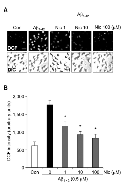
We examined the effects of nicotine on ROS production in fAβ1-42-stimulated microglia by measuring fluorescence signals from DCF-DA. Microglial cells were pre-treated with 1 µM, 10 µM, or 100 µM nicotine for 30 min and then stimulated with 0.5 µM fAβ1-42 for 2 h. Nicotine significantly decreased DCF fluorescence signals, in a dose-dependent manner (Figure 1). These results indicated that neuroprotective functions of nicotine might be mediated by suppressing fAβ1-42-induced ROS production in microglia.
Figure 1.
Effects of nicotine on reactive oxygen species production in fAβ1-42-stimulated microglia. (A) Primary rat microglia ere plated onto coverslips (3×10⁴ cells/coverslip), and then microglia were pretreated with nicotine for 30 min and stimulated with fAβ1-42(0.5 µM) for 2 h. The intracellular ROS production in microglial cells was determinded using 10 µM DCF as described in Materials and Methods. (B) DCF intensities of cells were counted using Imagegage 4.0 (Fujifilm). Fluorescence (DCF) images and differential interference contrast (DIC) images were taken using an IX71 confocal microscope (Olympus). Values are mean±SEM of 40-50 cells. *P <0.01 compared with Aβ1-42 alone. Scale bar, 20 µm.
Nicotine modulates ROS production via activation of nAChRs
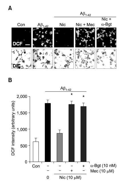
To determine whether nAChRs are involved in the nicotine inhibition of ROS production in microglia, we examined the effects of mecamylamine, a non-selective nAChR antagonist, and α-bungarotoxin, a selective α7 nAChR antagonist on the nicotine-induced decrease in ROS production. Nicotine treatment caused a marked reduction in fAβ1-42-induced ROS production. On the other hand, treatment with mecamylamine (10 µM) or α-bungarotoxin (10 nM) significantly eliminated this inhibitory activity of nicotine (Figure 2).
Figure 2.
Effects of nAChRs antagonists on fAβ1-42-induced ROS production from nicotine-treated microglia. (A) The cells were preincubated with nicotine (10 µM) for 30 min in the presence or absence of 10 µM mecamylamine (Mec) or 10 nM α-bungarotoxin (α-Bgt), and then treated with 0.5 µM fAβ1-42 for 2 h. The intracellular ROS production in microglial cells was determined using 10 µM DCF as described in Materials and Methods. (B) DCF intensities of cells were counted using Imagegage 4.0 (Fujifilm). Fluorescence (DCF) images and differential interference contrast (DIC) images were taken using an IX71 confocal microscope (Olympus). Values are mean±SEM of 40-50 cells. *P <0.01 compared with Aβ1-42plus nicotine. Scale bar, 20 µm.
fAβ1-42-induced NADPH oxidase activation is inhibited by nicotine
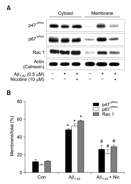
Because nicotine showed potent negative effects on ROS production, we sought to determine whether nicotine might inhibit NADPH oxidase activation by preventing the translocation of the NADPH oxidase cytoplasmic subunits p47phox, p67phox, and Rac 1 from the cytosol to the cell membrane after fAβ1-42 stimulation. Following fAβ1-42 treatment, the amounts of p47phox, p67phox, and Rac 1 in the membrane fraction increased, but the translocation of the cytosolic factors p47phox, p67phox, and Rac 1 to the plasma membrane was prevented by nicotine treatment (10 µM) (Figure 3). These results indicate that nicotine reduced fAβ1-42-induced ROS production through the inhibition of NADPH oxidase activation.
Figure 3.
Nicotine inhibits fAβ1-42-induced NADPH oxidase activation. NADPH oxidase was activated by fAβ1-42, as evidenced by the translocation of the p47phox, p67phox, Rac 1 subunits from the cytosol to the membrane; this translocation was inhibited by nicotine treatment. (A) The cells were treated with 10 µM nicotine for 30 min and stimulated with 0.5 µM fAβ1-42 for 90 min. Fractionated proteins were analyzed by SDS-PAGE and subjected to immunoblotting with anti-p47phox, anti-p67phox, anti-Rac 1 antibody. The blots were reprobed with antibodies against the calnexin membrane protein as loading controls to exhibit fractionation efficiency. (B) The histogram shows quantitation of p67phox, p47phox, Rac 1 levels expressed as the ratio of membrane fraction to total. The results represent the mean ± SEM of four to five separate experiments. *P < 0.01 compared with control, #P<0.01 compared with Aβ1-42 alone.
Nicotine inhibits fAβ1-42-induced ROS production in microglia by inhibition of ATP release from microglia, and not by blockade of P2X7R
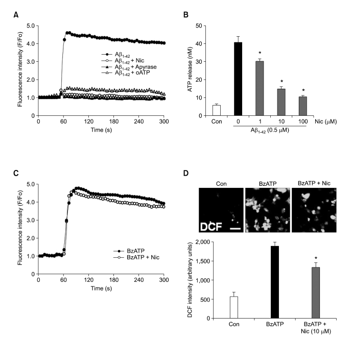
Our previous study demonstrated that fAβ1-42-stimulated ROS generation in microglial cells is regulated by ATP release-mediated Ca2+ influx through P2X7R, in an autocrine manner (Kim et al., 2007). Therefore, we investigated the effects of nicotine on ATP efflux and Ca2+ influx in fAβ1-42-stimulated microglia. Surprisingly, pretreatment of microglia with nicotine (10 µM) reduced fAβ1-42-induced Ca2+ influx to baseline levels in consistent with our previous study which showed the blockade of Ca2+ influx by pretreatment with apyrase (an ATP-hydrolyzing enzyme; 5 U/ml), or oATP (a P2X7R-specific antagonist; 100 µM) in fAβ1-42-stimulated microglia (Figure 4A). Moreover, ATP release from fAβ1-42-stimulated microglia was significantly suppressed by nicotine treatment (Figure 4B). At this point, because a previous study had reported superoxide generation after Ca2+ influx through P2X7R in microglia (Parvathenani et al., 2003), we investigated the effects of nicotine on the activation of P2X7R by examining the effects of the drug on Ca2+ influx in BzATP-stimulated microglia. Pretreatment of microglia with nicotine (10 µM) did not inhibit BzATP-induced Ca2+ influx (Figure 4C), but inhibited BzATP-induced ROS generation (Figure 4D), indicating that an inhibitory effect of nicotine lies downstream of signaling initiated from P2X7R. Taken together, these results suggest that the inhibitory effects of nicotine on fAβ1-42-induced ROS production are mediated by inhibition of ATP efflux from microglia, resulting in blockade of Ca2+ influx, and by prevention of P2X7R intracellular signaling.
Figure 4.
Effects of nicotine on Ca2+ influx in fAβ1-42- or BzATP-stimulated microglia and ATP efflux in fAβ1-42-stimulated microglia. Microglial cells were plated onto coverslips (3×10⁴ cells/coverslip), and pretreated with nicotine (10 µM) (A-D), apyrase (5 U/ml), or oATP (100 µM) for 30 min (A), and then treated with 0.5 µM fAβ1-42 (A and B) or 300 µM BzATP (C and D). (A and C) Intracellular Ca2+ concentration was measured by Fluo-3 as described in Materials and Methods, and represented by the ratio between the fluorescence intensity after treatment (F) and fluorescence in the resting state (F0). (B) Microglial cells (3×10⁴ cells/well) were plated into 96 well plate, and pretreated with nicotine (10 µM) for 30 min, and then treated with 0.5 µM fAβ1-42. ATP concentrations in the culture supernatants were determined at 1 h after fAβ1-42 stimulation. Values are mean ± SEM of triplicate samples. *P < compared with Aβ1-42. (D) Microglial cells were plated onto coverslips (3×10⁴ cells/coverslip), and pretreated with nicotine (10 µM) for 30 min, and then treated with 300 µM BzATP. Intracellular ROS levels were assayed 2 h after BzATP stimulation using 10 µM DCF. Fluorescence (DCF) images were taken using an IX71 confocal microscope (Olympus). Scale bar, 20 µm. DCF intensities of cells were counted using Imagegage 4.0 (Fujifilm). Values are mean ± SEM of 40-50 cells. *P < 0.01 compared with BzATP alone.
Discussion
The present study demonstrates the neuroprotective effects of nicotine against fAβ1-42-induced ROS production in rat microglial cultures. First, nicotine inhibits fAβ1-42-induced ROS production via activation of nAChRs and activation of fAβ1-42-induced microglial NADPH oxidase. Second, nicotine inhibits fAβ1-42-induced ROS production by blocking the Ca2+ influx that follows inhibition of ATP efflux, and by prevention of P2X7R downstream signaling.
fAβ1-42 has a direct toxic effect on neurons, but the accumulation of activated microglia at sites of fAβ1-42 deposits in AD indicates that activated microglia may also contribute to the progression of the disease (Akiyama et al., 2000). Recently, several lines of evidence have shown that oxidative stress plays an important role in inflammation-mediated neurodegeneration in AD (de la Monte and Wands, 2006; Sultana et al., 2006). Aβ stimulates ROS generation from cultured microglial cells via activation of NADPH oxidase (Bianca et al., 1999) and mediates neurotoxicity by stimulating production of ROS in mixed neuron-microglia cultures (Qin et al., 2002). Besides Aβ neurotoxicity, another feature of AD is the loss of cholinergic projections and decline of nAChRs from the early stage of AD (Oddo and LaFerla, 2006). In this regard, recent studies have reported the existence of a cholinergic control of microglial activation by showing that nicotine reduced LPS-induced production of TNF-α and IL-18, indicating that nicotine has immunosuppressive effects (Shytle et al., 2004; De Simone et al., 2005; Suzuki et al., 2006; Takahashi et al., 2006). On the other hand, nicotine treatment significantly increased the expression of COX-2 and the synthesis of PGE2 in LPS-stimulated microglia, and even enhanced TNF-α production in BzATP-stimulated microglia, suggesting a neuroprotective effect of nicotine at low concentration (De Simone et al., 2005). In this paper we expand the neuroprotective role of nicotine by showing that nicotine inhibits fAβ1-42-induced ROS production and NADPH oxidase activation in microglia. In line with our results, a recent study reported a neuroprotective effect of nicotine on dopaminergic neurons using LPS-induced in vitro and in vivo inflammation models (Park et al., 2007).
Our previous study showed that ROS generation in fAβ1-42-stimulated microglia is mediated by ATP release and subsequent Ca2+ influx, in a process involving the activation of P2X7R, in an autocrine manner (Kim et al., 2007). Interestingly, the present study demonstrated that inhibitory effects of nicotine on fAβ1-42-induced ROS production are mediated by decreasing Ca2+ influx to a basal level through inhibition of ATP release from microglia. On the other hand, pretreatment with nicotine did not inhibit BzATP-elicited Ca2+ influx. These results indicate that blockade of fAβ1-42-elicited Ca2+ influx by nicotine treatment is mediated by preventing ATP release from microglia, and not by either interference with ATP binding to P2X7R or by decreasing the activity of this channel. Recently, it has been recognized that ATP can be released from LPS- or glutamate-stimulated microglial cells (Ferrari et al., 1997; Seo et al., 2004; Liu et al., 2006), and ATP efflux from either astrocytes or microglia occurred via ATP binding cassette (ABC) proteins (Ballerini et al., 2002). Future work on identifying the mechanisms involved in nicotine-mediated inhibition of ATP release will provide a better understanding of the role of nicotine under the pathological conditions of AD.
Besides blockade of Ca2+ influx by inhibition of ATP efflux, nicotine-induced suppression of ROS production in fAβ1-42-stimulated microglia seems to also involve interference with P2X7R downstream signaling. Our present results showed that pretreatment of microglia with nicotine did not inhibit BzATP-induced Ca2+ influx but did inhibit BzATP-induced ROS generation. It has been shown that p38 MAPK and PI3 kinase (PI3-K) play key roles in the production of ROS in BzATP-stimulated microglia (Parvathenani et al., 2003). However, a recent study reported that nicotine did not affect the activation of p38 MAPK in BzATP-stimulated microglia (Suzuki et al., 2006), and it remains unclear whether nicotine inhibits PI3-K in BzATP-stimulated microglia.
In conclusion, our study provides evidence for the first time that nicotine can downregulate ROS production in fAβ1-42-stimulated microglia by inhibition of both ATP release and P2X7R signaling. The finding that nicotine prevents ROS production in microglia provides evidence for molecular links between Aβ, cholinergic dysfunction, and cognitive impairments, during the progress of AD.
Acknowledgment
This work was supported by the Korea Science and Engineering Foundation (KOSEF) through the Brain Disease Research Center at Ajou University, and the Korea Health 21 R&D Project, Ministry of Health and Welfare, Republic of Korea; Grant 03-PJ1-PG10-21300-0006.
Abbreviations
- ABC
ATP binding cassette
- Aβ
β amyloid peptide
- AD
Alzheimer's disease
- α-Bgt
α-bungarotoxin
- CREB
cAMP response element binding protein
- DCF-DA
dihydrodichlorofluorescein diacetate
- fAβ
fibrillar Aβ
- Mec
mecamylamine
- nAChR
nicotinic acetylcholine receptors
- oATP
oxidized ATP
- PI3-K
PI3 kinase
- PPADS
pyridoxal-phosphate-6-azophenyl-2',4'-disulfonate
- P2X7R
P2X7 receptor
- ROS
reactive oxygen species
References
- 1.Akiyama H, Barger S, Barnum S, Bradt B, Bauer J, Cole GM, Cooper NR, Eikelenboom P, Emmerling M, Fiebich BL. Inflammation and Alzheimer's disease. Neurobiol Aging. 2000;21:383–421. doi: 10.1016/s0197-4580(00)00124-x. [DOI] [PMC free article] [PubMed] [Google Scholar]
- 2.Ballerini P, Di Iorio P, Ciccarelli R, Nargi E, D'Alimonte I, Traversa U, Rathbone MP, Caciagli F. Glial cells express multiple ATP binding cassette proteins which are involved in ATP release. Neuroreport. 2002;13:1789–1792. doi: 10.1097/00001756-200210070-00019. [DOI] [PubMed] [Google Scholar]
- 3.Bianca VD, Dusi S, Bianchini E, Dal Pra I, Rossi F. β-amyloid activates the O-2 forming NADPH oxidase in microglia, monocytes, and neutrophils. A possible inflammatory mechanism of neuronal damage in Alzheimer's disease. J Biol Chem. 1999;274:15493–15499. doi: 10.1074/jbc.274.22.15493. [DOI] [PubMed] [Google Scholar]
- 4.Buisson B, Bertrand D. Nicotine addiction: the possible role of functional upregulation. Trends Pharmacol Sci. 2002;23:130–136. doi: 10.1016/S0165-6147(00)01979-9. [DOI] [PubMed] [Google Scholar]
- 5.Burghaus L, Schutz U, Krempel U, de Vos RA, Jansen Steur EN, Wevers A, Lindstrom J, Schroder H. Quantitative assessment of nicotinic acetylcholine receptor proteins in the cerebral cortex of Alzheimer patients. Brain Res Mol Brain Res. 2000;76:385–388. doi: 10.1016/s0169-328x(00)00031-0. [DOI] [PubMed] [Google Scholar]
- 6.Davies P, Maloney AJ. Selective loss of central cholinergic neurons in Alzheimer's disease. Lancet. 1976;2:1403. doi: 10.1016/s0140-6736(76)91936-x. [DOI] [PubMed] [Google Scholar]
- 7.de la Monte SM, Wands JR. Molecular indices of oxidative stress and mitochondrial dysfunction occur early and often progress with severity of Alzheimer's disease. J Alzheimers Dis. 2006;9:167–181. doi: 10.3233/jad-2006-9209. [DOI] [PubMed] [Google Scholar]
- 8.De Simone R, Ajmone-Cat MA, Carnevale D, Minghetti L. Activation of α7 nicotinic acetylcholine receptor by nicotine selectively up-regulates cyclooxygenase-2 and prostaglandin E2 in rat microglial cultures. J Neuroinflammation. 2005;2:4–13. doi: 10.1186/1742-2094-2-4. [DOI] [PMC free article] [PubMed] [Google Scholar]
- 9.Ferrari D, Chiozzi P, Falzoni S, Hanau S, Di Virgilio F. Purinergic modulation of interleukin-1β release from microglial cells stimulated with bacterial endotoxin. J Exp Med. 1997;185:579–582. doi: 10.1084/jem.185.3.579. [DOI] [PMC free article] [PubMed] [Google Scholar]
- 10.Gotti C, Clementi F. Neuronal nicotinic receptors: from structure to pathology. Prog Neurobiol. 2004;74:363–396. doi: 10.1016/j.pneurobio.2004.09.006. [DOI] [PubMed] [Google Scholar]
- 11.Kim KY, Kim MY, Choi HS, Jin BK, Kim SU, Lee YB. Thrombin induces IL-10 production in microglia as a negative feedback regulator of TNF-α release. Neuroreport. 2002;13:849–852. doi: 10.1097/00001756-200205070-00022. [DOI] [PubMed] [Google Scholar]
- 12.Kim SY, Moon JH, Lee HG, Kim SU, Lee YB. ATP released from β-amyloid-stimulated microglia induces reactive oxygen species production in an autocrine fashion. Exp Mol Med. 2007;39:820–827. doi: 10.1038/emm.2007.89. [DOI] [PubMed] [Google Scholar]
- 13.Liu GJ, Kalous A, Werry EL, Bennett MR. Purine release from spinal cord microglia after elevation of calcium by glutamate. Mol Pharmacol. 2006;70:851–859. doi: 10.1124/mol.105.021436. [DOI] [PubMed] [Google Scholar]
- 14.McGeer PL, McGeer EG, Suzuki J, Dolman CE, Nagai T. Aging, Alzheimer's disease, and the cholinergic system of the basal forebrain. Neurology. 1984;34:741–745. doi: 10.1212/wnl.34.6.741. [DOI] [PubMed] [Google Scholar]
- 15.Mousavi M, Hellstrom-Lindahl E, Guan ZZ, Shan KR, Ravid R, Nordberg A. Protein and mRNA levels of nicotinic receptors in brain of tobacco using controls and patients with Alzheimer's disease. Neuroscience. 2003;122:515–520. doi: 10.1016/s0306-4522(03)00460-3. [DOI] [PubMed] [Google Scholar]
- 16.Muir JL. Acetylcholine, aging, and Alzheimer's disease. Pharmacol Biochem Behav. 1997;56:687–696. doi: 10.1016/s0091-3057(96)00431-5. [DOI] [PubMed] [Google Scholar]
- 17.Newhouse PA, Potter A, Levin ED. Nicotinic system involvement in Alzheimer's and Parkinson's diseases. Implications for therapeutics. Drugs Aging. 1997;11:206–228. doi: 10.2165/00002512-199711030-00005. [DOI] [PubMed] [Google Scholar]
- 18.Oddo S, LaFerla FM. The role of nicotinic acetylcholine receptors in Alzheimer's disease. J Physiol Paris. 2006;99:172–179. doi: 10.1016/j.jphysparis.2005.12.080. [DOI] [PubMed] [Google Scholar]
- 19.O'Neill MJ, Murray TK, Lakics V, Visanji NP, Duty S. The role of neuronal nicotinic acetylcholine receptors in acute and chronic neurodegeneration. Curr Drug Targets CNS Neurol Disord. 2002;1:399–411. doi: 10.2174/1568007023339166. [DOI] [PubMed] [Google Scholar]
- 20.Park HJ, Lee PH, Ahn YW, Choi YJ, Lee G, Lee DY, Chung ES, Jin BK. Neuroprotective effect of nicotine on dopaminergic neurons by anti-inflammatory action. Eur J Neurosci. 2007;26:79–89. doi: 10.1111/j.1460-9568.2007.05636.x. [DOI] [PubMed] [Google Scholar]
- 21.Parvathenani LK, Tertyshnikova S, Greco CR, Roberts SB, Robertson B, Posmantur R. P2X7 mediates superoxide production in primary microglia and is up-regulated in a transgenic mouse model of Alzheimer's disease. J Biol Chem. 2003;278:13309–13317. doi: 10.1074/jbc.M209478200. [DOI] [PubMed] [Google Scholar]
- 22.Qin L, Liu Y, Cooper C, Liu B, Wilson B, Hong JS. Microglia enhance β-amyloid peptide-induced toxicity in cortical and mesencephalic neurons by producing reactive oxygen species. J Neurochem. 2002;83:973–983. doi: 10.1046/j.1471-4159.2002.01210.x. [DOI] [PubMed] [Google Scholar]
- 23.Seo DR, Kim KY, Lee YB. Interleukin-10 expression in lipopolysaccharide-activated microglia is mediated by extracellular ATP in an autocrine fashion. Neuroreport. 2004;15:1157–1161. doi: 10.1097/00001756-200405190-00015. [DOI] [PubMed] [Google Scholar]
- 24.Shimohama S, Tanino H, Kawakami N, Okamura N, Kodama H, Yamaguchi T, Hayakawa T, Nunomura A, Chiba S, Perry G. Activation of NADPH oxidase in Alzheimer's disease brains. Biochem Biophys Res Commun. 2000;273:5–9. doi: 10.1006/bbrc.2000.2897. [DOI] [PubMed] [Google Scholar]
- 25.Shytle RD, Mori T, Townsend K, Vendrame M, Sun N, Zeng J, Ehrhart J, Silver AA, Sanberg PR, Tan J. Cholinergic modulation of microglial activation by α7 nicotinic receptors. J Neurochem. 2004;89:337–343. doi: 10.1046/j.1471-4159.2004.02347.x. [DOI] [PubMed] [Google Scholar]
- 26.Sultana R, Perluigi M, Butterfield DA. Protein oxidation and lipid peroxidation in brain of subjects with Alzheimer's disease: insights into mechanism of neurodegeneration from redox proteomics. Antioxid Redox Signal. 2006;8:2021–2037. doi: 10.1089/ars.2006.8.2021. [DOI] [PubMed] [Google Scholar]
- 27.Suzuki T, Hide I, Matsubara A, Hama C, Harada K, Miyano K, Andra M, Matsubayashi H, Sakai N, Kohsaka S. Microglial α7 nicotinic acetylcholine receptors drive a phospholipase C/IP3 pathway and modulate the cell activation toward a neuroprotective role. J Neurosci Res. 2006;83:1461–1470. doi: 10.1002/jnr.20850. [DOI] [PubMed] [Google Scholar]
- 28.Takahashi HK, Iwagaki H, Hamano R, Yoshino T, Tanaka N, Nishibori M. Effect of nicotine on IL-18-initiated immune response in human monocytes. J Leukoc Biol. 2006;80:1388–1394. doi: 10.1189/jlb.0406236. [DOI] [PubMed] [Google Scholar]
- 29.Terry AV, Jr, Buccafusco JJ. The cholinergic hypothesis of age and Alzheimer's disease-related cognitive deficits: recent challenges and their implications for novel drug development. J Pharmacol Exp Ther. 2003;306:821–827. doi: 10.1124/jpet.102.041616. [DOI] [PubMed] [Google Scholar]
- 30.Wang H, Yu M, Ochani M, Amella CA, Tanovic M, Susarla S, Li JH, Wang H, Yang H, Ulloa L. Nicotinic acetylcholine receptor α7 subunit is an essential regulator of inflammation. Nature. 2003;421:384–388. doi: 10.1038/nature01339. [DOI] [PubMed] [Google Scholar]


