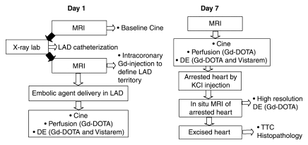
Figure 1:
Schematic diagram shows study design and imaging protocol. The study was performed with a hybrid system composed of an x-ray C-arm and MR imager. Black arrows = transfer of the animal with a floating table between the units. DE = delayed-enhanced MR imaging, Gd = gadolinium-based, Gd-DOTA = gadoterate meglumine, KCl = saturated potassium chloride, LAD = left anterior descending coronary artery, TTC = triphenyltetrazolium chloride.

