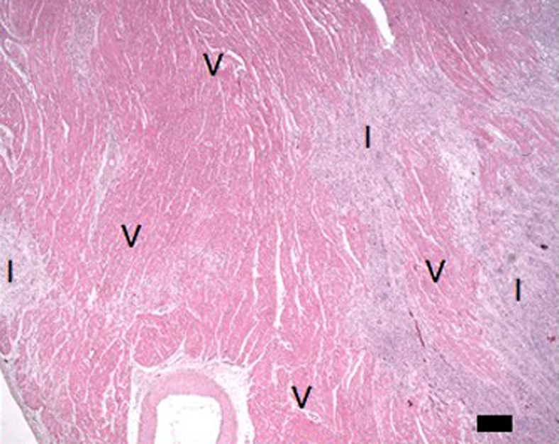
Figure 8b:

(a–d) Histologic slices obtained from the microinfarcted territory show microinfarction (I) surrounded by viable myocardium (V) and embolic agents (arrows) obstructing microvessels. (a) Several embolic agents are shown inside a vessel surrounded by microinfarction. (b) Stripes of infarcted tissue with inflammatory cells from the epicardium (left bottom corner) to the endocardium (out of field at right). (c) A thin stripe of healing microinfarction extends from epicardial surface (top right) to endocardium (bottom left) where the microinfarction is broader. (d) A longitudinally cut vessel with two embolic agents is surrounded by fibrin and organizing tissue, which are signs of inflammatory reaction within the vessel. Scale bars = 400 μm. (a and b, hematoxylin-eosin stain; original magnification, ×25.) (c and d, Masson trichrome stain; original magnification, ×100 for c and ×400 for d.)
