Figure 1.
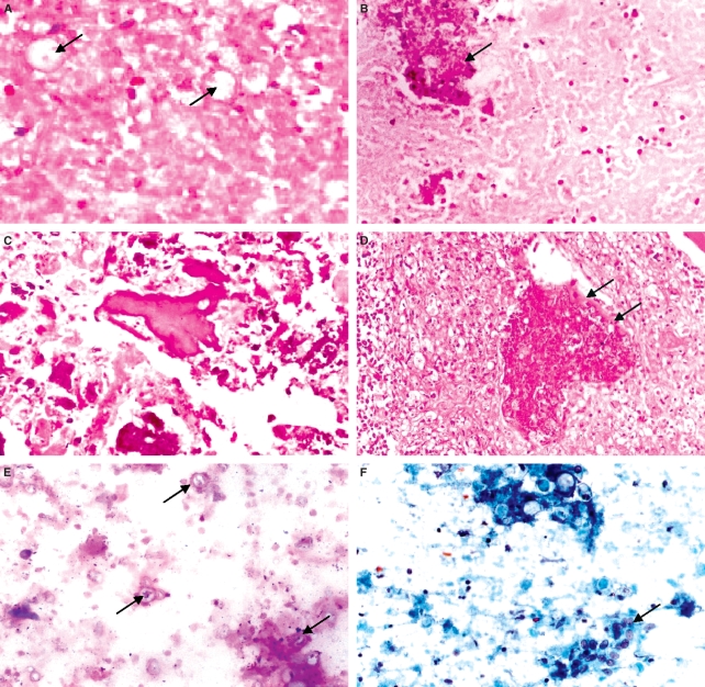
A, Case 1: Severe coagulative bone marrow necrosis (BMN) in an AIDS patient. Fungal cells can be observed within the ghosts of the dead hematopoietic cells (arrows). Bone marrow biopsy (BMB); H&E. B, Case 2: Extensive coagulative BMN. Bone trabecula was also affected (arrow). BMB; H&E. C, Case 5: Disarrangement of bone marrow architecture due to both parenchymal and trabecular severe BMN. BMB; H&E. D, Case 6: Necrotic bone trabecula in an extensive area of granulomatous reaction presenting numerous Paracoccidioides brasiliensis. Remnant of non-necrotic bone can be seen on its upper border (arrows). BMB; H&E. E, Case 7: Necrotic hematopoietic cells with a smudgy appearance. A stained smooth homogeneus background precipitate is present. Fungal cells can be seen (arrows). Aspirated smear; Giemsa. F, Case 8: Typical coagulative necrosis on aspirated smear. An agglomerate of histiocyte attempts to form a giant cell (arrow). Several P. brasiliensis are also present. Aspirated smear; Shorr.

