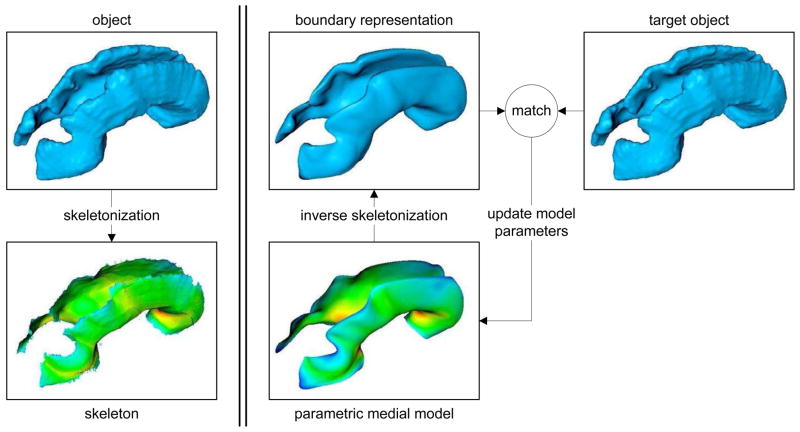
Fig. 1.
Skeletonization vs. medial modeling. Left: A 3D object and the skeleton derived by skeletonization. The color map on the skeleton is the “radius scalar field” R or, equivalently, the distance to the closest boundary point. Right: medial modeling, which is, essentially, the opposite of skeletonization. A deformable parametric medial model is defined as a surface or set of surfaces, and the boundary is derived analytically using “inverse skeletonization,” (see Eqn. 2). The model is then deformed to maximize fit between its boundary and the object of interest. The key difference between skeletonization and medial modeling is that the former computes exact skeletons, but does not guarantee that the branching topology of the skeletons is consistent across individuals; the latter computes approximate skeletons, but guarantees the same topology for all individuals, allowing effective statistical analysis.

