Abstract
Pyometra is an uncommon condition with an incidence of less than 1% in gynaecologic patients. Spontaneous rupture of pyometra in cervical cancer presenting as generalized peritonitis is very rare. Only four cases have been described in the English literature to the best of our knowledge and from a PubMed search. The index case is an elderly postmenopausal female who was diagnosed with cervical cancer, started on radiotherapy and presented with features of generalized peritonitis. Contrast-enhanced CT revealed uterine perforation at the fundus with multiple abdominal and pelvic collections. A brief review of all the cases of ruptured pyometra in cervical cancer in the literature and a discussion of the role of imaging is presented.
Keywords: uterine rupture, pyometra
Case report
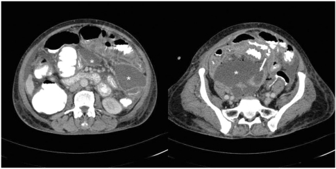
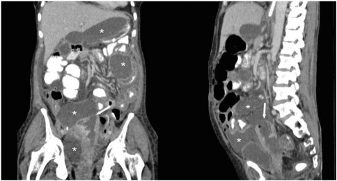
A 60-year-old female presented with a history of serosanginous discharge and bleeding (small amount) per vaginum for the last 4 months. She was postmenopausal for the last 10 years with P5+1+0+5. Local examination revealed hard infiltrative growth destroying both cervical lips and involving the vaginal wall. The upper third of the lateral and upper half of the anterior and posterior walls of the vagina were involved. Biopsy from the lesion was performed which showed adenocarcinoma (moderately differentiated). Hemogram (Hb = 11.4 gm%, TLC = 7600/μl) and renal function tests (urea = 24 mg/dl and creatinine = 0.81 mg/dl) were normal. Her intravenous pyelography (IVP) and cystoscopy studies were normal. Ultrasound of the abdomen and pelvis showed no intra-abdominal/pelvic collection or pyometra. With Carcinoma cervix III B (FIGO clinical staging), she was planned for external radiotherapy (46Gy in 23 fractions over 4.5 weeks). But after receiving 18Gy/9 fractions, she presented with pain in the abdomen and vomiting. Ultrasound showed a bulky and heterogeneous cervix with multiple intra-abdominal loculated collections. Contrast-enhanced CT showed multiple pelvic and intra-abdominal collections, a dilated endometrial cavity with breach at the uterine fundus and its continuation with large pelvic collection, suggestive of perforated pyometra. Sagittal and coronal reformats in multi-detector CT depicted the site and size of the uterine breach. The patient was managed with pigtail drainage of the larger pelvic and subhepatic collections, and with appropriate antibiotic cover (cefoperozone, sulbactam and amikacin). Pus culture and sensitivity showed Acinobacter. The pigtail drained approximately 300 ml of thick pus. She improved clinically and follow-up ultrasound after 1 week showed near complete resolution of the abscesses, and the catheter was taken out (Fig. 1 and 2).
Figure 1.
Contrast-enhanced axial CT images showing dilated endometrial cavity with site of the breach at uterine fundus (arrow). Pelvic collections (*) are seen with the abdominal/mesenteric extension and resultant intra-abdominal collections (*).
Figure 2.
Contrast-enhanced coronal and sagittal reformatted CT image depicting dilated endometrial cavity with site and size of the breach at uterine fundus (arrow). Multiple pelvic and intra-abdominal collections (*) are seen. The larger pelvic collection is seen in continuation with the ruptured uterus.
Discussion
Pyometra is an uncommon condition occurring mainly in elderly postmenopausal females, and results when natural drainage of the uterine cavity is compromised. It is an accumulation of purulent material/pus in the uterine cavity. The common causes of pyometra include malignant condition of the genital tract and sequele of their treatment, benign conditions such as infection and congenital cervical anomalies[1–2]. The benign or malignant conditions cause accumulation of secretions and gradual enlargement of the uterus, leading to thinned uterine walls which may be sloughed with spontaneous uterine rupture and causing generalized peritonitis. Spontaneous uterine perforation is rare and extensive review of the English literature has revealed only 26 such reported cases to date[1–7]. Furthermore, spontaneous rupture of pyometra in cervical cancer presenting as generalized peritonitis is extremely rare and only four cases have been described. We reviewed all the cases of spontaneous uterine perforation in cervical cancer and the findings are summarized in Table 1.
Table 1.
Cases of spontaneous uterine perforation in cervical cancer
| Ref. no. | Age | Symptoms | Provisional diagnosis | Perforation site | Histology | Treatment |
|---|---|---|---|---|---|---|
| 8 | 67 | AP, GB | GP | Fundus | Squamous cell carcinoma | Aspiration and drainage |
| 2 | 34 | AP | GP,PP | Left corneal region | Squamous cell carcinoma | Drainage and PL |
| 2 | 72 | AP | GP | Fundus | Squamous cell carcinoma | Drainage and PL |
| 1 | 60 | AP,F | GP | Fundus | Squamous cell carcinoma | TAH with BSO |
| Present case | 60 | AP,F,V | GP | Fundus | Adeno-carcinoma | Pigtail drainage |
AP, abdominal pain; BSO, bilateral salpingo-oophorectomy; F, fever; GB, genital bleeding; GP, generalized peritonitis; PL, peritoneal lavage; PP, perforated pyometra; TAH, total abdominal hysterectomy; V, vomiting.
All the patients presented with features of generalized peritonitis including the index case. Common presenting symptoms were abdominal pain, vomiting and fever. All the cases were elderly postmenopausal females except one[3]. The most common perforation site was the uterine fundus and in only one case it was the uterine cornual region[1]. Perforated pyometra was not the preoperative diagnosis in any of the cases. CT features of perforated pyometra have been described in only one case in which CT revealed the diagnosis and surgical intervention was performed[6]. In our case, the patient presented with features of generalized peritonitis, CT showed perforated pyometra at the uterine fundus with multiple pelvic and intra-abdominal collections. Sagittal and coronal reformats in multi-detector CT are very helpful in depicting the site and size of uterine breach, demonstrating the resultant intra-abdominal collections and staging of cervical cancer. The site of uterine breach can be missed on axial images as it is usually seen in fundus; hence, in elderly postmenopausal females with peritonitis sagittal and coronal reformats of uterus should be carefully evaluated. Sonography plays a limited role in the diagnosis of ruptured pyometra because of its inability to demonstrate the uterine breach and the limited sonographic window available due to perforation.
Treatment of ruptured pyometra in cervical cancer patients varies depending on the clinical condition of the patient and the preoperative diagnosis. In most cases drainage of the collections was carried out, except for one case in which total abdominal hysterectomy and bilateral salpingoophorectomy were peformed. The index case was managed by putting percutaneous pigtail catheters in pelvic and subhepatic collections.
Spontaneous rupture of pyometra in cervical cancer is very rare and should be considered in elderly postmenopausal women with cervical cancer presenting with an acute abdomen. Multi-detector CT with sagittal and coronal reformatted images could play an important role in the diagnosis of ruptured pyometra.
References
- 1.Lee SL, Huang LW, Seow KM, Hwang JL. Spontaneous perforation of a pyometra in a postmenopausal woman with untreated cervical cancer and "forgotten" intrauterine device. Taiwan J Obstet Gynecol. 2007;46:439–41. doi: 10.1016/s1028-4559(08)90021-8. PMid:18182356. [DOI] [PubMed] [Google Scholar]
- 2.Chan LY, Yu VS, Ho LC, Lok YH, Hui SK. Spontaneous uterine perforation of pyometra: A report of three cases. J Reprod Med. 2000;45:857–60. PMid:11077640. [PubMed] [Google Scholar]
- 3.Yildizhan B, Uyar E, Sismanoglu A, Gulluoglu G, Kavak ZN. Spontaneous perforation of pyometra: A case report. Infect Dis Obstet Gynecol. 2006;2006:26786. doi: 10.1155/IDOG/2006/26786. PMid:17093350. [DOI] [PMC free article] [PubMed] [Google Scholar]
- 4.Geranpayeh L, Fadaei-Araghi M, Shakiba B. Spontaneous uterine perforation due to pyometra presenting as acute abdomen. Infect Dis Obstet Gynecol. 2006;2006:60276. doi: 10.1155/IDOG/2006/60276. doi:10.1155/IDOG/2006/60276. PMid:17485806. [DOI] [PMC free article] [PubMed] [Google Scholar]
- 5.Nuamah NM, Hamaloglu E, Konan A. Spontaneous uterine perforation due to pyometra presenting as acute abdomen. Int J Gynaecol Obstet. 2006;92:145–146. doi: 10.1016/j.ijgo.2005.09.027. doi:10.1016/j.ijgo.2005.09.027. PMid:16325818. [DOI] [PubMed] [Google Scholar]
- 6.Chan KS, Tan CK, Mak CW, Chia CC, Kuo CY, Yu WL. Computed tomography features of spontaneously perforated pyometra: A case report. Acta Radiol. 2006;47:226–227. doi: 10.1080/02841850500480634. doi:10.1080/02841850500480634. PMid:16604973. [DOI] [PubMed] [Google Scholar]
- 7.Saha PK, Gupta P, Mehra R, Goel P, Huria A. Spontaneous perforation of pyometra presented as an acute abdomen: A case report. Medscape J Med. 2008;10:15. PMid:18324325. [PMC free article] [PubMed] [Google Scholar]
- 8.Imachi M, Tanaka S, Ishikawa S, Matsuo K. Spontaneous perforation of pyometra presenting as generalized peritonitis in a patient with cervical cancer. Gynecol Oncol. 1993;50:384–88. doi: 10.1006/gyno.1993.1231. doi:10.1006/gyno.1993.1231. PMid:8406207. [DOI] [PubMed] [Google Scholar]


