Abstract
Knowledge of the many mechanisms of vesicular stomatitis virus (VSV) transmission is critical for understanding of the epidemiology of sporadic disease outbreaks in the western United States. Migratory grasshoppers [Melanoplus sanguinipes (Fabricius)] have been implicated as reservoirs and mechanical vectors of VSV. The grasshopper-cattle-grasshopper transmission cycle is based on the assumptions that (i) virus shed from clinically infected animals would contaminate pasture plants and remain infectious on plant surfaces and (ii) grasshoppers would become infected by eating the virus-contaminated plants. Our objectives were to determine the stability of VSV on common plant species of U.S. Northern Plains rangelands and to assess the potential of these plant species as a source of virus for grasshoppers. Fourteen plant species were exposed to VSV and assayed for infectious virus over time (0 to 24 h). The frequency of viable virus recovery at 24 h postexposure was as high as 73%. The two most common plant species in Northern Plains rangelands (western wheatgrass [Pascopyrum smithii] and needle and thread [Hesperostipa comata]) were fed to groups of grasshoppers. At 3 weeks postfeeding, the grasshopper infection rate was 44 to 50%. Exposure of VSV to a commonly used grasshopper pesticide resulted in complete viral inactivation. This is the first report demonstrating the stability of VSV on rangeland plant surfaces, and it suggests that a significant window of opportunity exists for grasshoppers to ingest VSV from contaminated plants. The use of grasshopper pesticides on pastures would decrease the incidence of a virus-amplifying mechanical vector and might also decontaminate pastures, thereby decreasing the inter- and intraherd spread of VSV.
Vesicular stomatitis virus (VSV) is a highly transmissible rhabdovirus that causes economically important, Office of International Epizootics (OIE)-reportable disease outbreaks, primarily in horses and cattle of western U.S. rangelands. Vesicular stomatitis (VS) is endemic in portions of the southeastern United States, Mexico, and South America. Outbreaks in the western United States are sporadic, occurring every 2 to 9 years over the past 23 years, with the most recent outbreak in 2006. During outbreaks, clinically infected animals salivate excessively and shed copious amounts of virus (4 to 6 log units of virus per ml) (8). Virus-laden saliva contaminates facilities (e.g., water and feed troughs, stables, and corrals) as well as the environment (e.g., plants and soil), allowing extensive animal-to-animal transmission once the virus is in the herd (16). Insects are believed to play important roles in the initial introduction of the virus into a herd from undetermined natural reservoirs, as well as transmitting it across large distances between herds of similar or different species during animal movement quarantines (8).
The protocol of veterinary practitioners regarding VS is to control the spread of virus during outbreaks by keeping all animals on the premises; cleaning and disinfecting all personnel materials, instruments, equipment, vehicles, feed bunks, and water sources; and instructing personnel to shower and change clothing and boots when moving between herds. According to the OIE, soil and plants are suspected sources of virus, although no report to date confirms this. Therefore, decontamination of corrals and pastures is not a current recommendation. Sand flies, black flies, and biting midges (Culicoides sonorensis) have all been shown to be competent vectors, capable of transmitting the virus during blood feeding (2, 3, 12, 17). Thus, the control of biting insects in barns and other housing areas with screens and repellents is advised.
Although research has traditionally focused on these hematophagous insects as important VSV vectors, the migratory grasshopper Melanoplus sanguinipes (Fabricius) was recently shown to be an efficient amplifying reservoir and a possible mechanical vector for VSV (10). M. sanguinipes is distributed in North America from Alaska to Mexico and from coast to coast. It is a serious pest of both crops and grasslands, causing more crop damage than any other species of grasshopper in the United States (14). Grasshoppers are typically ingested by grazing animals when they are immobile during one of five molting stages. It is estimated that grazing cattle consume approximately 50 of these molting grasshoppers per day (10). In a previous study, grasshoppers were shown to amplify ingested VSV as much as 1,400-fold and to maintain high virus titers for at least 28 days (10). The route of VSV entry into cattle eating the infected grasshoppers was via scarifications on the tongue and gums, typically seen in cattle on rangeland pastures. Of significance to this study, the grasshopper-cattle-grasshopper transmission cycle is based on the assumptions that (i) virus shed from clinically infected animals would contaminate pasture plants and remain infectious on plant surfaces and (ii) grasshoppers would become infected by eating the virus-contaminated plants. To determine the stability of VSV on plants and the window of opportunity for grasshoppers to ingest viable VSV from those contaminated plants, we exposed rangeland plant species typically consumed by grasshoppers to VSV and determined the titer of virus over time. Several plant species harbored viable virus as long as 24 h. Grasshoppers were fed virus-contaminated plants, held for 21 days, and tested for virus. Current decontamination practices during VSV outbreaks do not address the viral contamination of pastures or the control of nonhematophagous virus-amplifying insect species such as grasshoppers. To that end, a commonly used no-withdrawal grasshopper pesticide was evaluated for its ability to inactivate VSV. A decontamination/deinfestation approach, as an additional VS control strategy for livestock in pastures, is discussed.
MATERIALS AND METHODS
Plant species.
A total of 14 monocot and dicot plant species were obtained as field clippings or were grown in laboratory and/or greenhouse settings. Monocot species were as follows: barley (Hordeum vulgare), blue grama (Bouteloua gracilis), Kentucky bluegrass (Poa pratensis), smooth brome (Bromus inermis), prairie junegrass (Koeleria macrantha), needleleaf sedge (Carex duriuscula), needle and thread (Hesperostipa comata), western wheatgrass (Pascopyrum smithii), and wheat (Triticum aestivum). Kentucky bluegrass and smooth brome are nonnative grasses that have become naturalized species. Barley and wheat are domesticated grasses commonly used as small grain crops. Dicot species were as follows: Dalmatian toadflax (Linaria dalmatica), dandelion (Taraxacum officinale), fringed sagewort (Artemisia frigida), scarlet globemallow (Sphaeralcea coccinea), and wild mustard (Brassica kaber). Dalmatian toadflax, dandelion, and wild mustard are weedy species in Northern Plains rangelands. All 14 species are known to be consumed by grasshoppers (13) and are prevalent in western U.S. rangelands where sporadic VSV outbreaks occur.
Cells and virus.
VSV-New Jersey (VSV-NJ) strain Hazelhurst was obtained from the American Type Culture Collection (Manassas, VA) and used throughout this study. For all experiments, virus isolation for the quantitation of infectious virus was performed by a standard plaque assay on African green monkey (Vero) MARU (Middle America Research Unit, Panama) cells (VM cells). Briefly, cells were grown in medium 199 with Earle's salts (M199-E; Sigma, St. Louis, MO) supplemented with antibiotics (100 U/ml penicillin, 100 μg/ml streptomycin sulfate) and 10% fetal bovine serum and were incubated at 37°C. Following adsorption for 1 h at 37°C, inocula were aspirated and monolayers were overlaid with a 1:1 mixture of 2× M199-E (including 20% fetal bovine serum, 200 U/ml penicillin, and 200 μg/ml streptomycin sulfate) and 12% methylcellulose. At 48 h, monolayers were fixed and stained with a crystal violet fixative stain (25% formaldehyde, 10% ethanol, 5% acetic acid, 1% crystal violet). Titers were reported as log10 PFU.
VSV stability in infected cow saliva versus medium for inoculum preparation.
Due to the limited supply of infected cow saliva for use as a plant inoculum, the stability of VSV in saliva versus cell culture medium was examined to determine whether the medium could be used as an equivalent diluent. Infected cow saliva containing 4.7 log10 PFU of VSV and M199-E spiked with 4.7 log10 PFU of VSV were incubated at room temperature and at 37°C, and titers were determined over time by a standard plaque assay on VM cells as described above. The inoculum titer was based on the titer detected in the saliva of a cow clinically infected by eating VSV-positive grasshoppers (10). A master inoculum was prepared, aliquoted, frozen at −80°C, and used throughout the experiments.
Application of VSV to plant species.
During two growing seasons, field clippings of plant species were put in small paper sacks and placed in a cooler, without ice, for approximately 2 h. Clippings (40 mg per species) were cut into pieces approximately 1 cm long for ease of handling and subsequent processing in microcentrifuge tubes. Cuttings were first placed in a petri dish, and 1 ml of viral inoculum (4.7 log10 PFU) was added. After 3 h, plant tissues were rinsed in the petri dish with sterile water to remove residual inocula and were transferred to a new dish to avoid potentially contaminating residual virus. Plants were then held in lidded dishes at room temperature for 3, 12, or 24 h postexposure (hpe), at which point plant cuttings were frozen at −80°C. Frozen tissues were then placed in microcentrifuge tubes, allowed to thaw at room temperature, and ground with sterile microcentrifuge tube pestles in 250 μl of fresh antibiotic/antimycotic M199-E containing penicillin (200 U/ml), streptomycin (200 μg/ml), gentamicin (100 μg/ml), neomycin (100 μg/ml), and amphotericin B (Fungizone; 5 μg/ml). Following centrifugation at 3,000 × g for 5 min to pellet plant debris, 200 μl of the supernatant was used for virus titrations by a standard plaque assay on VM cells as described above. Sterile filter paper was used as an inert cellulose substrate control with exposure and processing methods identical to those described above. The number of replicates was based on the availability of plant species. All plants were tested a minimum of three times on separate days during each field season except for six-weeks fescue, which was available for triplicate testing only during one season.
Grasshopper exposure to VSV-contaminated plants.
During a third field season, clippings from western wheatgrass and needle and thread were exposed to VSV as before and were held for 24 h. Sixty Melanoplus sanguinipes grasshoppers (S. Jaronski, ARS Northern Plains Agricultural Research Laboratory, Sidney, MT) were placed in individual cages with the contaminated plant cuttings (30 grasshoppers/plant species) and allowed 18 h to consume the meal. Positive controls (n = 15) were obtained by directly feeding 6 log10 PFU of VSV in M199-E to grasshoppers with a P20 Pipetman. Negative controls (n = 15) were obtained by directly feeding M199-E with a P20 Pipetman.
The four treatment groups (VSV-fed positive controls, medium-fed negative controls, western wheatgrass/VSV-fed grasshoppers, and needle and thread/VSV-fed grasshoppers) were placed in four large separate cages and maintained for 21 days postfeeding (dpf). Surviving grasshoppers were frozen at −80°C. Each grasshopper was tested for infectious VSV by virus isolation. Briefly, grasshopper exoskeletons were removed using a no. 11 scalpel blade, and viscera were homogenized in 500 μl of antibiotic/antimycotic M199-E as described above. Following adsorption of the cleared supernatant on VM monolayers for 1 h at 37°C, inocula were aspirated, and monolayers were rinsed twice with phosphate-buffered saline (pH 7.4) and overlaid with M199-E. Cultures were examined by phase-contrast microscopy at 24 and 48 h. Cultures showing cytopathic effects (CPE) were freeze-thawed for RNA extraction (RNAqueous kit; Ambion, Austin, TX). VSV-induced CPE was confirmed by reverse transcriptase PCR (RT-PCR) amplification of the nucleocapsid gene using primer pair VSVN-NJ-475F (GAAACTCCTGGACGGT)-VSVN-NJ-786R (AGTTCGTCTGCGACTT), which gives a predicted product of 311 bp. PCR products were separated by electrophoresis on a 1% agarose gel and were visualized and photographed under UV light.
Pesticide treatment of VSV.
The effect of a commonly used carbaryl-based grasshopper pesticide on VSV survivability was determined. A virus titer equivalent to an entire milliliter of contaminating infected saliva (4.7 log10 PFU) was added to a range of pesticide dilutions. The pesticide (Sevin XLR Plus; Bayer CropScience, Research Triangle Park, NC) was diluted in water according to the manufacturer's instructions. A 1:2 dilution is the recommended concentration for airplane dispersal, and a 1:41 dilution is the recommended concentration for ground rig application. Dilutions of 1:2, 1:4, 1:8, 1:16, 1:32, 1:41, and 1:64 were tested. Inoculated pesticide dilutions were mixed by vortexing, and the titer of infectious virus was determined by a standard plaque assay on VM cells as described above.
RESULTS
VSV stability in infected cow saliva versus medium as a diluent.
The limited supply of infected cow saliva for use as a plant inoculum necessitated an alternative diluent. The stability of VSV in saliva versus cell culture medium was examined to determine whether medium could be used as an equivalent diluent. No significant differences were seen in the survivability of VSV after 3 h at room temperature (93.3% versus 93.6%) or at 37°C (91.8% versus 92.5%) in the naturally infected cow saliva compared to spiked M199-E, respectively. Therefore, M199-E was used for plant inoculations.
Recovery of VSV from plants.
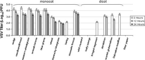
To determine the stability of VSV on plants and the window of opportunity for grasshoppers to ingest viable VSV from those contaminated plants, virus-exposed plant cuttings were tested and infectious virus quantitated by plaque assays. Following the initial inoculum (4.7 log10 PFU), the titer of virus was determined at 3, 12, and 24 hpe. At 3 hpe, virus was recovered from all plant species except wild mustard (Fig. 1), with virus titers ranging from 2.18 to 4.44 log10 PFU (46.4% to 94.5% of the initial inoculum) (Table 1). At 12 hpe, virus was recovered from all monocot species except barley, with titers ranging from 2.21 to 4.45 log10 PFU (47% to 94.7% of the initial inoculum). Of the dicot species, virus was recovered only from dandelion and scarlet globemallow, with titers of 3 log10 PFU (63.8%) and 2.57 log10 PFU (54.7%), respectively. At 24 hpe, virus was recovered from all monocot species except barley, with titers ranging from 1.86 to 3.44 log10 PFU (39.6% to 73.2%). No virus was recovered from any dicot species at 24 hpe. No virus was detected on the control filter paper after 1 hpe. No increase in titer was seen in any plant species tested.
FIG. 1.
Infectious VSV recovered from plants at 3, 12, and 24 hpe as measured by a plaque assay. Error bars, standard errors.
TABLE 1.
Titers of virus recovered from rangeland plant species at 3, 12, and 24 hpe
| Common name | Scientific name | VSV inoculum recovery (log10 PFU [% of total inoculum]) at:
|
||
|---|---|---|---|---|
| 3 h | 12 h | 24 h | ||
| Barley | Hordeumvulgare | 2.18 (46.4) | 0.00 (0) | 0.00 (0) |
| Blue grama | Boutelouagracilis | 4.11 (87.4) | 4.09 (87.0) | 3.41 (72.6) |
| Kentucky bluegrass | Poapratensis | 2.29 (48.7) | 2.21 (47.0) | 1.86 (39.6) |
| Smooth brome | Bromusinermis | 4.13 (87.9) | 3.92 (83.4) | 3.18 (67.7) |
| Prairie junegrass | Koeleriamacrantha | 3.86 (82.1) | 3.65 (77.7) | 3.44 (73.2) |
| Needleleaf sedge | Carexduriuscula | 4.25 (90.4) | 3.85 (81.9) | 3.28 (69.8) |
| Needle and thread | Hesperostipacomata | 4.44 (94.5) | 4.45 (94.7) | 3.35 (71.3) |
| Western wheatgrass | Pascopyrumsmithii | 4.31 (91.7) | 4.09 (87.0) | 3.35 (71.3) |
| Wheat | Triticumaestivum | 2.98 (63.4) | 2.80 (59.6) | 2.18 (46.4) |
| Dalmatian toadflax | Linariadalmatica | 3.40 (72.3) | 0.00 (0) | 0.00 (0) |
| Dandelion | Taraxacumofficinale | 3.18 (67.7) | 3.00 (63.8) | 0.00 (0) |
| Fringed sagewort | Artemisia frigida | 2.18 (46.4) | 0.00 (0) | 0.00 (0) |
| Scarlet globemallow | Sphaeralceacoccinea | 3.31 (70.4) | 2.57 (54.7) | 0.00 (0) |
| Wild mustard | Brassicakaber | 0.00 (0) | 0.00 (0) | 0.00 (0) |
Plant-to-grasshopper transmission.
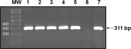
Two plant species with significant virus survival rates at 24 hpe were chosen for studies of grasshopper infection by ingestion. Infection rates for the four treatment groups (VSV-fed positive controls, medium-fed negative controls, western wheatgrass/VSV-fed grasshoppers, and needle and thread/VSV-fed grasshoppers) were determined by virus isolation at 21 dpf. Infection rates were zero for negative controls, 86% for positive controls, 44% for grasshoppers fed VSV-exposed western wheatgrass, and 50% for grasshoppers fed VSV-exposed needle and thread (Table 2). For all CPE-positive cultures, the presence of VSV was confirmed by RT-PCR (Fig. 2). There were no significant differences in grasshopper survival among the four treatment groups.
TABLE 2.
Infectivity of grasshoppers tested by virus isolation and RT-PCR
| Treatment group | Proportiona of grasshoppers:
|
||
|---|---|---|---|
| Surviving at 21 dpf | VSV+ by VIb at 21 dpf | For which the presence of VSV in CPE+ cultures was confirmed by RT-PCR | |
| Negative control | 8/15 (53) | 0/8 (0) | 0/8 (0) |
| Positive control | 7/15 (47) | 6/7 (86) | 6/6 (100) |
| Western wheatgrass/VSV | 18/30 (60) | 8/18 (44) | 8/8 (100) |
| Needle and thread/VSV | 16/30 (53) | 8/16 (50) | 8/8 (100) |
Given as the number of grasshoppers with the indicated characteristic/the total number of grasshoppers in the group (percentage).
VI, virus isolation assay.
FIG. 2.
Confirmation by RT-PCR of the presence of VSV in CPE-positive virus isolation cultures of grasshoppers. Lanes: MW, PCR molecular weight markers (Bio-Rad); 1 to 5, grasshopper cultures showing CPE; 6, negative-control grasshopper sample; 7, positive-control grasshopper sample.
Effect of a grasshopper pesticide on VSV viability.
A commonly used no-withdrawal grasshopper pesticide was evaluated for its ability to inactivate VSV. The range of pesticide dilutions tested included the 1:2 dilution recommended for application by air and the 1:41 dilution recommended for ground application. For the entire range of recommended dilutions (1:2 to 1:41), exposure of VSV to the pesticide resulted in complete inactivation of virus. At the highest dilution tested (1:64), 0.699 log10 PFU VSV was detected (data not shown).
DISCUSSION
Most rhabdoviruses utilize at least two natural hosts, one of which is an insect while the other is a plant or animal (5). Vesiculoviruses are known to naturally infect a variety of species from livestock to insects, and experimentally these viruses can infect an extremely wide host range. Additionally, selective pressures and the high rate of diversity among RNA viruses of the same strain allow for expansion to new and ever-changing niches for viral replication, survival, and possibly even hosts (15). Although laboratory transmission studies have been done on several suspected VSV vectors, it is probably unrealistic to expect a clear definition of a single arthropod vector for a virus that so readily replicates in a variety of insect species. It is possible that in nature, VSV exploits both hematophagous and herbivorous vector transmission mechanisms.
VSV has been shown to replicate in a variety of hematophagous insects (2, 3, 9, 11, 17), as well as in two nonhematophagous insects: leafhoppers (7) and grasshoppers (10). Although most of these insects do not travel distances great enough to explain the typical pattern of VSV spread to new and distant areas during outbreaks, the migratory grasshopper M. sanguinipes is an exception. Adult grasshoppers of this species have been reported to travel 48 km per day, as far as 925 km during their migration (14), and geospatial correlations of dates and locations of VS outbreaks and grasshopper infestations have been observed (10). No differences were seen in the behavior of infected versus uninfected grasshoppers, including general walking/hopping activity, feeding, mating, egg laying, or survival. This suggests that other behaviors, such as migration, may not be affected by VSV infection. The route of infection for herbivorous insects such as grasshoppers is likely the ingestion of virus-contaminated plants. It is understood that the excessive saliva of clinically infected animals contains copious amounts of virus, which contaminates not only the physical facilities of corrals, including feed and water troughs, but also plants and soil. It is likely that VSV in infected saliva is associated with sloughed cells and is therefore protected from the enzymatic activity of the saliva. Because the supply of infected saliva was limited, and spiking uninfected saliva (no cellular material) resulted in poor virus survival (data not shown), medium was used as the VSV diluent for plant exposure. Although quantitation of the volume of adherent material for either substrate was not possible due to the small volumes on plant surfaces, it is likely that the viscous nature of the saliva would increase drying times and therefore increase virus survival rates. Although the OIE states that plants and soil are believed to be sources of virus during outbreaks, to date there is no recommended practice for decontaminating those sources. There are no reports demonstrating the survivability of VSV in natural outside environments because the biosafety restrictions on working with this virus outside biosafety level 2 laboratories preclude field testing. Inactivation of VSV by direct exposure to an artificial source of UV radiation has been reported; the virus was placed in tissue culture dishes 10 cm from the UV light source and exposed to radiation at a wavelength of 254 nm and a dose rate of 85 ergs/mm2/s (18). However, there are no reports on what dose of UV irradiation from natural sunlight is required to inactivate VSV or on the level of penetration of sunlight into rangeland pastures of various densities. Most likely, gravity plays an important role in increasing the distribution of virus-contaminated saliva closer to the ground relative to that at the more UV exposed tops of plants, and many grasshoppers are geophilous, dwelling and feeding on or near the ground (13).
In this study we were able to show that many plant species are capable of harboring viable VSV for extended periods, even if the inoculum is in contact with the plants for only a few hours. Although no virus replication was observed in any of the plants tested, the survival of virus at 24 hpe (40 to 73%) on several plant species common to rangelands would provide a significant window of opportunity for grasshoppers to ingest infectious virus in contaminated pastures during VSV outbreaks. The number of monocot species harboring infectious virus at 12 and 24 hpe, compared to that of dicot species, may reflect host range limitations (4) and differences in plant anatomy. The monocot species used in this study have narrow leaves with large parallel veins and are mostly glabrous or have only a small number of hairs. The exception to the monocot-VSV survival trend is barley, which is known to have a complex wax layer on its surface (19). The poor recovery of virus from barley, compared to that for the rest of the monocots, may reflect the hydrophobicity of its waxy surface. The dicot species have broad leaves, net venation, and either large amounts of hair (e.g., scarlet globemallow, fringed sagewort) or waxy cuticles (e.g., dalmation toadflax), which may repel the viral inoculum. We draw the inference that the amount of VSV that can use leaf material as a suitable surface host is reduced with dicot species. The rapid decline of viable virus on filter paper is likely attributable to the rate of drying compared to that on plant surfaces.
One of the most commonly used carbaryl-based pesticides for grasshoppers is Sevin XLR Plus (Bayer CropScience). A 1:2 dilution of this pesticide in water is recommended for application by air, and a dilution of 1:41 is recommended for ground rig application. This “no-withdrawal” chemical pesticide is used without removal of livestock from pastures during its application. It is predicted that 90% of grasshoppers are killed within 3 days following application. For all but the highest dilution tested, exposure of VSV to the pesticide resulted in complete inactivation of virus. Mixing of virus inocula with pesticide dilutions represents optimum pesticide coverage on the maximum virus titer (per milliliter of saliva) one might expect to be shed from an infected cow. The exact mechanism of virus inactivation was not investigated, since the inactivation itself was the critical aspect of pesticide exposure. To date there are no reports regarding any antiviral properties of this pesticide.
Although no definitive natural reservoir for VSV has been determined, it is believed that small grass-eating rodents, such as cotton rats (6) and deer mice (1), may play a role in viral maintenance. It is not known whether the source of virus for these rodents is the environment (plants, soil) or the bites of VSV-infected insects.
As with all laboratory infection models, laboratory conditions cannot substitute for field conditions. Thus, in the absence of actual field testing, inferences from these laboratory results, as they apply to field practices, must be drawn conservatively. However, these findings may shape the disinfection protocols used during outbreaks. Currently there are no recommendations for decontaminating pastures or eliminating grasshoppers, which have been shown to amplify the virus and to infect cattle when eaten. Spraying pastures with a grasshopper pesticide during VSV outbreaks in cattle and horse herds would eliminate this amplifying mechanical vector and decontaminate the plant species in pastures, thereby eliminating the viral source for both virus-amplifying herbivorous insects and grazing animals.
Acknowledgments
We thank S. Schell, K. VanDyke, J. Lowy, and K. Bennett for reviewing earlier versions of this article; S. Clapp for providing field-collected plant species; C. Alford, S. Miller, and A. Kniss for providing greenhouse plant species; and S. Schell for providing expertise in grasshopper biology and pesticide use.
Funding for this research was provided by the United States Department of Agriculture, Veterinary, Medical and Urban Entomology National Program, project 5410-32000-014, and the Animal Health National Program, project 5410-32000-016.
Mention of trade names or commercial products in this publication is solely for the purpose of providing specific information and does not imply recommendation or endorsement by the U.S. Department of Agriculture.
Footnotes
Published ahead of print on 13 March 2009.
REFERENCES
- 1.Cornish, T. E., D. E. Stallknecht, C. C. Brown, B. S. Seal, and E. W. Howerth. 2001. Pathogenesis of experimental vesicular stomatitis virus (New Jersey serotype) infection in the deer mouse (Peromyscus maniculatus). Vet. Pathol. 38:396-406. [DOI] [PubMed] [Google Scholar]
- 2.Cupp, E. W., C. J. Mare, M. S. Cupp, and F. B. Ramberg. 1992. Biological transmission of vesicular stomatitis virus (New Jersey) by Simulium vittatum (Diptera: Simuliidae). J. Med. Entomol. 29:137-140. [DOI] [PubMed] [Google Scholar]
- 3.Drolet, B. S., C. L. Campbell, M. A. Stuart, and W. C. Wilson. 2005. Vector competence of Culicoides sonorensis (Diptera: Ceratopogonidae) for vesicular stomatitis virus. J. Med. Entomol. 42:409-418. [DOI] [PubMed] [Google Scholar]
- 4.Götz, R., and E. Maiss. 2002. The complete sequence of the genome of Cocksfoot streak virus (CSV), a grass-infecting Potyvirus. Arch. Virol. 147:1573-1583. [DOI] [PubMed] [Google Scholar]
- 5.Hogenhout, S. A., M. G. Redinbaugh, and E.-D. Ammar. 2003. Plant and animal rhabdovirus host range: a bug's view. Trends Microbiol. 11:264-271. [DOI] [PubMed] [Google Scholar]
- 6.Jimenez, A. E., F. Vargas Herrera, M. Salman, and M. V. Herrero. 2000. Survey of small rodents and hematophagous flies in three sentinel farms in a Costa Rican vesicular stomatitis endemic region. Ann. N. Y. Acad. Sci. 916:453-463. [DOI] [PubMed] [Google Scholar]
- 7.Lastra, J. R., and J. Esparza. 1976. Multiplication of vesicular stomatitis virus in the leafhopper Peregrinus maidis (Ashm.), a vector of a plant rhabdovirus. J. Gen. Virol. 32:139-142. [DOI] [PubMed] [Google Scholar]
- 8.Letchworth, G. J., L. L. Rodriguez, and J. D. C. Barrera. 1999. Vesicular stomatitis. Vet. J. 157:239-260. [DOI] [PubMed] [Google Scholar]
- 9.Liu, I. K., and Y. C. Zee. 1976. The pathogenesis of vesicular stomatitis virus, serotype Indiana, in Aedes aegypti mosquitoes. I. Intrathoracic injection. Am. J. Trop. Med. Hyg. 25:177-185. [DOI] [PubMed] [Google Scholar]
- 10.Nunamaker, R. A., J. A. Lockwood, C. E. Stith, C. L. Campbell, S. P. Schell, B. S. Drolet, W. C. Wilson, D. M. White, and G. J. Letchworth. 2003. Grasshoppers (Orthoptera: Acrididae) could serve as reservoirs and vectors of vesicular stomatitis virus. J. Med. Entomol. 40:957-963. [DOI] [PubMed] [Google Scholar]
- 11.Nunamaker, R. A., A. A. Perez de Leon, C. C. Campbell, and S. M. Lonning. 2000. Oral infection of Culicoides sonorensis (Diptera: Ceratopogonidae) by vesicular stomatitis virus. J. Med. Entomol. 37:784-786. [DOI] [PubMed] [Google Scholar]
- 12.Perez de Leon, A. A., and W. J. Tabachnick. 2006. Transmission of vesicular stomatitis New Jersey virus to cattle by the biting midge Culicoides sonorensis (Diptera: Ceratopogonidae). J. Med. Entomol. 43:323-329. [DOI] [PubMed] [Google Scholar]
- 13.Pfadt, R. E. 2002. Field guide to common western grasshoppers, 3rd ed. University of Wyoming, Laramie.
- 14.Pfadt, R. E. 1994. Field guide to common western grasshoppers, 2nd ed. USDA, ARS, Wyoming Agricultural Experiment Station, Laramie.
- 15.Roossinck, M. J. 1997. Mechanisms of plant virus evolution. Annu. Rev. Phytopathol. 35:191-209. [DOI] [PubMed] [Google Scholar]
- 16.Stallknecht, D. E., E. W. Howerth, C. L. Reeves, and B. S. Seal. 1999. Potential for contact and mechanical vector transmission of vesicular stomatitis virus New Jersey in pigs. Am. J. Vet. Res. 60:43-48. [PubMed] [Google Scholar]
- 17.Tesh, R. B., B. N. Chaniotis, and K. M. Johnson. 1971. Vesicular stomatitis virus, Indiana serotype: multiplication in and transmission by experimentally infected phlebotomine sandflies (Lutzomyia trapidoi). Am. J. Epidemiol. 93:491-495. [DOI] [PubMed] [Google Scholar]
- 18.Weck, P. K., A. R. Carroll, D. M. Shattuck, and R. R. Wagner. 1979. Use of UV irradiation to identify the genetic information of vesicular stomatitis virus responsible for shutting off cellular RNA synthesis. J. Virol. 30:746-753. [DOI] [PMC free article] [PubMed] [Google Scholar]
- 19.Wisniewska, S. K., J. Nalaskowski, E. Witka-Jezewska, J. Hupka, and J. D. Miller. 2003. Surface properties of barley straw. Colloids Surfaces B 29:131-142. [Google Scholar]


