

Abstract
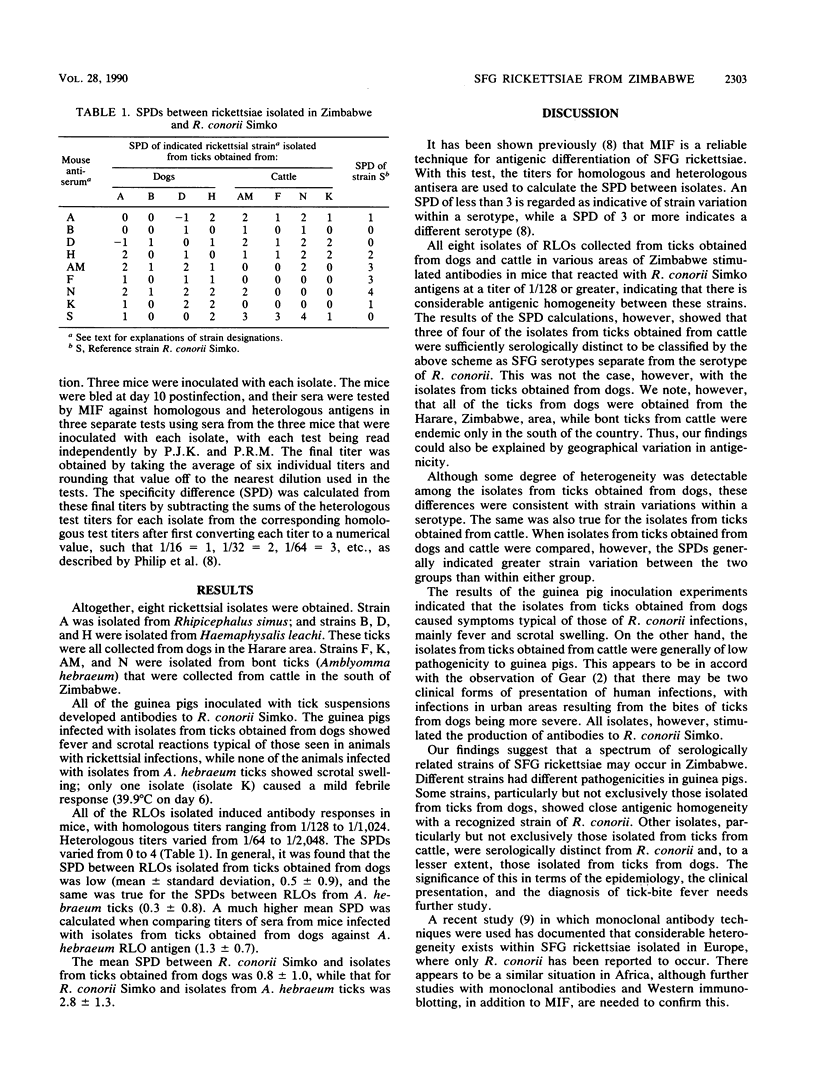
Eight rickettsialike organisms were isolated in tissue culture from ticks of dogs and cattle from various areas of Zimbabwe. These isolates and a reference strain, Rickettsia conorii Simko, were tested by microimmunofluorescence against homologous and heterologous antisera raised in mice. From the titers obtained by this method, specificity differences (SPDs) were calculated between each of the rickettsiae. Only small serological differences were detected among the isolates from ticks obtained from dogs (mean SPD, 0.5) and also among the isolates from ticks obtained from cattle (mean SPD, 0.3). However, when isolates from ticks obtained from dogs and cattle were compared, the serological differences were greater (mean SPD, 1.3). The isolates from ticks obtained from dogs were found to be very similar serologically to the Simko strain of R. conorii (mean SPD, 0.8), while three of four isolates from ticks obtained from cattle were different enough (SPD, greater than or equal to 3) to be identified as separate serotypes. These findings indicate that there is a high degree of antigenic heterogeneity among the tick-transmitted spotted fever group rickettsiae in Zimbabwe.
Full text
PDF
Selected References
These references are in PubMed. This may not be the complete list of references from this article.
- Burgdorfer W. Hemolymph test. A technique for detection of rickettsiae in ticks. Am J Trop Med Hyg. 1970 Nov;19(6):1010–1014. [PubMed] [Google Scholar]
- GIMENEZ D. F. STAINING RICKETTSIAE IN YOLK-SAC CULTURES. Stain Technol. 1964 May;39:135–140. doi: 10.3109/10520296409061219. [DOI] [PubMed] [Google Scholar]
- Gear J. H., Miller G. B., Martins H., Swanepoel R., Wolstenholme B., Coppin A. Tick-bite fever in South Africa. The occurrence of severe cases on the Witwatersrand. S Afr Med J. 1983 May 21;63(21):807–810. [PubMed] [Google Scholar]
- Goldwasser R. A., Steiman Y., Klingberg W., Swartz T. A., Klingberg M. A. The isolation of strains of rickettsiae of the spotted fever group in Israel and their differentiation from other members of the group by immunofluorescence methods. Scand J Infect Dis. 1974;6(1):53–62. doi: 10.3109/inf.1974.6.issue-1.10. [DOI] [PubMed] [Google Scholar]
- Philip C. B., Hoogstraal H., Reiss-Gutfreund R., Clifford C. M. Evidence of rickettsial disease agents in ticks from Ethiopian cattle. Bull World Health Organ. 1966;35(2):127–131. [PMC free article] [PubMed] [Google Scholar]
- Philip R. N., Casper E. A., Burgdorfer W., Gerloff R. K., Hughes L. E., Bell E. J. Serologic typing of rickettsiae of the spotted fever group by microimmunofluorescence. J Immunol. 1978 Nov;121(5):1961–1968. [PubMed] [Google Scholar]
- Vitale G., Di Stefano R., Damiani G., Mansueto S. Characterization of Sicilian strains of spotted fever group rickettsiae by using monoclonal antibodies. J Clin Microbiol. 1989 May;27(5):1081–1085. doi: 10.1128/jcm.27.5.1081-1085.1989. [DOI] [PMC free article] [PubMed] [Google Scholar]
- Weiss E., Coolbaugh J. C., Williams J. C. Separation of viable Rickettsia typhi from yolk sac and L cell host components by renografin density gradient centrifugation. Appl Microbiol. 1975 Sep;30(3):456–463. doi: 10.1128/am.30.3.456-463.1975. [DOI] [PMC free article] [PubMed] [Google Scholar]