Abstract
The beneficial effects of probiotic Enterococcus spp. in different hosts, such as mice and humans, have previously been reported in several studies. However, studies of large domestic animals, as well as challenge studies with pathogenic microorganisms, are very rare. Here, we investigated the influence of oral treatment of pigs with the probiotic bacterium Enterococcus faecium NCIMB 10415 on Salmonella enterica serovar Typhimurium DT104 infections in weaning piglets. Clinical symptoms, fecal excretion, the organ distribution of Salmonella, and the humoral immune response (immunoglobulin G [IgG], IgM, and IgA levels) in serum were examined. A pool of 89 piglets was randomly divided into probiotic and control groups. The probiotic group received a feed supplement containing E. faecium starting on day 14 postpartum prior to challenge with Salmonella serovar Typhimurium DT104 at 28 days postpartum. After challenge with Salmonella serovar Typhimurium DT104, piglets in both groups showed no severe clinical signs of salmonellosis. However, fecal excretion and colonization of Salmonella in organs were significantly greater in piglets fed E. faecium. Likewise, the humoral immune response against Salmonella (serum IgM and IgA levels) was significantly greater in the probiotic group animals than in control animals. The results of this study suggest that E. faecium NCIMB 10415 treatment enhanced the course of infection in weaning piglets challenged with Salmonella serovar Typhimurium DT104. However, the probiotic treatment also appeared to result in greater production of specific antibodies against Salmonella serovar Typhimurium DT104.
The problem of increasing microbial resistance to antibiotics resulting from years of overuse and the resulting ban on the use of antibiotics in animal production have led to increased interest in alternatives to antibiotics in animal production. In recent years, probiotic bacteria have been considered as an alternative means of reducing pathogen loads in animal breeding and production units. However, while a number of studies have focused on the mode of action of probiotics, the mode of action these bacteria is not fully understood yet.
A recent interdisciplinary research study of the modes of action of probiotics in swine showed that Enterococcus faecium NCIMB 10415 reduced the pathogenic bacterial load of healthy piglets (20, 26, 30, 36). In vitro studies further demonstrated that this E. faecium probiotic strain decreased the rate of invasion of a porcine intestinal epithelial cell line by Salmonella enterica serovar Typhimurium. To determine whether probiotics also provide a measure of protection during infections, experimental challenge studies with pathogenic bacteria at a defined infectious dose and under comparable conditions seem to be necessary. Field studies could be more representative of the real situation; however, the infection pressure is too low and difficult to define, and systematic sampling cannot be done.
Studies of larger domestic and production animals are rare. Most such studies deal with the mode of action of probiotics in the healthy host, and only a few studies have investigated the mode of action in the context of infections with pathogenic bacteria, such as Salmonella.
In a related study, weaned piglets were fed a mixture of five probiotic strains (one Pediococcus strain and four Lactobacillus strains) and challenged with Salmonella serovar Typhimurium (7). In that study, reduced incidence, severity, and duration of diarrhea and a reduced microbiological load of Salmonella were observed. Fedorka-Cray et al. (11) observed reduced numbers of Salmonella bacteria in cecal contents and at the ileocolic junction in S. enterica serovar Choleraesuis-challenged weaning piglets fed a competitive exclusion culture. In vitro investigations showed that Enterococcus strains have inhibitory effects on the growth of S. enterica serovar Enteritidis, and these effects were explained by both enterotoxin and nonenterotoxin factors (37). Other studies showed that E. faecium may be beneficial to the adhesion and colonization of Clostridium jejuni in the canine intestine (29) and reduced the rate of carryover infections with obligate intracellular pathogens from infected sows in piglets (26). E. faecium has also been shown to influence the composition of the bacterial community in the avian, swine, and canine gastrointestinal tracts (25, 29, 36).
Infections with S. enterica are some of the most important sources of human gastroenteritis (39). In Germany, 52,563 human salmonellosis cases were reported in 2006 (http://www3.rki.de/SurvStat). The consumption of contaminated pork and pork products was found to be associated with 20% of human salmonellosis cases in Germany (33), indicating the importance of meat or meat products as a potential source of infection for consumers. Salmonella serovar Typhimurium, especially phage type DT104, is the Salmonella serotype most frequently isolated from pork (27), and it is of particular concern because of its acquisition of multiple antibiotic resistance (1, 38).
In this study, we investigated the effect of E. faecium NCIMB 10415 on the infection dynamics of Salmonella serovar Typhimurium DT104, fecal shedding, and the patterns of Salmonella distribution in internal organs, as well as on the humoral immune response to Salmonella in weaning piglets. To the best of our knowledge, this is the first experimental study of the mode of action of a probiotic strain of E. faecium in which dissemination to different internal organs was investigated using weaned piglets experimentally infected with Salmonella.
MATERIALS AND METHODS
Animal study.
A total of 89 weaning piglets (10 litters) were investigated using the following study design. A group of 10 sows (crossbred Landrace and Duroc) were randomly divided into two groups (probiotic group and control group). The sows in the probiotic group were treated with the probiotic strain E. faecium NCIMB 10415 (Cylactin; Roche) starting on day 25 of gestation. The piglets of the sows in the probiotic group (n = 43) had free access to supplemented prestarter feed from day 14 until day 28 and to supplemented starter feed from day 29 to day 56. The sows in the control group and their litters were not treated with the probiotic. Piglets in both the probiotic (n = 43) and control (n = 46) groups were weaned at 28 days postpartum and challenged with Salmonella serovar Typhimurium DT104 on day 29 postpartum by intragastric application using a stomach tube. Each piglet was sedated by intramuscular application of 1.0 mg/kg azaperon prior to infection.
Two piglets from each litter were randomly selected and sacrificed at 3, 24, and 72 h postinfection (p.i.); the remaining animals were monitored for 4 weeks and sacrificed at day 28 p.i. During necropsy, the liver, the spleen, the kidneys, the palatine tonsils, the mandibular lymph nodes, segments of the colon, the jejunal lymph nodes, and muscle samples from the forelimb, hind limb, and diaphragm were collected, and the presence and numbers of Salmonella serovar Typhimurium DT104 bacteria were determined.
During the observation period, the following clinical parameters were monitored: (i) the general condition and fecal consistency, which was measured daily using a macroscopic score from 1 to 6 (firm to liquid); (ii) the rectal temperature, which was taken daily; (iii) the prevalence and numbers of Salmonella bacteria in feces, which were determined daily for the first 5 days p.i. and then twice a week; (iv) the prevalence of specific immunoglobulins in blood samples obtained before infection and then at weekly intervals; and (v) weight, which was determined for all pigs twice a week.
Bacterial strains.
The E. faecium NCIMB 10415 strain used was in a microencapsulated form in a commercial batch of the European Commission-authorized probiotic feed additive Cylactin LBC ME10 (lots C00020-P00009, C00013-P00006, and C00001; Cerbios-Pharma, Barbengo, Switzerland). Cylactin LBC ME 10 was included in the diets of sows, suckling piglets, and weaned piglets at levels of 100 ppm, 25 ppm, and 30 ppm, respectively. The diets for sows and weaned piglets were pelleted at 50°C (measured in the conditioner).
An isolate of a multiresistant Salmonella serovar Typhimurium DT104 strain (BB 440) obtained from a swine with sepsis was used for animal challenge; this strain was also nalidixic acid resistant. The Salmonella strain was cultured in Luria broth at 37°C overnight. Portions (1 ml) of the precultures were used to inoculate 100 ml of prewarmed Luria broth and grown at 37°C to an optical density of 2.0, corresponding to an average bacterial count of approximately 3 × 10⁹ cells/ml. Each pig was infected with 2 ml of this culture mixed with 8 ml buffered peptone water (BPW) (catalog no. 1.12535; Merck, Darmstadt, Germany).
Detection of Salmonella in feces.
Approximately 10 g of rectal fecal samples was inoculated into 1% BPW and homogenized using a Stomacher 400 (Seward, London, United Kingdom) for 2 min at high speed. During the first 10 days p.i., quantitative detection was performed using a spiral plater (Whitley, Meintrup DWS, Germany) with a detection limit of 100 CFU/g because of the expected high rate of excretion of Salmonella in feces. After this, enumeration of Salmonella was carried out by direct plating to decrease the detection limit (10 CFU/g). One milliliter from the first dilution was streaked onto 3 XLD (xylose-lysine-deoxycholate; catalog no. 1.05287; Merck) agar plates (catalog no. CM 469; Oxoid, Hampshire, United Kingdom) supplemented with 50 μg/ml nalidixic acid and incubated at 37°C for 48 h.
For qualitative analysis, the remaining liquid was used for enrichment cultures that were incubated at 37°C for 24 h, and the following day 0.1 ml was inoculated onto MSRV (modified semisolid Rappaport-Vassiliadis medium; catalog no. CM 900100; SR161E; Oxoid) agar plates and incubated at 42°C for 24 h.
Detection of Salmonella in internal organs.
Tissue samples (10 g) were immersed in 95% ethanol, flamed, minced aseptically, and homogenized with BPW (1:10) using a stomacher for 2 min at high speed. All samples were examined for Salmonella serovar Typhimurium quantitatively by direct plating and qualitatively as described above.
Blood samples and serological examination.
Blood samples for preparation of serum were obtained from the cranial vena cava, coagulated overnight at 4°C, and centrifuged, and the serum fraction was collected and stored at −20°C until analysis. Swine sera were analyzed for the presence of anti-Salmonella antibodies using the Salmotype pig STM-WCE enzyme-linked immunosorbent assay test system according to the manufacturer's instructions (Labordiagnostik Leipzig, Leipzig, Germany). The test kit is designed to discriminate between Salmonella-specific immunoglobulin M (IgM), IgA, and IgG antibodies. The results were calculated using a reference standard method and are expressed below in enzyme-linked immunosorbent assay units per ml.
Statistical analysis.
Statistical analysis was conducted using the software SPSS 12.0 (SPSS, Inc., Chicago, IL). Multivariate analysis of variance was used to evaluate the influence of the probiotic diet and of the time after infection on clinical signs, fecal excretion, and the patterns of distribution of Salmonella in internal organs and the humoral immune response (IgG, IgM, and IgA titers).
Differences between the probiotic and control groups 3 h, 24 h, 72 h, and 28 days p.i. were also evaluated using quantitative results. The Student t test was used for normal distributions. When nonparametric analysis was required, the Mann-Whitney U test was used. The χ2 test was performed to calculate the differences between the two feeding groups for qualitative results, such as the number of Salmonella-positive samples. Differences were considered significant if the P value was <0.05. For graphic representation of results, box-whisker plots were used. The boxes indicate the medians (horizontal lines) and the lower and upper quartiles (lower and upper sides of the boxes). Outliers, numerically distant from the rest of the data, were included for determination of the statistical significance.
RESULTS
Clinical signs.
During the postinfection observation period (28 days), the body weights of animals in the two feeding groups increased continuously and showed similar patterns that corresponded to the normal development of body weight in healthy weaned piglets. Elevated body temperatures over 40.0°C were observed in the control and probiotic groups for 31% (5/16) and 69% (9/13) of the animals, respectively. When the data for the two groups were compared, no significant difference was detected.
When fecal consistency was examined, diarrhea was observed in 3% of the animals in both the control and probiotic groups within the first 3 days p.i. During the 4-week observation period, 38% (6/16) of the animals in the control group and 62% (8/13) of the animals in the probiotic group exhibited diarrhea, whereas 50% of the animals in the probiotic group showed severe diarrhea (score, 6 [liquid feces]). Statistical analysis using the χ2 test revealed no significant differences in the development of fecal consistency between the two feeding groups.
Fecal shedding of Salmonella serovar Typhimurium DT104.
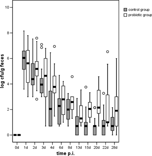
Salmonella serovar Typhimurium DT104 was isolated from feces of all animals 1 day p.i., and all animals in the probiotic group excreted Salmonella in their feces until the end of the study, except for one animal which was Salmonella negative at day 28 p.i. For the control group, Salmonella was detected in feces as early as day 13 p.i. in 31% of the animals (5/16). This tendency was observed until the end of the experiment. At days 13 (P = 0.048), 15 (P = 0.020), 20 (P = 0.020), and 22 (P = 0.048) p.i., the numbers of animals shedding Salmonella in their feces were significantly higher for the probiotic group than for the control group.
The levels of Salmonella shed in feces during the first 3 days p.i. varied from <log 3 to log 8.1 CFU/g for the control group and from <log 3 to log 7.8 CFU/g for the probiotic group. A continuous decrease in Salmonella excretion was observed for both groups starting at day 1 (Fig. 1). The level of Salmonella excreted in the feces of 88% (14/16) of the animals in the control group was under the detection limit (log 1 CFU/g). At days 4 (P = 0.040), 13 (P = 0.036), 15 (P = 0.001), 20 (P = 0.000), 22 (P = 0.019), and 28 (P = 0.010) p.i., the numbers of Salmonella bacteria in fecal samples were significantly higher for animals in the probiotic group than for animals in the control group.
FIG. 1.
Salmonella serovar Typhimurium DT104 counts in feces of animals in the probiotic (n = 13) and control (n = 16) groups over the entire study period (28 days). ○, mild outliers.
Dissemination of Salmonella serovar Typhimurium DT104 in internal organs.
Salmonella serovar Typhimurium was recovered from all tissue samples investigated. For both the control and probiotic groups the highest detection rates were the detection rates for the tonsils and the colon, followed by the jejunal lymph nodes, but Salmonella was detected only infrequently in other organs.
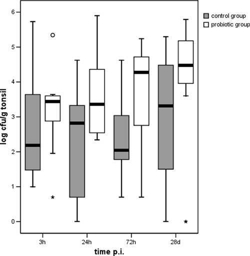
Salmonella was isolated from the tonsils of all animals in both diet groups sacrificed 3, 24, and 72 h p.i., and the maximum concentrations were 5.7 log CFU/g for the control group and 5.3 log CFU/g for the probiotic group, which were found as early as 3 h p.i. (Fig. 2). Two animals in the probiotic group and one animal in the control group had no Salmonella in their tonsils after the 4-week observation period. The comparison of bacterial colonization for the different sacrifice days revealed no significant differences between the two feeding groups when t tests were used, although animals in the probiotic group tended to have a higher Salmonella count on each day investigated. However, the numbers of Salmonella bacteria recovered from the tonsils were significantly higher for the animals in the probiotic group as a whole than for the animals in the control group (P = 0.003).
FIG. 2.
Salmonella serovar Typhimurium DT104 counts in tonsils of piglets in the probiotic and control groups sacrificed 3 h, 24 h, 72 h, and 28 days p.i. ○, mild outlier; *, extreme outliers.
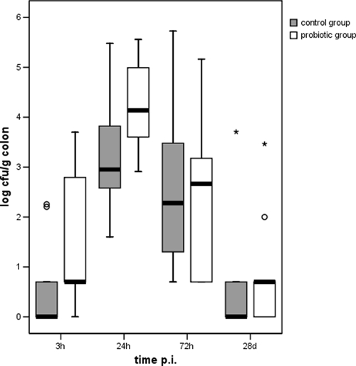
In the colon samples, Salmonella was detected 3 h p.i. in 30% (3/10) and 80% (8/10) of the animals in the control and probiotic groups, respectively. At 24 and 72 h p.i., Salmonella was found in all colon tissue samples from animals in both diet groups, and after the 4-week observation period, colon samples were positive for 50% (8/16) of the control animals and for 69% (9/13) of the probiotic group animals. Statistical analysis using the χ2 test revealed that at 3 h p.i. significantly more Salmonella-positive colon samples were detected for animals in the probiotic group than for animals in the control group (P = 0.025). The highest levels of Salmonella serovar Typhimurium were detected in colon samples from animals in both diet groups at 24 and 72 h p.i. (Fig. 3), in which again the levels of Salmonella were significantly higher at 3 h p.i. in animals in the probiotic group than in animals in the control group (P = 0.045).
FIG. 3.
Salmonella serovar Typhimurium DT104 counts in colon tissue of piglets in the probiotic and control groups sacrificed 3 h, 24 h, 72 h, and 28 days p.i. ○, mild outliers; *, extreme outliers.
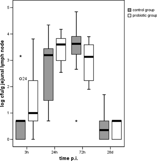
Salmonella was isolated from the jejunal lymph nodes for 60% (6/10) of animals in the control group and for 90% (9/10) of animals in the probiotic group (Fig. 4). Similar to the findings for the colon samples, Salmonella was detected in all jejunal lymph node samples from animals in both diet groups at 24 and 72 h p.i., and at day 28 p.i., 50% (8/16) of control animals and 69% (9/13) of probiotic group animals were still positive for Salmonella. However, in contrast to the data for the colon samples, no statistical differences were observed between the control and probiotic-treated groups.
FIG. 4.
Salmonella serovar Typhimurium DT104 counts in jejunal lymph nodes of piglets in the probiotic and control groups sacrificed 3 h, 24 h, 72 h, and 28 days p.i. ○, mild outlier; *, extreme outliers.
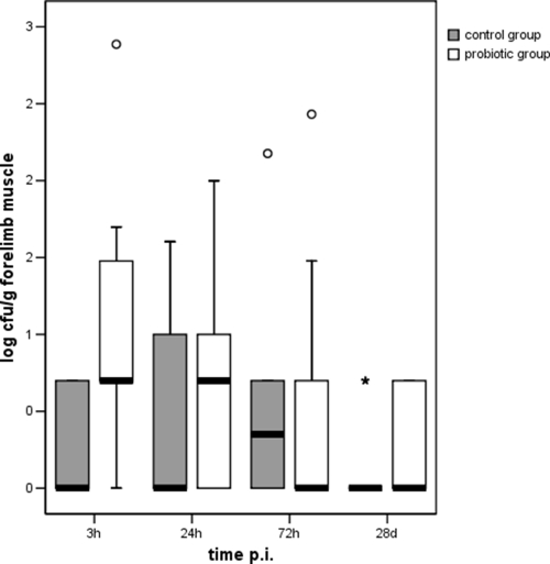
Salmonella counts were generally below the detection limit in the spleen, liver, kidney, and mandibular lymph nodes for both diet groups (data not shown). However, Salmonella counts in the foreleg could be determined for seven animals in the probiotic group (up to log 2.4 CFU/g) and for two animals in the control group (up to log 2.2 CFU/g) (Fig. 5). Statistically, the number of Salmonella bacteria in the forelimb muscle samples was significantly higher at 3 h p.i. for the probiotic group (P = 0.036) than for the control group. Furthermore, when all data were analyzed irrespective of the date of sacrifice, the results for hind limb muscle samples showed that there was a significantly higher number of Salmonella-positive animals in the probiotic group (P = 0.020).
FIG. 5.
Salmonella serovar Typhimurium DT104 counts in forelimb muscle (M. flexor digitalis superficialis) of piglets in the probiotic and control groups sacrificed 3 h, 24 h, 72 h, and 28 days p.i. ○, mild outliers; *, extreme outlier.
Serological results.
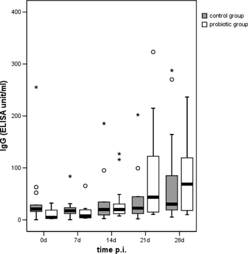
Anti-Salmonella IgG levels increased from days 21 and 28 p.i. in animals in the probiotic and control groups, respectively (Fig. 6). In general, the mean serum IgG levels for the probiotic group remained higher than those for the control animals; however, no significant differences were found between the two groups.
FIG. 6.
Salmonella IgG antibody detected in serum of piglets in the probiotic and control groups over the entire study period (28 days). ○, mild outliers; *, extreme outliers. ELISA, enzyme-linked immunosorbent assay.
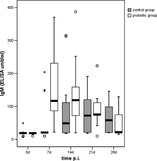
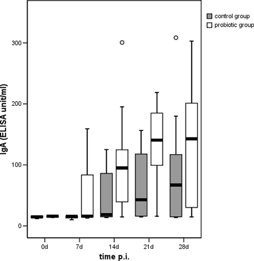
As early as 1 week p.i., the anti-Salmonella IgM levels were significantly higher (P = 0.009) in animals in the probiotic group than in animals in the control group, whereas the IgM levels in both diet groups were similarly low at days 21 and 28 p.i. (Fig. 7). Similar to the IgM levels, the anti-Salmonella IgA levels increased in the probiotic group, but a statistical analysis indicated that the IgA levels in animals in the probiotic group were significantly higher than those in animals in the control group on days 7 (P = 0.024), 14 (P = 0.029), 21 (P = 0.005), and 28 (P = 0.0016) p.i. (Fig. 8).
FIG. 7.
Salmonella IgM antibody detected in serum of piglets in the probiotic and control groups over the entire study period (28 days). ○, mild outliers; *, extreme outliers. ELISA, enzyme-linked immunosorbent assay.
FIG. 8.
Salmonella IgA antibody detected in serum of piglets in the probiotic and control groups over the entire study period (28 days). ○, mild outliers. ELISA, enzyme-linked immunosorbent assay.
DISCUSSION
The results of our study showed that addition of E. faecium NCIMB 10415 to the diet of weaned piglets that were challenged orally with 109 CFU/pig of Salmonella serovar Typhimurium DT104 did not result in significant differences in clinical symptoms compared to the symptoms of animals that did not receive probiotic treatment. However, we observed moderate increases in the incidence of liquid feces and elevated body temperature in the probiotic group. This result contrasts with the results of another challenge study, in which a five-strain probiotic combination was used and a decreased incidence of liquid feces in pigs was reported (7). Other studies of pigs treated with E. faecium but not subjected to a pathogen challenge also demonstrated that the incidence of postweaning diarrhea was lower in the probiotic group (23, 36). The observed clinical symptoms induced by Salmonella serovar Typhimurium DT104 after oral exposure to 109 CFU/pig are consistent with several previous studies of Salmonella infection in swine (16, 31, 43). The data for body weight gain in piglets did not show any significant differences between the two diet groups. Similar findings were obtained in other studies of the effects of E. faecium in noninfected pigs (6, 36). However, in 35% of 40 peer-reviewed studies of supplementation of feed of pigs with probiotics, a significant positive effect on the average daily weight gain was observed (32). However, one should consider the fact that the probiotics used in these trials consisted of different strains and were administered to animals in different preparations. Several studies have suggested that the growth-promoting effects of probiotics become more effective as the environmental and nutritional challenges confronting the animal are exacerbated. Thus, it is possible that the conditions used were not severe enough to discriminate positive performance effects (6, 22, 36).
The results of our study further revealed that the amount of Salmonella excreted in feces on several days (days 4, 13, 15, 20, 22, and 28 p.i.) was significantly larger for the probiotic group than for the control group. In comparison, the results of the few previous challenge trials with different probiotic strains, various pathogens, and animal models show that there is either similar fecal excretion in probiotic and control groups or decreased fecal excretion in the probiotic group (7, 10, 13, 19, 29, 45).
After Salmonella infection, pathogenesis is characterized by three phases: first, colonization of intestines; second, invasion of enterocytes; and third, bacterial dissemination to lymph nodes and organs (4). Several organs, including the tonsils, serve as important sites for persistence and dissemination of Salmonella (10, 21). The results of our study are consistent with other reports which showed that all organs investigated were affected after Salmonella serovar Typhimurium DT104 infection in pigs (4, 15, 41, 42). In addition, the tonsils were identified as the most highly colonized organs, followed by the gastrointestinal tract and the gut-associated lymphatic tissue. Corresponding with findings of other studies (31, 43), high concentrations of Salmonella were detected in tonsils 3 h and 28 days p.i. for both diet groups. One possible explanation for the high levels of Salmonella in these organs could be the continuous contact with other infected pigs and their feces (i.e., continuous oral-nasal reinfection from fecal shedding of infected animals). Based on the observation that the amount of Salmonella in feces of animals in the probiotic group was larger than that the amount in the control group, the reinfection level of tonsils in the probiotic group would also be higher. According to previous studies, Salmonella was able to invade the tonsils within 30 min after oral uptake or contact with the contamination source and within a few hours (2 to 3 h p.i.) colonized the mandibular lymph nodes, colon, cecum, and ileocaecal lymph nodes (5, 14, 21). Results of our investigations show that both the qualitative and quantitative manifestations of Salmonella in the tonsils and colon were significantly greater for the probiotic group than for the control group, but only the tonsil samples showed a continued high Salmonella concentration at the end of this study (Fig. 2). In contrast, most of the Salmonella counts for the spleen, liver, kidney, and mandibular lymph nodes and for various muscle samples were less than the detection limit for both diet groups, indicating that these organs play a minor role in bacterial colonization. However, the results for forelimb and hind limb muscle tissues showed that the number of Salmonella bacteria in animals in the probiotic group was significantly higher.
The immune status of the host is determined by a number of complex interactions, including factors such as immune cell interactions with bacteria and their products (2). In several studies with swine challenged with Salmonella, the development of the humoral immune response in serum (IgG, IgM, and IgA) was described (8, 18, 35). The results of this study indicated that there were earlier and higher IgM and IgA levels in the serum of probiotic-treated animals than in the serum of the control group animals. Benyacoub et al. (3) observed similar effects in the serum of newborn dogs fed E. faecium but not subjected to a challenge infection. In the present study at 1 week p.i. probiotic-treated animals showed high IgM and IgA activity, whereas control animals remained negative for these antibody classes, and for all days during the investigation the IgA titer in the probiotic-treated animals was significantly higher than that in control animals. No significant differences in the IgG titers between the two groups were observed. However, the average values for the probiotic-treated animals were higher than those for the control group animals at 3 and 4 weeks after infection. The curve for the IgG titers throughout the study (until 4 weeks after infection) indicated that no plateau was reached, suggesting that the IgG titers were still increasing. Therefore, the possibility that a significant difference might have occurred at a later time point cannot be excluded.
Although the intestinal mucosa is an effective barrier consisting of populations of intestinal epithelial and immune cells that are adapted to the presence of normal intestinal flora (28), it is also one of the most common locations of infections. Competitive exclusion is one of the most important proposed beneficial health effects of probiotics, and a number of studies have indicated that probiotic bacteria have suppressive effects on pathogens. In vitro tests with E. faecium revealed an antagonistic effect against Escherichia coli K88 (12) and negative effects on the growth of Salmonella in vitro (37). Other studies reported a reduction in the level of pathogenic intestinal bacteria after application of Enterococcus spp. to test animals (24, 26, 34) and a reduction in diarrhea in piglets (36, 44). However, negative effects on the intestinal flora have also been reported. Vahjen and Männer observed a significant increase in the level of Salmonella and Campylobacter after E. faecium was fed to dogs (40), and E. faecium was found to enhance the adhesion of Campylobacter to immobilized mucus isolates from dogs (29).
Stimulation of the immune system by probiotic bacteria is another beneficial effect suggested by some authors, including authors who describe immune stimulation after the application of Enterococcus spp. (3, 17). However, an additional consideration could be that the observed immune stimulation could also be the result of a stronger immunological challenge resulting from a change in the microbial status in the intestine (i.e., an increased pathogen load). Only a few animal studies have considered both changes in the immune parameters and effects on the microbial colonization of the gut. Two recent studies support the necessity for such approaches for clarifying the interaction between probiotics and the immune system. Sows and their litters given feed supplemented with E. faecium showed a reduction in Chlamydia infections (26) and lower frequencies of pathogenic E. coli serovars, but this was coupled with a reduced fraction of CD8+ cells in the intestinal epithelium (30). In the current animal study the impact of the probiotic E. faecium on the population of CD8+ intraepithelial lymphocytes was also monitored. A significant reduction in the number of cytotoxic T cells was detected in the jejunum of the probiotic-treated piglets (L. Scharek-Tedin, unpublished data). However, in contrast to the previous studies, the sizes of the naturally occurring E. coli and Chlamydia populations were reduced; feeding animals E. faecium in this study appeared to favor the invasion and dissemination of Salmonella, which was followed by an elevated antibody response against the pathogen. In the current animal infection study with weaned piglets challenged with Salmonella, the sows were shown to have been free of antibodies against the pathogen. The piglets were therefore completely dependent on functional cellular immunity during this first contact with the pathogen. The reduction in the CD8+ level in this situation could have been a critical disadvantage, leading to a more severe infection. An interesting observation that supports this suggestion was reported in a study with feedlot steers. E. faecium induced an inflammatory response against a probiotic yeast strain when both microbes were administered together (9). These observations suggest that during an acute infection or challenge, treatment with E. faecium prepares the ground for later infection with intestinal microbes.
In summary, the E. faecium probiotic-treated animals were found to be less able to defend themselves against a Salmonella infection than the animals in the control group, as shown by increased excretion of Salmonella in the feces, as well as greater colonization of organs. However, the elevated intestinal bacterial load and the levels of Salmonella in tissues and lymphoid organs also resulted in earlier and more intense development of humoral immunity. Whether the humoral response (i.e., IgA, IgM, and/or IgG titers) represented an enhanced immune response as a result of the probiotic treatment or the natural immune response to elevated Salmonella loads in tissues requires further study.
Acknowledgments
This work was supported by Deutsche Forschungsgemeinschaft grant FOR 438/1 to an interdisciplinary research group (German Research Foundation Research Unit 438) involving multiple partner institutes.
We thank the German National Reference Laboratory of Salmonella and Dieter Wolff and his team at the Center for Animal Experiments of the Federal Institute for Risk Assessment for their support. We also acknowledge P. Bahn, W. Barownick, C. Goellner, E. Luge, and G. Schindhelm for excellent technical assistance and M. Filter for help with statistical analysis.
Footnotes
Published ahead of print on 6 March 2009.
REFERENCES
- 1.Baggesen, D. L., and F. M. Aarestrup. 1998. Characterisation of recently emerged multiple antibiotic-resistant Salmonella enterica serovar Typhimurium DT 104 and other multiresistant phage types from Danish pig herds. Vet. Rec. 25:95-97. [DOI] [PubMed] [Google Scholar]
- 2.Bailey, M., F. J. Plunkett, H. J. Rothkotter, M. A. Vega-Lopez, K. Haverson, and C. R. Stokes. 2001. Regulation of mucosal immune responses in effector sites. Proc. Nutr. Soc. 60:427-435. [DOI] [PubMed] [Google Scholar]
- 3.Benyacoub, J., G. L. Czarnecki-Maulden, C. Cavadini, T. Sauthier, R. E. Anderson, E. J. Schiffrin, and T. von der Weid. 2003. Supplementation of food with Enterococcus faecium (SF68) stimulates immune functions in young dogs. J. Nutr. 133:1158-1162. [DOI] [PubMed] [Google Scholar]
- 4.Berends, B. R., H. A. Urlings, J. M. Snijders, and F. Van Knapen. 1996. Identification and quantification of risk factors in animal management and transport regarding Salmonella spp. in pigs. Int. J. Food Microbiol. 30:37-53. [DOI] [PubMed] [Google Scholar]
- 5.Blaha, T., G. Solano-Aguillar, and C. Pijoan. 1997. The early colonization pattern of Salmonella Typhimurium in pigs after oral intake, p. 71-73. In Proceedings of the 2nd International Symposium on the Epidemiology and Control of Salmonella in Pork, Copenhagen, Denmark.
- 6.Broom, L. J., H. M. Miller, K. G. Kerr, and J. S. Knapp. 2006. Effects of zinc oxide and Enterococcus faecium SF68 dietary supplementation on the performance, intestinal microbiota and immune status of weaned piglets. Res. Vet. Sci. 80:45-54. [DOI] [PubMed] [Google Scholar]
- 7.Casey, P. G., G. E. Gardiner, G. Casey, B. Bradshaw, P. G. Lawlor, P. B. Lynch, F. C. Leonard, C. Stanton, R. P. Ross, G. F. Fitzgerald, and C. Hill. 2007. A five-strain probiotic combination reduces pathogen shedding and alleviates disease signs in pigs challenged with Salmonella enterica serovar Typhimurium. Appl. Environ. Microbiol. 73:1858-1863. [DOI] [PMC free article] [PubMed] [Google Scholar]
- 8.Ehlers, J., M. Alt, D. Trepnau, and J. Lehmann. 2006. Anwendung neuer Immunglobulin-isotypspezifischer ELISA-Systeme zur Erkennung von Salmonelleninfektionen bei Schweinen. Berl. Muench. Tieraerztl. Wochenschr. 119:461-466. [PubMed] [Google Scholar]
- 9.Emmanuel, D. G., A. Jafari, K. A. Beauchemin, J. A. Leedle, and B. N. Ametaj. 2007. Feeding live cultures of Enterococcus faecium and Saccharomyces cerevisiae induces an inflammatory response in feedlot steers. J. Anim. Sci. 85:233-239. [DOI] [PubMed] [Google Scholar]
- 10.Fedorka-Cray, P. J., S. C. Whipp, R. E. Isaacson, N. Nord, and K. Lager. 1994. Transmission of Salmonella typhimurium to swine. Vet. Microbiol. 41:333-344. [DOI] [PubMed] [Google Scholar]
- 11.Fedorka-Cray, P. J., J. S. Bailey, N. J. Stern, N. A. Cox, S. R. Ladely, and M. Musgrove. 1999. Mucosal competitive exclusion to reduce Salmonella in swine. J. Food Prot. 62:1376-1380. [DOI] [PubMed] [Google Scholar]
- 12.García-Galaz, A., R. Perez-Morales, M. Diaz-Cinco, and E. Acedo-Felix. 2004. Resistance of Enterococcus strains isolated from pigs to gastrointestinal tract and antagonistic effect against Escherichia coli K88. Rev. Latinoam. Microbiol. 46:5-11. [PubMed] [Google Scholar]
- 13.Genovese, K. J., R. C. Anderson, R. B. Harvey, T. R. Callaway, T. L. Poole, T. S. Edrington, P. J. Fedorka-Cray, and D. J. Nisbet. 2003. Competitive exclusion of Salmonella from the gut of neonatal and weaned pigs. J. Food Prot. 66:1353-1359. [DOI] [PubMed] [Google Scholar]
- 14.Hurd, H. S., J. K. Gailey, J. D. McKean, and M. H. Rostagno. 2001. Experimental rapid infection in market swine following exposure to a Salmonella contaminated environment. Berl. Muench. Tieraerztl. Wochenschr. 114:382-384. [PubMed] [Google Scholar]
- 15.Jung, B. Y., W. W. Lee, H. S. Lee, O. K. Kim, and B. H. Kim. 2001. Prevalence of Salmonella in slaughtered pigs in South Korea, p. 202-204. In P. S. Van der Wolf (ed.), Proceedings of the 4th International Symposium on the Epidemiology and Control of Salmonella and Other Food Borne Pathogens in Pork, Leipzig, Germany. Enke Verlag, Stuttgart, Germany.
- 16.Kampelmacher, E. H., W. Edel, P. A. M. Guinee, and L. M. van Noorle Jansen. 1969. Experimental Salmonella infections in pigs. J. Vet. Med. Ser. B 16:717-724. [DOI] [PubMed] [Google Scholar]
- 17.Kanasugi, H., T. Hasegawa, Y. Goto, H. Ohtsuka, S. Makimura, and T. Yamamoto. 1997. Single administration of enterococcal preparation (FK-23) augments nonspecific immune responses in healthy dogs. Int. J. Immunopharmacol. 19:655-659. [DOI] [PubMed] [Google Scholar]
- 18.Lehmann, J., T. Lindner, M. Naumann, T. Kramer, G. Steinbach, T. Blaha, J. Ehlers, H.-J. Selbitz, J. Gabert, and U. Roesler. 2004. Application of a novel pig immunoglobulin-isotype-specific enzyme-linked immunosorbent assay for detection of Salmonella enterica serovar Typhimurium antibodies in serum and meat juice, p. 383. In Proceedings of the 18th International Pig Veterinary Society World Congress, Hamburg, vol. 1. Bear's Verlag, Hamburg, Germany. [Google Scholar]
- 19.Lema, M., L. Williams, and D. R. Rao. 2001. Reduction of fecal shedding of enterohemorrhagic Escherichia coli O157:H7 in lambs by feeding microbial feed supplement. Small Ruminant Res. 39:31-39. [DOI] [PubMed] [Google Scholar]
- 20.Lodemann, U., K. Hubener, N. Jansen, and H. Martens. 2006. Effects of Enterococcus faecium NCIMB 10415 as probiotic supplement on intestinal transport and barrier function of piglets. Arch. Anim. Nutr. 60:35-48. [DOI] [PubMed] [Google Scholar]
- 21.Loynachan, A. T., J. M. Nugent, M. M. Erdman, and D. L. Harris. 2004. Acute infection of swine by various Salmonella serovars. J. Food Prot. 67:1484-1488. [DOI] [PubMed] [Google Scholar]
- 22.Madec, F., N. Bridoux, S. Bounaix, and A. Jestin. 1998. Measurement of digestive disorders in the piglet at weaning and related risk factors. Prev. Vet. Med. 35:53-72. [DOI] [PubMed] [Google Scholar]
- 23.Männer, K., and A. Spieler. 1997. Probiotics in piglets—an alternative to traditional growth promoters. Micoecol. Ther. 26:243-256. [Google Scholar]
- 24.Marcinakova, M., M. Simonova, V. Strompfova, and A. Laukova. 2006. Oral application of Enterococcus faecium strain EE3 in healthy dogs. Folia Microbiol. 51:239-242. [DOI] [PubMed] [Google Scholar]
- 25.Netherwood, T., H. J. Gilbert, D. S. Parker, and A. G. O'Donnell. 1999. Probiotics shown to change bacterial community structure in the avian gastrointestinal tract. Appl. Environ. Microbiol. 65:5134-5138. [DOI] [PMC free article] [PubMed] [Google Scholar]
- 26.Pollmann, M., M. Nordhoff, A. Pospischil, K. Tedin, and L. H. Wieler. 2005. Effects of a probiotic strain of Enterococcus faecium on the rate of natural chlamydia infection in swine. Infect. Immun. 73:4346-4353. [DOI] [PMC free article] [PubMed] [Google Scholar]
- 27.Poppe, C., K. Ziebell, L. Martin, and K. Allen. 2002. Diversity in antimicrobial resistance and other characteristics among Salmonella typhimurium DT104 isolates. Microb. Drug Resist. 8:107-122. [DOI] [PubMed] [Google Scholar]
- 28.Raibaud, P. 1992. Bacterial interactions in the gut, p. 9-28. In R. Fuller (ed.), Probiotics. Chapman & Hall, London, England.
- 29.Rinkinen, M., K. Jalava, E. Westermarck, S. Salminen, and A. C. Ouwehand. 2003. Interaction between probiotic lactic acid bacteria and canine enteric pathogens: a risk factor for intestinal Enterococcus faecium colonization? Vet. Microbiol. 92:111-119. [DOI] [PubMed] [Google Scholar]
- 30.Scharek, L., J. Guth, K. Reiter, K. D. Weyrauch, D. Taras, P. Schwerk, P. Schierack, M. F. Schmidt, L. H. Wieler, and K. Tedin. 2005. Influence of a probiotic Enterococcus faecium strain on development of the immune system of sows and piglets. Vet. Immunol. Immunopathol. 105:151-161. [DOI] [PubMed] [Google Scholar]
- 31.Scherer, K., I. Szabó, U. Roesler, B. Appel, A. Hensel, and K. Nöckler. 2008. Time course of infection with Salmonella Typhimurium and its influence on fecal shedding, distribution in inner organs and antibody response in fattening pigs. J. Food Prot. 4:699-705. [DOI] [PubMed] [Google Scholar]
- 32.Simon, O., W. Vahjen, and D. Taras. 2007. Potentials of probiotics in pig nutrition. Feed Mix. 15:2-3. [Google Scholar]
- 33.Steinbach, G., and M. Hartung. 1999. An attempt to estimate the share of human cases of salmonellosis attributable to Salmonella originating from swine. Berl. Muench. Tieraerztl. Wochenschr. 112:296-300. (In German.) [PubMed] [Google Scholar]
- 34.Strompfova, V., M. Marcinakova, M. Simonova, S. Gancarcikova, Z. Jonecova, L. Scirankova, J. Koscova, V. Buleca, K. Cobanova, and A. Laukova. 2006. Enterococcus faecium EK13—an enterocin a-producing strain with probiotic character and its effect in piglets. Anaerobe 12:242-248. [DOI] [PubMed] [Google Scholar]
- 35.Szabó, I., K. Scherer, U. Roesler, B. Appel, K. Nöckler, and A. Hensel. 2008. Comparative examination and validation of ELISA test systems for Salmonella Typhimurium diagnosis of slaughtering pigs. Int. J. Food Microbiol. 124:65-69. [DOI] [PubMed] [Google Scholar]
- 36.Taras, D., W. Vahjen, M. Macha, and O. Simon. 2006. Performance, diarrhea incidence, and occurrence of Escherichia coli virulence genes during long-term administration of a probiotic Enterococcus faecium strain to sows and piglets. J. Anim. Sci. 84:608-617. [DOI] [PubMed] [Google Scholar]
- 37.Theppangna, W., K. Otsuki, and T. Murase. 2006. Inhibitory effects of Enterococcus strains obtained from a probiotic product on in vitro growth of Salmonella enterica serovar Enteritidis strain IFO3313. J. Food Prot. 69:2258-2262. [DOI] [PubMed] [Google Scholar]
- 38.Threlfall, E. J., J. A. Frost, L. R. Ward, and B. Rowe. 1994. Epidemic in cattle and humans of Salmonella Typhimurium DT 104 with chromosomally integrated multiple drug resistance. Vet. Rec. 134:577. [DOI] [PubMed] [Google Scholar]
- 39.Tokumaru, M., H. Konuma, M. Umesako, S. Konno, and K. Shinagawa. 1991. Rates of detection of Salmonella and Campyobacter in meats in response to the sample size and the infection level of each species. Int. J. Food Microbiol. 13:41-46. [DOI] [PubMed] [Google Scholar]
- 40.Vahjen, W., and K. Männer. 2003. The effect of a probiotic Enterococcus faecium product in diets of healthy dogs on bacteriological counts of Salmonella spp., Campylobacter spp. and Clostridium spp. in faeces. Arch. Tierernaehr. 57:229-233. [DOI] [PubMed] [Google Scholar]
- 41.Vieira-Pinto, M., M. Oliveira, F. Bernardo, and C. Martins. 2005. Evaluation of fluorescent in situ hybridization (FISH) as a rapid screening method for detection of Salmonella in tonsils of slaughtered pigs for consumption: a comparison with conventional culture method. J. Food Saf. 25:109-119. [Google Scholar]
- 42.Vieira-Pinto, M., R. Tenreiro, and C. Martins. 2006. Unveiling contamination sources and dissemination routes of Salmonella sp. in pigs at a Portuguese slaughterhouse through macrorestriction profiling by pulsed-field gel electrophoresis. Int. J. Food Microbiol. 110:77-84. [DOI] [PubMed] [Google Scholar]
- 43.Wood, R. L., and R. Rose. 1992. Populations of Salmonella typhimurium in internal organs of experimentally infected carrier swine. Am. J. Vet. Res. 53:653-658. [PubMed] [Google Scholar]
- 44.Zeyner, A., and E. Boldt. 2006. Effects of a probiotic Enterococcus faecium strain supplemented from birth to weaning on diarrhoea patterns and performance of piglets. J. Anim. Physiol. Anim. Nutr. 90:25-31. [DOI] [PubMed] [Google Scholar]
- 45.Zhao, T., M. P. Doyle, B. G. Harmon, C. A. Brown, P. O. Mueller, and A. H. Parks. 1998. Reduction of carriage of enterohemorrhagic Escherichia coli O157:H7 in cattle by inoculation with probiotic bacteria. J. Clin. Microbiol. 36:641-647. [DOI] [PMC free article] [PubMed] [Google Scholar]


