Abstract
Botulinum neurotoxins are known to be among the most toxic known substances. They produce severe paralysis by preventing the release of acetylcholine at the neuromuscular junction. Thus, new strategies for efficient production of safe and effective anti-botulinum neurotoxin antisera have been a high priority. Here we describe the use of DNA electrotransfer into the skeletal muscle to enhance antiserum titers against botulinum toxin serotypes A, B, and E in mice. We treated animals with codon-optimized plasmid DNA encoding the nontoxic but highly immunogenic C-terminal heavy chain fragment of the toxin. By employing both codon optimization and the electrotransfer procedure, the immune response and corresponding neutralizing antiserum titers were markedly increased. The cellular localization of the antigen and the immunization regimens were also shown to increase neutralizing titers to >100 IU/ml. This study demonstrates that DNA electrotransfer is an effective procedure for raising neutralizing antiserum titers to remarkably high levels.
Botulinum neurotoxins (BoNTs) are among the most toxic known substances and have been characterized as the most potent substances known. They have accounted for several food poisoning cases in humans and animals (1, 24). Among the seven serologically distinct types of BoNTs (types A to G), BoNT types A, B, E, and F are commonly linked to human disease. BoNT/A is the deadliest of the seven toxins, with a very high potency; the theoretical lethal dose is estimated to be on the order of 1 nanogram per kilogram of body weight (1, 31).
BoNT consists of a poorly active single polypeptide chain of 150 kDa, which is proteolytically cleaved to an active double chain comprised of a light subunit (about 50 kDa) and a heavy subunit (about 100 kDa) linked by a disulfide bridge. The toxin is composed of three functional domains (50). The C-terminal half of the heavy chain (fragment C [Fc]) mediates binding to the target neurons, which triggers the internalization of the whole toxin into endocytic vesicles. The N-terminal half of the heavy chain mediates the translocation of the light chain, which is the intracellular active domain, into the cytoplasm of the neuron. In motor nerve endings and autonomic cholinergic junctions, BoNTs cleave one of three SNARE (soluble NSF attachment protein receptor) proteins, synaptobrevin, SNAP-25, and syntaxin, which constitute the synaptic fusion complex and have a determinant role in neuroexocytosis. Thus, BoNTs block the release of acetylcholine, leading to flaccid paralysis (36).
Botulism is naturally a relatively rare disease in humans. However, based on their high toxicity, BoNTs are considered potential biological weapons via aerosols, which could raise the necessity to develop a vaccine against these toxins. However, on the other hand, BoNTs are currently used as FDA-approved therapeutic agents for the treatment of numerous diseases, such as dystonias and strabismus, or for cosmetic surgery (8); multiple novel applications (not FDA approved) are currently being used for the treatment of various disorders in a variety of medical fields (26). Because of these implications, the use of toxoid vaccine may not be suitable, and thus, better strategies to neutralize BoNTs, including the production of safe and effective anti-BoNT antisera, are needed.
Current therapies for botulism consist mainly of supportive care, active vaccination, and passive immunization with anti-BoNT antibodies. Although these antibodies will not reverse existing paralysis, they prevent additional nerve intoxication if given before all circulating toxins bind to the neuromuscular junction. Antitoxin antibodies used in adults are of equine origin, including the bivalent equine botulinum antitoxin for serotypes A and B and equine botulinum antitoxin type E. The U.S. Army has developed an investigational heptavalent botulinum antitoxin (serotypes A to G). However, its efficacy in humans is not yet known (1).
Genetic immunization by intramuscular DNA electrotransfer is a cost-effective and widely used technique involving the application of electrical pulses after intramuscular injection of plasmid DNA encoding antigens to enhance immunogenicity and vaccine efficiency (3, 35, 48). This technique requires only plasmid DNA, which can easily be produced under good manufacturing production conditions. Furthermore, intramuscular electrotransfer leads to sustained production in muscles for more than several months, with secretion into the blood circulation (5). Thus, long-lasting antibody production is expected in treated animals.
In this study, we investigated the possibility of antiserum production using in vivo intramuscular DNA electrotransfer. We focused on the production of antisera against BoNT/A, BoNT/B, and BoNT/E, which are the most potent forms of BoNT identified so far (38). We treated animals with plasmid DNA encoding the nontoxic C-terminal heavy chain fragment of each toxin. This fragment is responsible for the interaction of BoNTs with the extracellular membrane and has been described as the best minimal part of the protein to elicit efficient production of neutralizing antibodies (20, 47).
MATERIALS AND METHODS
Plasmids.
All plasmids used in this study are based on a pVax1 vector backbone (Invitrogen) in which the original cytomegalovirus (CMV) promoter has been replaced with the CMV promoter of the pCMVβ plasmid (Clontech), as already described (22).
Plasmid pVax-FcBoNT/A is an expression vector encoding the nontoxic C-terminal fragment of BoNT/A (FcBoNT/A) under the control of a CMV promoter. This fragment was obtained by PCR amplification from the complete Clostridium botulinum A DNA (strain NCTC2916), using primers 5′-GGATCCAATATTATTAATACTTCTATATTGAATTT-3′ and 5′-GTCGACTTACAGTGGCCTTTCTCCCCA-3′. All codon-optimized sequences were designed and resynthesized chemically by Geneart. The secretion signal SecEpo sequence was derived from GenBank (accession no. M12930), based on the erythropoietin sequence, and was also synthesized by oligonucleotide assembly via Geneart (together with the construction of the codon-optimized fragment). The final fragment contained unique restriction sites (BamHI site at the 5′ end, XhoI site at the 3′ end, and EcoRI site between the secretion signal and the Fc fragment) to facilitate subsequent cloning into the expression vector pVax1.
Plasmid pVax-Fc*BoNT/A contains an optimized synthetic DNA sequence encoding the C-terminal fragment of BoNT/A (Fc*BoNT/A), based on mouse high-frequency codons, under the control of the CMVβ promoter (Clontech). The Fc*BoNT/A sequence was synthesized chemically (Geneart) and cloned into pVax1. The encoded amino acid sequence is identical to the initial FcBoNT/A sequence.
Plasmid pVax-SecEpo-Fc*BoNT/A contains an optimized synthetic DNA sequence encoding the C-terminal fragment of BoNT/A with an amino-terminal fusion to the murine erythropoietin secretion signal (SecEpo). The resulting fragment is under the control of the CMVβ promoter (Clontech).
Plasmids pVax-Fc*BoNT/B and pVax-Fc*BoNT/E are expression vectors containing optimized synthetic DNA sequences encoding the C-terminal fragments of BoNT/B and BoNT/E, based on mouse high-frequency codons (initial sequences were obtained from the genomic DNAs of strain 472.00 for BoNT/B [from CNR BAT, Pasteur Institute] and strain P34 for BoNT/E, and sequences were resynthesized chemically by Geneart and named Fc*BoNT/B and Fc*BoNT/E, respectively), under the control of the CMVβ promoter (Clontech).
Plasmids pVax-SecEpo-Fc*BoNT/B and pVax-SecEpo-Fc*BoNT/E encode the synthetic C-terminal fragments of BoNT/B and BoNT/E, respectively, with an amino-terminal fusion to the murine erythropoietin secretion signal (SecEpo). The resulting fragments are under the control of the CMVβ promoter (Clontech).
Plasmid DNAs were transformed into and produced in Escherichia coli strain DH5α and then further purified using Qiagen Megaprep endotoxin-free kits (Qiagen). All dilutions were done in saline (0.9% NaCl).
In vitro expression.
Expression of FcBoNT/A was demonstrated in vitro following transfection of B16 melanoma cells (mouse melanoma; ATCC RL-6323), routinely used in the laboratory to check the expression of plasmid DNA vectors because these cells are easy to transfect. Transfection was performed using RPR209120 cationic lipid (6). Cells were analyzed for expression 24 h after transfection, as described below. Cells were fixed in 4% paraformaldehyde for 30 min at room temperature, washed in phosphate-buffered saline (PBS), and incubated for 30 min in PBS containing 0.2% bovine serum albumin and 0.05% saponin. Primary antibody (2.16 mg/ml monoclonal anti-FcBoNT/A [TB5]; a gift from H. Volland, Commissariat à l'Energie Atomique) was diluted 1:200 in PBS-bovine serum albumin-saponin and incubated for 2 h with fixed cells at room temperature. After the cells were washed, anti-mouse immunoglobulin G (IgG) conjugated to fluorescein (F2653; Sigma) was added, and the cells were subjected to further incubation for 1 hour at room temperature. Finally, the cells were washed and nuclei were stained with DAPI (4′,6-diamidino-2-phenylindole) for 30 min.
RT-PCR.
Total RNA (5 μg) was reverse transcribed in a final volume of 20 μl containing 4 μl 5× reverse transcription (RT) buffer, 2 μl of 0.1 M dithiothreitol, 1 μl containing a 10 mM concentration of each deoxynucleoside triphosphate, 200 U Superscript II (Gibco BRL, Life Technologies), and 500 ng of specific primers. Specific primer sets were as follows: for the native FcBoNT/A sequence, forward primer 5′-TGCATCACAGGCAGGCGTAG and reverse primer 5′-CCCATGAGCAACCCAAAGTCC; and for the optimized Fc*BoNT/A sequence, forward primer 5′-GCCTGAACTACGGCGAGATCATCTGG and reverse primer 5′-GATCTCCAGGGCGCTCAGGATCTT. Reaction mixtures were incubated at 42°C for 60 min, and reverse transcriptase was inactivated by being heated at 70°C for 15 min and cooled at 4°C for 5 min. A PCR was then performed on 2 μl cDNA: for each tube, 2.5 μl forward primer (20 nM), 2.5 μl reverse primer (20 nM), 5.5 μl RNase-free DNase-free water, and 12.5 μl PCR master mix (Applied Biosystems) were added.
Titration of FcBoNT/A protein in the supernatant.
Microtiter plates were coated with polyclonal rabbit anti-FcBoNT/A (1 μg/ml in PBS; 100 μl/well) overnight at room temperature and were blocked with PBS-0.1% Tween 20-0.2% gelatin (30 min, room temperature). The plates were washed three times with PBS-0.1% Tween 20-0.2% gelatin, and dilutions of cell culture supernatant in PBS-0.1% Tween 20-0.2% gelatin were then added (100 μl/well). The plates were incubated for 2 h at 37°C and washed three times. Biotinylated rabbit anti-FcBoNT/A diluted to 1 μg/ml in PBS-0.1% Tween 20-0.2% gelatin was then added for 1 h at 37°C. The plates were incubated for 30 min with 1:400 streptavidin conjugated to peroxidase (ExtrAvidin; Sigma) in PBS. The revelation was done with orthophenylenediamine (Sigma) and H2O2, and the reaction was stopped with 3 M HCl (50 μl/well). The plates were read with a microplate reader (490 nm).
Plasmid DNA injection and delivery of electrical pulses.
In vivo experiments were carried out on 6-week-old Swiss female mice (Janvier, France). Electrotransfer experiments were carried out as previously described (33). Briefly, mice were anesthetized by intraperitoneal injection of 0.3 ml of a mix of ketamine (8.66 mg/ml) and xylazine (0.27 mg/ml) in 150 mM NaCl. Hind legs were shaved. Forty micrograms of plasmid DNA in 30 μl 0.9% NaCl was injected longitudinally, using a syringe, into the tibial cranial muscle. After injection, transcutaneous electrical pulses were applied by two stainless steel external plate electrodes placed about 5 mm apart, at each side of the leg. Electrical contact with the leg skin was ensured by application of conductive gel. Eight square-wave electric pulses of 200 V/cm and 20-ms duration were generated at a frequency of 2 Hz by a Genetronix BTX ECM 830 instrument.
For multivalent serum production, mice were treated with 60 μg of DNA (20 μg of each construct) per leg in both legs.
Experiments were conducted according to the NIH recommendations for animal experimentation.
Protein boosting.
When necessary, mice were injected intraperitoneally with 1 μg of recombinant protein (recombinant FcBoNTA was produced in E. coli strain BL21 and homogenized in 1 mg aluminum hydroxide [Alugel; Serva]) (47). Production of recombinant BoNT/A complex was performed according to a procedure described by Shone and Tranter (42).
Titration of antibodies against FcBoNT/A in serum.
To quantify antibody responses, sera were collected at various time points by retro-orbital puncture and stored at −20°C before being assayed.
Microtiter plates were coated with recombinant FcBoNTA, as previously described (http://www.pasteur.fr/sante/clre/cadrecnr/anaer/anaer-rapport2006.pdf) (1 μg/ml in 15 mM Na2CO3, 36 mM NaHCO3, pH 9.8, 100 μl/well), overnight at room temperature and were blocked with PBS-0.1% Tween 20-0.2% gelatin (30 min, room temperature). The plates were washed three times with PBS-0.1% Tween 20-0.2% gelatin, and serial twofold dilutions of mouse serum samples in PBS-0.1% Tween 20-0.2% gelatin (starting at 1:100) were then added (100 μl/well). The plates were then incubated for 2 h at 37°C and washed three times. Peroxidase-conjugated anti-mouse immunoglobulin (1:2,000; 100 μl/well) (Amersham Biotech) was added, and the plates were incubated for 1 h at 37°C and washed. For IgG1 and IgG2a assays, biotin-conjugated anti-mouse IgG1 and IgG2a (LO-MG1-2 biotin and LO-MG2a-9 biotin, respectively; Abcys) were used at 1:4,000 and 1:2,000 dilutions, respectively. The revelation was done with orthophenylenediamine (Sigma) and H2O2, and the reaction was stopped with 3 M HCl (50 μl/well). The plates were read with a microplate reader (490 nm).
Absorbance readings were plotted against the reciprocal of the dilution. The antibody titer of a serum was determined graphically and calculated as the reciprocal of the dilution where the absorbance of the serum was 0.3 optical density unit above that of the control serum.
The same protocol was used for titration of antibodies against FcBoNT/B and FcBoNT/E.
Mouse protection assays.
Sera (around 50 μl of serum from each mouse) were pooled for mouse protection assays. Tenfold serial dilutions of these pooled sera were made in the incubation buffer and were incubated for 30 min at 37°C with BoNT/A at 10 mouse lethal doses/ml (MLD/ml). One milliliter of the diluted toxin (corresponding to 10 MLD) was injected intraperitoneally into Swiss male mice (20 to 25 g). The mice were observed for 4 days, and the total numbers of deaths and survivors were recorded.
Statistical analyses.
All results throughout the report are expressed as means ± standard errors of the means (SEM). The values of the measured parameters were subjected to variance analysis, and comparisons between treatments were analyzed with an analysis of variance (ANOVA) test, followed by Fisher's test. In the figures, the following symbols apply: *, P < 0.1; **, P < 0.01; and ***, P < 0.001.
RESULTS
In vitro expression of different forms of FcBoNT/A.
Three different plasmids were constructed to contain the following different cDNAs encoding the C-terminal heavy chain fragment of neurotoxin A: (i) the native cDNA sequence encoding FcBoNT/A; (ii) a codon-optimized version of the antigen sequence, denoted Fc*BoNT/A; and (iii) a secreted form of Fc*BoNT/A obtained by fusion of Fc*BoNT/A to the murine erythropoietin secretion signal (SecEpo). These plasmids were termed pVax-FcBoNT/A, pVax-Fc*BoNT/A, and pVax-SecEpo-Fc*BoNT/A, respectively.
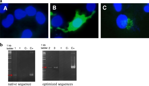
Detection of the antigen by immunostaining following transfection of B16 melanoma cells confirmed the expression and cellular localization of FcBoNT/A with the two codon-optimized plasmids. Fc*BoNT/A expression was cytoplasmic, as expected, whereas SecEpo-Fc*BoNT/A expression was switched from the cytoplasm (Fig. 1a) to mainly the Golgi apparatus (data not shown), which may indicate preparation for subsequent extracellular secretion. None of the protein could be detected after native FcBoNT/A cDNA transfection, although RT-PCR analysis confirmed the presence of both plasmids, namely, the native and codon-optimized constructs, at a transcriptional level (Fig. 1b). This suggests that the native Clostridium botulinum codons are poorly translated in mammalian cells.
FIG. 1.
(a) Immunofluorescence analysis of B16 melanoma cells transfected with either pVax-FcBoNT/A (A), pVax-Fc*BoNT/A (B), or pVax-SecEpo-Fc*BoNT/A (C), stained as described in Materials and Methods, and visualized by fluorescence microscopy. (b) RT-PCR of FcBoNT/A, Fc*BoNT/A, and SecEpo-Fc*BoNT/A mRNAs 48 h after transfection of B16 cells. Lane 1, FcBoNT/A; lane 2, Fc*BoNT/A; lane 3, SecEpo-Fc*BoNT/A; lanes −, RNA alone; lanes C−, control PCR on water; lanes C+, control PCR on plasmid. Specific primers used were as follows: for FcBoNT/A (259-bp product), 5′-TGCATCACAGGCAGGCGTAG-3′ (forward) and 5′-CCCATGAGCAACCCAAAGTCC-3′; and for Fc*BoNT/A and SecEpo-Fc*BoNT/A (703-bp product), 5′-GCCTGAACTACGGCGAGATCATCTGG-3′ (forward) and 5′-GATCTCCAGGGCGCTCAGGATCTT-3′ (reverse). The red arrows indicate the expected band size (259 bp or 703 bp).
An enzyme-linked immunosorbent assay (ELISA) was performed to assess the efficiency of the SecEpo secretion signal and to quantify the amount of secreted antigen in the supernatant of B16 cells after transfection with pVax-SecEpo-Fc*BoNT/A. This secretion signal produced a basal secretion level of 35.08 ± 1.72 ng/ng total protein of FcBoNT/A. We employed the current secretion signal sequence for our subsequent studies, based on a pilot experiment comparing it to a previously reported sequence (derived from the human secreted alkaline phosphatase cDNA) which showed a significantly lower concentration of FcBoNT/A (28.01 ± 0.85 ng/ng total protein; P < 0.001) by comparison.
Improvement of immunization with codon optimization and electrotransfer procedure.
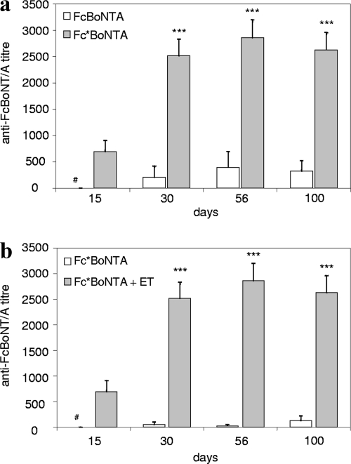
To determine the effect of codon optimization of the FcBoNT/A sequence, mice were immunized with one intramuscular injection of the mentioned sequence, followed by electrotransfer with either pVax-FcBoNT/A or pVax-Fc*BoNT/A plasmid. Antibodies against FcBoNT/A were detected in both groups of mice at 1 month posttreatment (Fig. 2a); however, the antibody titer was 10-fold higher for the codon-optimized sequence (2,518 ± 315 at 1 month) than that obtained with the native sequence (210 ± 210 at 1 month). Moreover, antibody titers remained stable for more than 3 months after a single DNA treatment (324 ± 197 with pVax-FcBoNT/A and 2,629 ± 331 with pVax-Fc*BoNT/A at 100 days).
FIG. 2.
(a) Antibody responses of mice injected with plasmid pVax-FcBoNT/A or pVax-Fc*BoNT/A, with subsequent electrotransfer (b). Antibody responses were measured in mice that were injected only (Fc*BoNT/A) or injected followed by electrotransfer (Fc*BoNT/A+ET) with plasmid pVax-Fc*BoNT/A. Swiss mice (n = 4) were treated with 40 μg of plasmid DNA. Antibody titers were determined by ELISA on serum samples at different time points after treatment. Results show means ± SEM. ***, P < 0.001; #, the titer is below the limit of detection.
In order to determine the effectiveness of the electrotransfer procedure, we evaluated the antibody titer obtained after a single intramuscular injection of pVax-FcBoNT/A or pVax-Fc*BoNT/A without administration of any electrical pulses. No antibody could be detected with a single injection of pVax-FcBoNT/A (data not shown), although the antibody titers with the codon-optimized sequence were detectable, but at a level 20-fold lower than that with electrotransfer (as shown in the example of 125 ± 96 at 100 days) (Fig. 2b).
Effect of secretion signal.
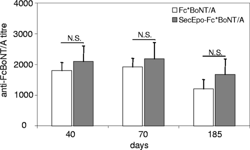
We hypothesized that the addition of a secretion signal to Fc*BoNT/A would enhance the immune response. To test this possibility, mice were treated by intramuscular injection followed by electrotransfer with the codon-optimized constructs with and without a secretion signal fused to the 5′ end, i.e., pVax-Fc*BoNT/A and pVax-SecEpo-Fc*BoNT/A, respectively (Fig. 3). Although antibodies against FcBoNT/A could be detected in both groups of mice, both titers were determined to be similar and there was no statistical difference (1,800 ± 265 with Fc*BoNT/A and 2,100 ± 505 with SecEpo-Fc*BoNT/A at 40 days). Both titers remained quite stable for at least 6 months after a single treatment.
FIG. 3.
Antibody responses of mice injected with plasmid pVax-Fc*BoNT/A or pVax-SecEpo-Fc*BoNT/A, with subsequent electrotransfer. Swiss mice (n = 4) were treated with 40 μg of plasmid DNA. Antibody titers were determined by ELISA on serum samples at different time points after treatment. Results show means ± SEM. N.S., no significant difference between the two groups.
In order to verify that 40 μg was an optimal dose for injection, we performed a dose titration of 20 μg, 40 μg, and 80 μg per mouse with the pVax-SecEpo-Fc*BoNT/A construct. We observed a corresponding trend showing response at 20 μg < response at 40 μg < response at 80 μg, although the immune responses obtained were not statistically different (analysis of variance; data not shown). Thus, we decided to continue using 40 μg per mouse for subsequent experiments. Based on our experience with plasmid DNA electrotransfer and reports from the literature (reviewed in reference 45), this amount of DNA optimally yielded sustained expression of the encoded gene.
Characterization of antisera.
To determine whether the raised antibodies could passively protect against BoNT/A, we used the most sensitive and accurate in vivo mouse neutralization assay to quantify the neutralizing titer. In this assay, 10 MLD of native BoNT/A-producing organisms were initially preincubated with 10-fold serial dilutions of sera from immunized mice, as already described (47), and we then challenged naïve Swiss mice via an intraperitoneal injection with this mixture (Table 1).
TABLE 1.
Protection against BoNT/A with antisera from mice treated with several plasmid constructs and/or several proceduresa
| Expt | Toxin construct | No. of living mice/total no. of mice after 4 days for antiserum dilution against 10 MLD
|
Neutralization titer (IU/ml) | ||||
|---|---|---|---|---|---|---|---|
| 1/100 | 1/1,000 | 1/10,000 | 1/50,000 | 1/100,000 | |||
| Single electrotransfer | Fc*BoNT/A | 6/6 | 0/6 | ND | ND | ND | 0.2 |
| SecEpo-Fc*BoNT/A | 2/2 | 6/6 | 3/6 | ND | ND | 8 | |
| Multivalent antiserum | Monovalent SecEpo-Fc*BoNT/A | 2/2 | 2/2 | 1/2 | ND | ND | 8 |
| Multivalent SecEpo-Fc*BoNT/A, -B, and -E | 2/2 | 2/2 | 1/2 | ND | ND | 0.8 | |
| Double electrotransfer | Fc*BoNT/A | 2/2 | 2/2 | 2/2 | ND | 0/2 | 8 |
| SecEpo-Fc*BoNT/A | 2/2 | 2/2 | 2/2 | ND | 0/2 | 20 | |
| Protein boost | Fc*BoNT/A | ND | 2/2 | 2/2 | 5/5 | 3/7 | 130 |
| SecEpo-Fc*BoNT/A | ND | 2/2 | 2/2 | 5/5 | 3/7 | 142 | |
Multivalent refers to the coinjection and electrotransfer of three plasmids, pVax-SecEpo-Fc*BoNT/A, pVax-SecEpo-Fc*BoNT/B, and pVax-SecEpo-Fc*BoNT/E. Swiss mice were challenged with 10 MLD of BoNT/A mixed with serial dilutions of antiserum. The final neutralizing titer was determined by a mathematical formula (barycenter calculation) using the two highest dilutions, knowing that 1 IU corresponds to neutralization of 5,000 MLD. ND, not done.
Sera from mice immunized with the secretion signal-containing pVax-SecEpo-Fc*BoNT/A construct showed much higher levels of neutralizing antibodies, with a neutralization titer of 8 IU/ml (n = 6 per dilution), than did sera from mice immunized with pVax-Fc*BoNT/A, with a titer of 0.2 IU/ml (n = 6 per dilution).
In order to better characterize the observed immune response, we performed an ELISA for IgG1 and IgG2a levels. We observed an IgG1 increase and an IgG2a decrease with the secreted-form SecEpo-Fc*BoNT/A construct compared to the non-secreted-form Fc*BoNT/A construct (Fig. 4). This suggests a Th2-shifted response with a secreted antigen, which is more similar to an immune response observed with a recombinant protein injection (Fig. 4, dotted lines).
FIG. 4.
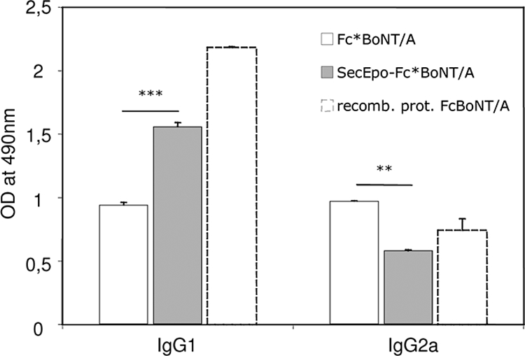
IgG1 and IgG2a responses to FcBoNT/A in mice injected with pVax-Fc*BoNT/A or pVax-SecEpo-Fc*BoNT/A, with subsequent electrotransfer, or immunized with three intraperitoneal injections of 10 μg of recombinant FcBoNT/A protein at 2-week intervals. Swiss mice (n = 4) were treated with 40 μg of plasmid DNA or 1 μg of recombinant FcBoNT/A homogenized in 1 mg aluminum hydroxide. IgG1 and IgG2a responses were determined by ELISA on serum samples at day 40. Results show means ± SEM. ***, P < 0.001; **, P < 0.01. OD, optical density.
Multivalent antiserum against BoNT/A, BoNT/B, and BoNT/E after electrotransfer.
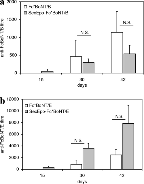
Similar immunization experiments were conducted with plasmids containing codon-optimized antigen sequences encoding the C-terminal fragments of neurotoxins B and E (Fc*BoNT/B and Fc*BoNT/E, respectively). In comparing immunizations between pVax-Fc*BoNT/B and pVax-SecEpo-Fc*BoNT/B and between pVax-Fc*BoNT/E and pVax-SecEpo-Fc*BoNT/E, the results were inconclusive. The antibodies against FcBoNT/B (Fig. 5a) and FcBoNT/E (Fig. 5b) were detected in all groups of mice, with similar titers, with or without a secretion signal. However, this could be attributed to the limits of ELISA; since the ELISA sensitivities for serotypes A, B, and E are different, only antibody titers in response to the secreted and nonsecreted forms of the same serotype, but not across serotypes, can be distinguished.
FIG. 5.
Antibody responses of mice injected with plasmids pVax-Fc*BoNT/B and pVax-SecEpo-Fc*BoNT/B (a) or pVax-Fc*BoNT/E and pVax-SecEpo-Fc*BoNT/E (b), followed by electrotransfer. Swiss mice (n = 4) were treated with 40 μg of plasmid DNA. Antibody titers were determined by ELISA on serum samples at different time points after treatment. Results show means ± SEM. N.S., no significant difference between the two groups.
Ideally, it would be useful to have a single multivalent antiserum against fragments for all three toxins, i.e., FcBoNT/A, FcBoNT/B, and FcBoNT/E. In order to test this possibility, mice were immunized with a mixture of three plasmids, pVax-SecEpo-Fc*BoNT/A, pVax-SecEpo-Fc*BoNT/B, and pVax-SecEpo-Fc*BoNT/E, in both hind legs to limit the amount of DNA injected per muscle. Antibodies against all FcBoNT/A, FcBoNT/B, and FcBoNT/E fragments could be detected in the sera of these mice. The average titers obtained in the multivalent sera with respect to those obtained with the homologous neurotoxins remained similar to those observed with a monovalent antiserum for FcBoNT/A or FcBoNT/B and lower than those observed for FcBoNT/E (Table 2).
TABLE 2.
Antibody responses of mice injected with plasmids pVax-Fc*BoNT/A, pVax-Fc*BoNT/B, and pVax-Fc*BoNT/E, with electrotransfer, as a monovalent or multivalent seruma
| Antigen | Antibody titer at 45 days
|
|
|---|---|---|
| Multivalent | Monovalent | |
| FcBoNT/A | 2,280 ± 648 | 1,759 ± 571 (NS) |
| FcBoNT/B | 1,155 ± 462 | 1,816 ± 845 (NS) |
| FcBoNT/E | 7,876 ± 3,056 | 1,330 ± 377** |
Multivalent refers to the coinjection and electrotransfer of three plasmids, pVax-SecEpo-Fc*BoNT/A, pVax-SecEpo-Fc*BoNT/B, and pVax-SecEpo-Fc*BoNT/E. Swiss mice (n = 4) were treated with 40 μg of each plasmid DNA. Antibody titers were determined by ELISA on serum samples at day 45. Results show means ± SEM. **, P < 0.01; NS, no significant difference between the two groups.
We then used a neutralization assay with native BoNT/A, as described previously, to compare the neutralization titers of monovalent and multivalent sera against BoNT/A (Table 1). The multivalent neutralization titer against BoNT/A appeared to be 10-fold lower (0.8 IU/ml; n = 2 per dilution) than the corresponding monovalent neutralization titer (8 IU/ml; n = 2 per dilution).
Immunization regimens.
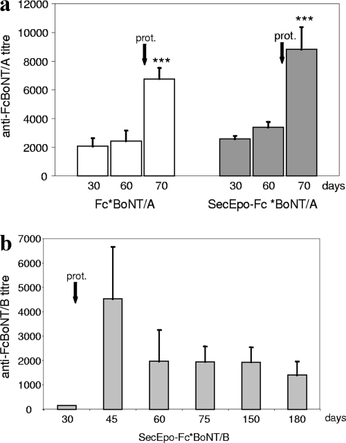
In order to increase the antibody response and the corresponding neutralizing antibody titer, we evaluated various immunization regimens. We first tested the effect of performing two DNA electrotransfers at 1-month intervals. This showed that performing a second electrotransfer increased the antibody response and the resulting neutralizing antibody titers (20 IU/ml [n = 2 per dilution] for SecEpo-Fc*BoNT/A and 8 IU/ml [n = 2 per dilution] for Fc*BoNT/A) (Table 1). We also tested the combination of one DNA electrotransfer followed by a booster injection of the recombinant protein (1 μg of FcBoNT/A in 1 mg of Alugel, given intraperitoneally). With both pVax-Fc*BoNT/A and pVax-SecEpo-Fc*BoNT/A constructs, the regimen of one DNA electrotransfer followed by a protein boost dramatically increased antibody titers 10 days after the protein injection (Fig. 6a). A neutralization assay performed on both antisera at day 70 showed increased neutralizing titers of 130 IU/ml (n = 5 or 7 per dilution) for Fc*BoNT/A and 142 IU/ml (n = 5 or 7 per dilution) for SecEpo-Fc*BoNT/A. This procedure allows up to a 500-fold increase in neutralizing antibody titer compared to that obtained with a single DNA electrotransfer procedure (Table 1).
FIG. 6.
(a) Antibody responses of mice injected with plasmid pVax-Fc*BoNT/A or pVax-SecEpo-Fc*BoNT/A, with electrotransfer, at day 0 and boosted with a recombinant protein injection (prot.) at day 60. Swiss mice (n = 4) were treated with 40 μg of plasmid DNA and 1 μg of recombinant FcBoNT/A homogenized in 1 mg aluminum hydroxide. Antibody titers were determined by ELISA on serum samples at different time points after treatment. Results show means ± SEM. ***, P < 0.001 (significant difference between day 70 and previous titers). (b) Antibody responses of mice injected with plasmid pVax-SecEpo-Fc*BoNT/B with electrotransfer at day 0 and boosted with a recombinant protein injection at day 30. Swiss mice (n = 4) were treated with 40 μg of plasmid DNA and 1 μg of recombinant FcBoNT/B homogenized in 1 mg aluminum hydroxide. Antibody titers were determined by ELISA on serum samples at different time points after treatment. Results show means ± SEM.
In another experiment, mice were treated with pVax-SecEpo-Fc*BoNT/B plasmid and boosted with a recombinant protein injection at day 30. Interestingly, very few antibodies could be detected before the protein boost (Fig. 6b). However, a high peak of anti-BoNT/B antibodies was detected 15 days after the protein boost (day 45), which was similar to the scenario for BoNT/A 10 days after the protein boost. Two additional samples, at day 60 and day 75, showed only about 50% of the maximum antibody titer. This titer was stabilized for about 6 months (day 180) and then declined at day 180.
DISCUSSION
In the early 1990s, the administration of a naked DNA plasmid encoding an antigen was shown to elicit both humoral (46) and cellular (51) immune responses. Since then, numerous publications have reported the effectiveness of DNA vaccines in a variety of preclinical models, including both infectious (either viral, bacterial, or parasitic) and acquired diseases, such as cancer (reviewed in references 19 and 44). Although DNA vaccination results for small animals were robust, they were ineffective in nonhuman primates or clinical trials. One possible explanation might be that the level of protein expression is insufficient to elicit a strong immune response. Consequently, a number of methods have been developed to increase DNA uptake efficiency in order to increase antigen expression. Among the resulting innovative methods, electrotransfer (or in vivo electroporation) has proven to be one of the most efficient means for gene delivery, particularly in skeletal muscle (48), where high levels of protein expression can be achieved. The delivery of electrical pulses enhances DNA uptake, resulting in a 100- to 1,000-fold increase in gene expression (in comparison with naked DNA administration) as well as an enhanced immune response toward the transgene, including in large animals (5, 35, 40). DNA immunization in a tissue rich in dendritic cells (for example, skin) leads more toward a Th2-type immune response, whereas intramuscular DNA immunization results in a more Th1 humoral response (17). The latter provides another use of intramuscular DNA immunization (which is different from full protection, which favors usage toward vaccination), namely, the production of a high-titer neutralizing antiserum for protection against a particular toxin. This idea has been shown in the production of specific antibodies against snake venom toxin (16). Plasmid-based DNA immunization to protect mice from BoNT/A (9, 43) and BoNT/F (4, 18) has previously been evaluated in an intramuscular administration protocol with naked DNA, but the protective response obtained was highly variable, even after several high doses of DNA injections.
In the present study, we describe the use of DNA electrotransfer into skeletal muscle to efficiently raise a high-titer antiserum against various BoNT serotypes in mice. The encoded antigens are the nontoxic C-terminal fragments (Fc) of the heavy chains of toxins A, B, and E; these regions are responsible for the interactions of BoNTs with receptors of the motoneuron (27, 39). Fc has been described as the best minimal part of the protein required to elicit efficient production of neutralizing antibodies (20, 47). As largely reported in the literature, the immune response is dependent on the level of antigen expression (32). Therefore, prior to vaccination, it is imperative that the best possible DNA expression vector is engineered to yield a high level of antigen expression. Clostridium botulinum has a very different codon bias from that for mammals. Using the Graphical Codon Usage Analyser (Geneart), we observed that the difference between Mus musculus and Clostridium botulinum codon usage was 28.8%, whereas the difference between Escherichia coli and Mus musculus codon usage was only 12.5% and that between Mus musculus and Homo sapiens codon usage was 1.3%. This bias may result in low expression of Clostridium botulinum genes in mammalian cells by use of native DNA sequences. We therefore designed a synthetic FcBoNT/A cDNA by codon optimization, retaining the natural amino acid sequences but using the preferred mouse codons. Such a strategy has already been shown to enhance expression of Plasmodium falciparum-encoded proteins in mammalian cells (34). It has also been employed for vaccination purposes (10, 11), for optimal antigen expression directly into targeted cells after “naked” DNA injection (4, 45), and for antiserum production, such as the optimal E. coli production of FcBoNT/A (9). We have observed that reengineering the FcBoNT/A gene dramatically improved the expression of FcBoNT/A after transfection of B16 melanoma cells, as shown in Fig. 1b. In contrast, no protein was detected by immunostaining with the native cDNA sequence. Thus, codon optimization appears to be necessary for this antigen.
We also hypothesized that the cellular localization and eventual extracellular secretion of the antigen might be crucial parameters in controlling the magnitude of the immune response, due to the influence of antigen presentation. Skeletal muscle contains an abundant vascular supply (25), which facilitates the produced secreted proteins being transported easily to the blood circulation (13). Strong immune responses have been obtained with transmembrane, secreted, or intracellular proteins. Several studies have indicated that a secreted form of the antigen leads to a stronger immune response and more neutralizing antibodies (2, 12), although conflicting results have also been reported (43). Since the FcBoNT/A fragment is not naturally secreted, we fused the murine erythropoietin or human secreted alkaline phosphatase secretion signal 5′ of the FcBoNT/A cDNA sequence. Since the murine erythropoietin secretion signal led to better expression than that with the human secreted phosphatase alkaline, as revealed by an in vitro ELISA (data not shown), only the former was considered in this study.
The three plasmids encoding either the native antigen FcBoNT/A, the codon-optimized version of the antigen (Fc*BoNT/A), or a secreted form of Fc*BoNT/A with the murine erythropoietin secretion signal were then evaluated for the ability to elicit a humoral immune response in Swiss mice after intramuscular immunization. Both codon optimization and electrotransfer dramatically improved the blood antibody titer for more than 3 months. While the explanation for the codon optimization effect is most likely higher expression of the antigen, the mechanism of enhanced ELISA titer in response to electrotransfer might involve several factors, including (i) more antigen expression in the tissue, (ii) the possibility of transfecting resident mononuclear cells (antigen-presenting cells) (15), and (iii) the effect of the related inflammatory response induced by the electrotransfer procedure (37). Furthermore, the electrotransfer procedure seems to play an adjuvant role in the Th1 response, as reported by Grovenik et al. (14).
Interestingly, while there was no difference in total antibody titers between the nonsecreted and secreted proteins (Fig. 3), the neutralizing titers showed a higher level in the latter case after a single immunization (titer of 8 IU/ml versus 0.2 IU/ml) (Table 1). Immunization experiments conducted with the Fc fragments of toxins B and E led to similar results with respect to the antibody titers in blood for the secreted and nonsecreted forms of each serotype (Fig. 5 and Table 2).
To better characterize the induced immune response, IgG1 and IgG2a titers in the blood of immunized mice were analyzed. It is agreed that high IgG1 induction is related to a Th2-type immune response, while high IgG2a expression is more related to a Th1 response (7, 30). It appeared that the cellular localization of the antigen influenced the immune response (Fig. 4), as the secreted forms induced a strong Th2 response characterized by the main presence of IgG1, similar to that obtained by immunization with the recombinant protein. In contrast, the nonsecreted form elicited a more balanced immune response of both Th1 and Th2 types. The two immune responses are thus different, as shown by the IgG1/IgG2a analysis and protective antibody titers. This may be due to different presentation of the antigen or to an altered conformation with the secretion signal, which may have increased the exposure of protective epitopes. However, our data do not provide an explanation for this difference. The mechanism by which DNA immunization elicits an efficient immune response has not been elucidated and is currently under intense investigation. The current notion of how DNA immunization produces antigen-specific immunity has been described fully previously (19).
In order to increase the neutralizing antibody titers, additional regimens were evaluated; these consisted of a second DNA electrotransfer boost or a recombinant protein boost after the first DNA electrotransfer. Interestingly, the electrotransfer boost increased the neutralizing titer from 0.2 IU/ml to 8 IU/ml for the nonsecreted antigen FcBoNT/A and from 8 IU/ml to 20 IU/ml for the secreted form of the antigen, leading to a highly neutralizing antiserum. A protein boost after DNA electrotransfer has already been shown to improve the humoral response in mice (21) and in nonhuman primates (23). In this work, a single protein boost instead of an electrotransfer boost dramatically increased the neutralizing titer, leading to a titer of about 130 to 140 IU/ml for both the nonsecreted and secreted proteins (Table 1). Focusing on the kinetics of antibody production, the protein boost allows a transitory increase of the antibody titer. A titer peak is observed from day 10 to day 15 after the boost, and then the titer stabilizes for several months at about 50% of the maximum value. If a maximal antibody titer is required, then a new protein boost would be necessary at the desired time point.
Finally, it has been reported that coinjection and electrotransfer of several plasmids into skeletal muscle are feasible for the expressions of various transgenes (28, 49). We thus evaluated the possibility of obtaining a multivalent neutralizing antiserum against all three toxin serotypes via the electrotransfer procedure. After coelectrotransfer of the three antigen-encoding plasmids, we obtained an antiserum against serotypes A, B, and E. The total antibody titers were equivalent to those obtained with a one-plasmid treatment for toxins A and B but not for toxin E (Table 2). In addition, the neutralizing titer against toxin A was dramatically lower than that with the corresponding monovalent antiserum (0.8 IU/ml versus 8 IU/ml) (Table 1). This result is in accord with a recent report by Sedegah et al. after coinjection of nine plasmids encoding nine different antigens of Plasmodium falciparum. A dramatic decrease in the immune response against each antigen, from 8- to 2,500-fold, was observed compared to that obtained with individual injections (41). Several hypotheses were suggested to explain this effect, such as lower expression of each antigen due to competition at a transcriptional level or translational level in each muscle cell or competition for antigen presentation.
In summary, this work confirms that the expression level, the intracellular fate, and the method of DNA delivery are important for optimizing DNA immunization. Though botulism is a rare disease, BoNTs are commonly used for therapeutic and cosmetic purposes and are known to present a potential threat as a biological weapon. For these reasons, there is high demand for the development of a prophylactic or therapeutic treatment. Although several strategies can be developed to counter these toxins, passive immunotherapy using botulinum antitoxin is the only effective postexposure therapy (29). Producing an antitoxin is a time-consuming and expensive process, and a technique that would not require the production and purification of a recombinant protein would thus be of great importance.
In conclusion, we have shown in this work that the injection and electrotransfer of an optimized DNA construct encoding the Fc fragments of the heavy chains of BoNTs lead to a sustainable high titer of neutralizing antiserum. These results may be of interest for the production of anti-BoNT antisera in larger animal species. Furthermore, since electrotransfer is a fast and easy procedure, the proposed genetic immunization procedure represents a useful tool to map a particular protein and screen for immunodominant epitopes in order to accelerate vaccine development.
Acknowledgments
This work was supported by the Institut de Médecine Aérospatiale du Service de Santé des Armées (IMASSA).
We also thank Hervé Volland (CEA) for kindly providing us with the monoclonal antibody TB5. We thank Andrew Ho for critically reading the manuscript.
Editor: S. R. Blanke
Footnotes
Published ahead of print on 23 February 2009.
REFERENCES
- 1.Arnon, S. S., R. Schechter, T. V. Inglesby, D. A. Henderson, J. G. Bartlett, M. S. Ascher, E. Eitzen, A. D. Fine, J. Hauer, M. Layton, S. Lillibridge, M. T. Osterholm, T. O'Toole, G. Parker, T. M. Perl, P. K. Russell, D. L. Swerdlow, and K. Tonat. 2001. Botulinum toxin as a biological weapon: medical and public health management. JAMA 2851059-1070. [DOI] [PubMed] [Google Scholar]
- 2.Ashok, M. S., and P. N. Rangarajan. 2002. Protective efficacy of a plasmid DNA encoding Japanese encephalitis virus envelope protein fused to tissue plasminogen activator signal sequences: studies in a murine intracerebral virus challenge model. Vaccine 201563-1570. [DOI] [PubMed] [Google Scholar]
- 3.Bachy, M., F. Boudet, M. Bureau, Y. Girerd-Chambaz, P. Wils, D. Scherman, and C. Meric. 2001. Electric pulses increase the immunogenicity of an influenza DNA vaccine injected intramuscularly in the mouse. Vaccine 191688-1693. [DOI] [PubMed] [Google Scholar]
- 4.Bennett, A. M., S. D. Perkins, and J. L. Holley. 2003. DNA vaccination protects against Botulinum neurotoxin type F. Vaccine 213110-3117. [DOI] [PubMed] [Google Scholar]
- 5.Bettan, M., F. Emmanuel, R. Darteil, J. M. Caillaud, F. Soubrier, P. Delaere, D. Branelec, A. Mahfoudi, N. Duverger, and D. Scherman. 2000. High-level protein secretion into blood circulation after electric pulse-mediated gene transfer into skeletal muscle. Mol. Ther. 2204-210. [DOI] [PubMed] [Google Scholar]
- 6.Bollerot, K., D. Sugiyama, V. Escriou, R. Gautier, S. Tozer, D. Scherman, and T. Jaffredo. 2006. Widespread lipoplex-mediated gene transfer to vascular endothelial cells and hemangioblasts in the vertebrate embryo. Dev. Dyn. 235105-114. [DOI] [PubMed] [Google Scholar]
- 7.Boyle, J. S., C. Koniaras, and A. M. Lew. 1997. Influence of cellular location of expressed antigen on the efficacy of DNA vaccination: cytotoxic T lymphocyte and antibody responses are suboptimal when antigen is cytoplasmic after intramuscular DNA immunization. Int. Immunol. 91897-1906. [DOI] [PubMed] [Google Scholar]
- 8.Chertow, D. S., E. T. Tan, S. E. Maslanka, J. Schulte, E. A. Bresnitz, R. S. Weisman, J. Bernstein, S. M. Marcus, S. Kumar, J. Malecki, J. Sobel, and C. R. Braden. 2006. Botulism in 4 adults following cosmetic injections with an unlicensed, highly concentrated botulinum preparation. JAMA 2962476-2479. [DOI] [PubMed] [Google Scholar]
- 9.Clayton, J., and J. L. Middlebrook. 2000. Vaccination of mice with DNA encoding a large fragment of botulinum neurotoxin serotype A. Vaccine 181855-1862. [DOI] [PubMed] [Google Scholar]
- 10.Doria-Rose, N. A., and N. L. Haigwood. 2003. DNA vaccine strategies: candidates for immune modulation and immunization regimens. Methods 31207-216. [DOI] [PubMed] [Google Scholar]
- 11.Frelin, L., G. Ahlen, M. Alheim, O. Weiland, C. Barnfield, P. Liljestrom, and M. Sallberg. 2004. Codon optimization and mRNA amplification effectively enhances the immunogenicity of the hepatitis C virus nonstructural 3/4A gene. Gene Ther. 11522-533. [DOI] [PubMed] [Google Scholar]
- 12.Geissler, M., V. Bruss, S. Michalak, B. Hockenjos, D. Ortmann, W. B. Offensperger, J. R. Wands, and H. E. Blum. 1999. Intracellular retention of hepatitis B virus surface proteins reduces interleukin-2 augmentation after genetic immunizations. J. Virol. 734284-4292. [DOI] [PMC free article] [PubMed] [Google Scholar]
- 13.Goldspink, G. 2003. Skeletal muscle as an artificial endocrine tissue. Best Pract. Res. Clin. Endocrinol. Metab. 17211-222. [DOI] [PubMed] [Google Scholar]
- 14.Gronevik, E., I. Mathiesen, and T. Lomo. 2005. Early events of electroporation-mediated intramuscular DNA vaccination potentiate Th1-directed immune responses. J. Gene Med. 71246-1254. [DOI] [PubMed] [Google Scholar]
- 15.Gronevik, E., S. Tollefsen, L. I. Sikkeland, T. Haug, T. E. Tjelle, and I. Mathiesen. 2003. DNA transfection of mononuclear cells in muscle tissue. J. Gene Med. 5909-917. [DOI] [PubMed] [Google Scholar]
- 16.Harrison, R. A. 2004. Development of venom toxin-specific antibodies by DNA immunisation: rationale and strategies to improve therapy of viper envenoming. Vaccine 221648-1655. [DOI] [PubMed] [Google Scholar]
- 17.Harrison, R. A., and A. E. Bianco. 2000. DNA immunization with Onchocerca volvulus genes, Ov-tmy-1 and OvB20: serological and parasitological outcomes following intramuscular or GeneGun delivery in a mouse model of onchocerciasis. Parasite Immunol. 22249-257. [DOI] [PubMed] [Google Scholar]
- 18.Jathoul, A. P., J. L. Holley, and H. S. Garmory. 2004. Efficacy of DNA vaccines expressing the type F botulinum toxin Hc fragment using different promoters. Vaccine 223942-3946. [DOI] [PubMed] [Google Scholar]
- 19.Kutzler, M. A., and D. B. Weiner. 2008. DNA vaccines: ready for prime time? Nat. Rev. Genet. 9776-788. [DOI] [PMC free article] [PubMed] [Google Scholar]
- 20.LaPenotiere, H. F., M. A. Clayton, and J. L. Middlebrook. 1995. Expression of a large, nontoxic fragment of botulinum neurotoxin serotype A and its use as an immunogen. Toxicon 331383-1386. [DOI] [PubMed] [Google Scholar]
- 21.Law, M., R. M. Cardoso, I. A. Wilson, and D. R. Burton. 2007. Antigenic and immunogenic study of membrane-proximal external region-grafted gp120 antigens by a DNA prime-protein boost immunization strategy. J. Virol. 814272-4285. [DOI] [PMC free article] [PubMed] [Google Scholar]
- 22.Leblond, J., N. Mignet, C. Largeau, M. V. Spanedda, J. Seguin, D. Scherman, and J. Herscovici. 2007. Lipopolythioureas: a new non-cationic system for gene transfer. Bioconjug. Chem. 18484-493. [DOI] [PubMed] [Google Scholar]
- 23.Li, Z., H. Zhang, X. Fan, Y. Zhang, J. Huang, Q. Liu, T. E. Tjelle, I. Mathiesen, R. Kjeken, and S. Xiong. 2006. DNA electroporation prime and protein boost strategy enhances humoral immunity of tuberculosis DNA vaccines in mice and non-human primates. Vaccine 244565-4568. [DOI] [PubMed] [Google Scholar]
- 24.Lindstrom, M., M. Nevas, J. Kurki, R. Sauna-aho, A. Latvala-Kiesila, I. Polonen, and H. Korkeala. 2004. Type C botulism due to toxic feed affecting 52,000 farmed foxes and minks in Finland. J. Clin. Microbiol. 424718-4725. [DOI] [PMC free article] [PubMed] [Google Scholar]
- 25.Lu, Q. L., G. Bou-Gharios, and T. A. Partridge. 2003. Non-viral gene delivery in skeletal muscle: a protein factory. Gene Ther. 10131-142. [DOI] [PubMed] [Google Scholar]
- 26.Mahajan, S. T., and L. Brubaker. 2007. Botulinum toxin: from life-threatening disease to novel medical therapy. Am. J. Obstet. Gynecol. 1967-15. [DOI] [PubMed] [Google Scholar]
- 27.Mahrhold, S., A. Rummel, H. Bigalke, B. Davletov, and T. Binz. 2006. The synaptic vesicle protein 2C mediates the uptake of botulinum neurotoxin A into phrenic nerves. FEBS Lett. 5802011-2014. [DOI] [PubMed] [Google Scholar]
- 28.Martel-Renoir, D., V. Trochon-Joseph, A. Galaup, C. Bouquet, F. Griscelli, P. Opolon, D. Opolon, E. Connault, L. Mir, and M. Perricaudet. 2003. Coelectrotransfer to skeletal muscle of three plasmids coding for antiangiogenic factors and regulatory factors of the tetracycline-inducible system: tightly regulated expression, inhibition of transplanted tumor growth, and antimetastatic effect. Mol. Ther. 8425-433. [DOI] [PubMed] [Google Scholar]
- 29.Mayers, C. N., J. L. Holley, and T. Brooks. 2001. Antitoxin therapy for botulinum intoxication. Rev. Med. Microbiol. 1229-37. [Google Scholar]
- 30.McClements, W. L., M. E. Armstrong, R. D. Keys, and M. A. Liu. 1997. The prophylactic effect of immunization with DNA encoding herpes simplex virus glycoproteins on HSV-induced disease in guinea pigs. Vaccine 15857-860. [DOI] [PubMed] [Google Scholar]
- 31.Middlebrook, J. L. 2005. Production of vaccines against leading biowarfare toxins can utilize DNA scientific technology. Adv. Drug Deliv. Rev. 571415-1423. [DOI] [PubMed] [Google Scholar]
- 32.Miller, M., G. Rekas, K. Dayball, Y. H. Wan, and J. Bramson. 2004. The efficacy of electroporated plasmid vaccines correlates with long-term antigen production in vivo. Vaccine 222517-2523. [DOI] [PubMed] [Google Scholar]
- 33.Mir, L. M., M. F. Bureau, J. Gehl, R. Rangara, D. Rouy, J. M. Caillaud, P. Delaere, D. Branellec, B. Schwartz, and D. Scherman. 1999. High-efficiency gene transfer into skeletal muscle mediated by electric pulses. Proc. Natl. Acad. Sci. USA 964262-4267. [DOI] [PMC free article] [PubMed] [Google Scholar]
- 34.Narum, D. L., S. Kumar, W. O. Rogers, S. R. Fuhrmann, H. Liang, M. Oakley, A. Taye, B. K. Sim, and S. L. Hoffman. 2001. Codon optimization of gene fragments encoding Plasmodium falciparum merozoite proteins enhances DNA vaccine protein expression and immunogenicity in mice. Infect. Immun. 697250-7253. [DOI] [PMC free article] [PubMed] [Google Scholar]
- 35.Otten, G., M. Schaefer, B. Doe, H. Liu, I. Srivastava, J. zur Megede, D. O'Hagan, J. Donnelly, G. Widera, D. Rabussay, M. G. Lewis, S. Barnett, and J. B. Ulmer. 2004. Enhancement of DNA vaccine potency in rhesus macaques by electroporation. Vaccine 222489-2493. [DOI] [PubMed] [Google Scholar]
- 36.Pellizzari, R., O. Rossetto, G. Schiavo, and C. Montecucco. 1999. Tetanus and botulinum neurotoxins: mechanism of action and therapeutic uses. Philos. Trans. R. Soc. Lond. B 354259-268. [DOI] [PMC free article] [PubMed] [Google Scholar]
- 37.Peng, B., Y. Zhao, L. Xu, and Y. Xu. 2007. Electric pulses applied prior to intramuscular DNA vaccination greatly improve the vaccine immunogenicity. Vaccine 252064-2073. [DOI] [PubMed] [Google Scholar]
- 38.Rainey, G. J., and J. A. Young. 2004. Antitoxins: novel strategies to target agents of bioterrorism. Nat. Rev. Microbiol. 2721-726. [DOI] [PubMed] [Google Scholar]
- 39.Rummel, A., T. Eichner, T. Weil, T. Karnath, A. Gutcaits, S. Mahrhold, K. Sandhoff, R. L. Proia, K. R. Acharya, H. Bigalke, and T. Binz. 2007. Identification of the protein receptor binding site of botulinum neurotoxins B and G proves the double-receptor concept. Proc. Natl. Acad. Sci. USA 104359-364. [DOI] [PMC free article] [PubMed] [Google Scholar]
- 40.Scheerlinck, J. P., J. Karlis, T. E. Tjelle, P. J. Presidente, I. Mathiesen, and S. E. Newton. 2004. In vivo electroporation improves immune responses to DNA vaccination in sheep. Vaccine 221820-1825. [DOI] [PubMed] [Google Scholar]
- 41.Sedegah, M., Y. Charoenvit, L. Minh, M. Belmonte, V. F. Majam, S. Abot, H. Ganeshan, S. Kumar, D. J. Bacon, A. Stowers, D. L. Narum, D. J. Carucci, and W. O. Rogers. 2004. Reduced immunogenicity of DNA vaccine plasmids in mixtures. Gene Ther. 11448-456. [DOI] [PubMed] [Google Scholar]
- 42.Shone, C. C., and H. S. Tranter. 1995. Growth of clostridia and preparation of their neurotoxins. Curr. Top. Microbiol. Immunol. 195143-160. [DOI] [PubMed] [Google Scholar]
- 43.Shyu, R. H., M. F. Shaio, S. S. Tang, H. F. Shyu, C. F. Lee, M. H. Tsai, J. E. Smith, H. H. Huang, J. J. Wey, J. L. Huang, and H. H. Chang. 2000. DNA vaccination using the fragment C of botulinum neurotoxin type A provided protective immunity in mice. J. Biomed. Sci. 751-57. [DOI] [PubMed] [Google Scholar]
- 44.Srivastava, I. K., and M. A. Liu. 2003. Gene vaccines. Ann. Intern. Med. 138550-559. [DOI] [PubMed] [Google Scholar]
- 45.Stratford, R., G. Douce, L. Zhang-Barber, N. Fairweather, J. Eskola, and G. Dougan. 2000. Influence of codon usage on the immunogenicity of a DNA vaccine against tetanus. Vaccine 19810-815. [DOI] [PubMed] [Google Scholar]
- 46.Tang, D. C., M. DeVit, and S. A. Johnston. 1992. Genetic immunization is a simple method for eliciting an immune response. Nature 356152-154. [DOI] [PubMed] [Google Scholar]
- 47.Tavallaie, M., A. Chenal, D. Gillet, Y. Pereira, M. Manich, M. Gibert, S. Raffestin, M. R. Popoff, and J. C. Marvaud. 2004. Interaction between the two subdomains of the C-terminal part of the botulinum neurotoxin A is essential for the generation of protective antibodies. FEBS Lett. 572299-306. [DOI] [PubMed] [Google Scholar]
- 48.Trollet, C., C. Bloquel, D. Scherman, and P. Bigey. 2006. Electrotransfer into skeletal muscle for protein expression. Curr. Gene Ther. 6561-578. [DOI] [PubMed] [Google Scholar]
- 49.Trollet, C., M. Ibanez-Ruiz, C. Bloquel, G. Valin, D. Scherman, and P. Bigey. 2004. Regulation of gene expression using a conditional RNA antisense strategy. J. Genome Sci. Technol. 31-13. [Google Scholar]
- 50.Turton, K., J. A. Chaddock, and K. R. Acharya. 2002. Botulinum and tetanus neurotoxins: structure, function and therapeutic utility. Trends Biochem. Sci. 27552-558. [DOI] [PubMed] [Google Scholar]
- 51.Ulmer, J. B., J. J. Donnelly, S. E. Parker, G. H. Rhodes, P. L. Felgner, V. J. Dwarki, S. H. Gromkowski, R. R. Deck, C. M. DeWitt, A. Friedman, et al. 1993. Heterologous protection against influenza by injection of DNA encoding a viral protein. Science 2591745-1749. [DOI] [PubMed] [Google Scholar]


