Abstract
Myxoma virus (MYXV) is a poxvirus pathogenic only for European rabbits, but its permissiveness in human cancer cells gives it potential as an oncolytic virus. A recombinant MYXV expressing both the tdTomato red fluorescent protein and interleukin-15 (IL-15) (vMyx-IL-15-tdTr) was constructed. Cells infected with vMyx-IL-15-tdTr secreted bioactive IL-15 and had in vitro replication kinetics similar to that of wild-type MYXV. To determine the safety of this virus for future oncolytic studies, we tested its pathogenesis in European rabbits. In vivo, vMyx-IL-15-tdTr no longer causes lethal myxomatosis. Thus, ectopic IL-15 functions as an antiviral cytokine in vivo, and vMyx-IL-15-tdTr is a safe candidate for animal studies of oncolytic virotherapy.
Myxoma virus (MYXV) is a poxvirus from the Leporipoxvirus genus (6) which causes lethal myxomatosis in European rabbits (Oryctolagus cuniculus). Myxomatosis is characterized by disseminated cutaneous and mucocutaneous lesions. During infection, MYXV expresses and releases a wide range of host-targeted immunomodulators (11, 30), leading to a dramatic compromise in the ability of the rabbit host to mount an effective immune response (4). Recent studies using targeted gene knockouts of diverse MYXV open reading frames (10) have provided a better understanding of the disease mechanisms that contribute to lethal myxomatosis. Studies of determinants of the MYXV host range outside of rabbit cells have led to investigations into the oncolytic potential of MYXV against human cancer cells (2, 10, 28, 31). Also, genetic manipulation of MYXV continues to produce next-generation recombinant MYXVs with reduced virulence and enhanced oncolytic potential (2, 26).
Interleukin-15 (IL-15) is a key immunoregulatory cytokine and has been shown to antagonize tumor growth in vivo (3, 7, 8, 21, 33, 37) by recruiting and stimulating immune effector cells, especially NK cells, which participate in tumor clearance (12). IL-15 binds to a receptor complex formed with IL-15Rα (a unique subunit for IL-15 signaling), IL-2R/IL-15Rβ (shared with IL-2), and common γc subunits. The subsequent receptor signaling induces unique biological responses. IL-15 is important for supporting the proliferation of CD8+ memory T cells as well as for enhancing high-avidity T-cell responses (14, 38). These functions have led to the development of IL-15 as an adjuvant for vaccine development (22, 24) and for immunotherapy against cancer (20, 32). To study the immunoregulatory effects of IL-15 in cancer therapy and test for a potential synergism of this cytokine during MYXV oncolytic virotherapy, we constructed a recombinant MYXV expressing murine IL-15 (vMyx-IL-15-tdTr). This construct will be useful in murine tumor models, and we expect that this IL-15 would also be biologically active in the rabbit (36). Here, we investigated the effect of this vMyx-IL-15-tdTr construct on virulence in the rabbit host.
Cells infected with vMyx-IL-15-tdTr secrete IL-15 and support normal virus replication.
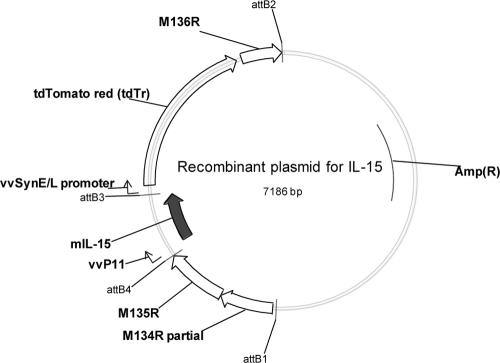
vMyx-IL-15-tdTr was constructed by inserting an IL-15-expressing cassette containing the murine IL-15 coding sequence (NM_008357; Open Biosystem) under the control of the poxvirus p11 late promoter at an intergenic location between the M135 and M136 genes in the wild-type (wt) MYXV strain Laussane (vMyx-Lau) genome. An expression cassette for a synthetic fluorescent protein, tdTomato red (tdTr) (AY678269), was inserted immediately downstream of the IL-15 expression cassette, and its expression was driven by a poxvirus synthetic early/late promoter (Fig. 1). The tdTr serves as a fluorescent marker for MYXV replication in vitro and in vivo, as MYXV infection can be monitored by live imaging of tdTr expression (35). MultiSite Gateway Pro (Invitrogen) (19) technology was used to clone three DNA fragments into a destination vector for the insertion of IL-15 into the MYXV genome (Fig. 1). A wt control virus vMyx-tdTr was also constructed with only the tdTr expression cassette inserted in the same M135-M136 intergenic locus. The IL-15 and tdTr or tdTr-only expression cassettes were inserted into the MYXV genome by infecting BGMK cells with vMyx-Lau and then transfecting the appropriate recombination plasmids. Multiple rounds of focus purification were conducted to obtain pure stocks of the recombinant viruses, and the purity was confirmed by PCR.
FIG. 1.
Recombinant plasmid used to construct vMyx-IL-15-tdTr. Murine IL-15 (mIL-15) expression is driven by the vaccinia virus p11 late promoter (vvP11), followed by the tdTr expression cassette. The expression of tdTr, a strong red fluorescent protein, is driven by a poxvirus synthetic early/late promoter (vvSynE/L). Two recombination sites containing sequences from the MYXV M134 and M135 (upstream) and M136 (downstream) genes were used to insert the expression cassettes at an intergenic location between the M135 and M136 genes in the genome of vMyx-Lau.
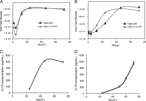
Rabbit RK-13 (ATCC CCL-37) and murine melanoma B16F10 (CRL-6475) cells were used to characterize the replication of recombinant MYXVs by a one-step growth curve, and similar growth was observed for vMyx-IL-15-tdTr and vMyx-tdTr (Fig. 2A and B). These viruses also replicated with identical kinetics in other cell lines, including BGMK, human Namalwa (CRL-1432), and Ramos (CRL-1596) cells (data not shown).
FIG. 2.
Growth kinetics and IL-15 expression from vMyx-IL-15-tdTr. (A) One-step growth of vMyx-IL-15-tdTr in RK-13 cells. RK-13 cells were infected with either vMyx-tdTr or vMyx-IL-15-tdTr at a multiplicity of infection (MOI) of 3. Viral yields from cell lysates were determined at different hours postinfection (Hrs p.i.) in BSC40 cells. (B) One-step growth of vMyx-IL-15-tdTr in murine melanoma B16F10 cells. B16F10 cells were infected with either vMyx-tdTr or vMyx-IL-15-tdTr at an MOI of 3. Viral yields from cell lysates were determined as described in panel A. (C) Concentration of IL-15 in the supernatant of RK-13 cells infected with vMyx-IL-15-tdTr. RK-13 cells were infected at an MOI of 3, and supernatants were collected at 24, 46, and 72 h p.i. for the detection of IL-15 by ELISA (eBioscience) according to the manufacturer's instructions. (D) Concentrations of IL-15 in the supernatant of BGMK cells infected with vMyx-IL-15-tdTr. BGMK cells were infected at an MOI of 10 and supernatants were collected at 24, 50, 60, and 72 h p.i. for detection of IL-15 by ELISA. In panels C and D, the average optical density reading from mock-infected samples was subtracted from the optical density readings of samples infected with vMyx-IL-15-tdTr or vMyx-tdTr. No detectable levels of IL-15 were observed from samples infected with vMyx-tdTr at the latest time point.
At various time points after infection (of RK-13 or BGMK cells) with vMyx-IL-15-tdTr, supernatants were collected for an enzyme-linked immunosorbent assay (ELISA; eBioscience) to estimate the expression of secreted IL-15 (Fig. 2C and D). Different kinetics of IL-15 accumulation were observed with time, which could be due to differences in the efficiency of IL-15 secretion and differences in the response to infection in different cell lines.
MYXV-IL-15-tdTr is attenuated in European rabbits.
This animal study was approved by the Institutional Animal Care and Usage Committee (IACUC) at the University of Florida. MYXV pathogeneses in New Zealand White (NZW) rabbits (Charles River Laboratories International) were compared for four treatment groups inoculated with phosphate-buffered saline (PBS), vMyx-Lau, vMyx-tdTr, or vMyx-IL-15-tdTr. NZW rabbits were inoculated with either PBS or 1,000 focus-forming units (ffu) of the corresponding MYXV in the left thigh intradermally (1). Clinical scores that reflected the rabbits' overall physical conditions and clinical signs of myxomatosis were obtained daily (18). Statistic evaluations were conducted using SigmaStat3.1.1. One-way analysis of variance was applied to compare daily lesion measurements among groups, and a P value of <0.05 was defined as statistically significant.
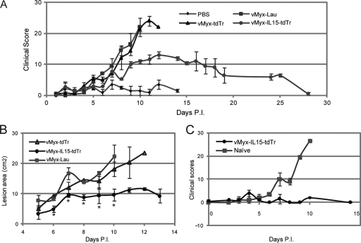
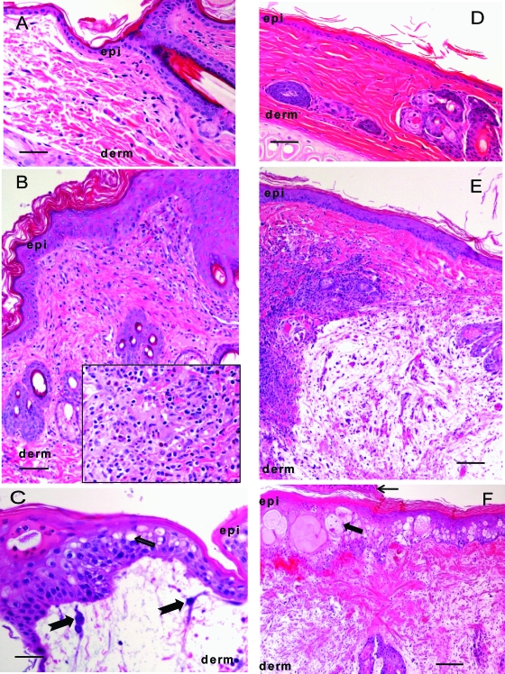
vMyx-IL-15-tdTr was significantly attenuated and failed to induce lethal myxomatosis in NZW rabbits, while the control vMyx-tdTr infection exhibited the same virulence, clinical signs, and lethality as did vMyx-Lau infection. All the rabbits infected with vMyx-IL-15-tdTr survived with clinical scores significantly lower than those of animals infected with control MYXVs (Fig. 3A). Primary lesions caused by vMyx-IL-15-tdTr were significantly smaller than those caused by vMyx-Lau and vMyx-tdTr (Fig. 3B), and scabs formed between 10 and 14 days postinfection (dpi); this was followed by complete recovery at the time of challenge. Secondary lesions were observed in the vMyx-IL-15-tdTr group, which resulted in an increase in the daily clinical scores, but they were visible 1 to 2 days later than were those from the vMyx-Lau or vMyx-tdTr group. The clinical scores in these rabbits remained above 10 for approximately 10 days. After this period, healing of the primary and secondary lesions was noted, resulting in a gradual decrease in the clinical score. Eventually, rabbits infected with vMyx-IL-15-tdTr fully recovered, and their clinical scores went back to normal range. One animal infected with vMyx-IL-15-tdTr was sacrificed to collect tissue for histology at 14 dpi, when all animals infected with vMyx-Lau and vMyx-tdTr had reached euthanasia criteria. Hematoxylin and eosin staining of primary lesions from animals with lethal myxomatosis (vMyx-Lau and vMyx-tdTr groups) showed severe edema, widespread collagenous degeneration, myxomatous stroma infiltration and epidermal necrosis, ulceration of the epithelium, and vasculitis. In the tissues collected from the vMyx-IL-15-tdTr-infected animal, dense mixed inflammatory cells (e.g., macrophages and heterophils) were evident in the dermis, and severe inflammatory infiltrates were observed adjacent to sites of ulceration or myxomatous cells (Fig. 4A to C). In most lymphoid tissues (e.g., Peyer's patch, thymus, submandibular lymph node, and spleen) collected from vMyx-Lau- and vMyx-tdTr-infected animals, significant necrosis of lymphocytes was evident and was associated with a dense macrophage infiltrate containing cellular debris. In these tissues, highly dense inflammatory cell infiltrates also filled subcapsular sinuses and medullary cords. In the vMyx-IL-15-tdTr-infected rabbit, lymphoid tissues showed only mild necrosis compared to vMyx-Lau and vMyx-tdTr controls, and normal lymph node architecture was observed. However, very similar damages in the popliteal lymph node were noted among rabbits infected with vMyx-Lau, vMyx-tdTr, and vMyx-IL-15-tdTr. This damage may be caused by similar initial viral replications from the primary lesion between vMyx-IL-15-tdTr and control viruses. In secondary lesions collected from the ear of the vMyx-IL-15-tdTr-infected animal, dense mixed inflammatory cells were observed surrounding lesion areas, while in the vMyx-Lau or vMyx-tdTr group, similar severe pathologies were detected in the tissues (Fig. 4D to F). Animals previously infected with vMyx-IL-15-tdTr were challenged with a lethal dose of vMyx-Lau (1,000 ffu) 28 days post-initial infection. They exhibited complete resistance to the challenge, with no reaction observed at the site of inoculation, and they remained healthy. However, control naïve rabbits did not survive beyond 10 dpi (Fig. 3C).
FIG. 3.
Pathogenesis of vMyx-IL-15-tdTr in NZW rabbits. (A) Pathogenesis of vMyx-IL-15-tdTr in NZW rabbits. NZW rabbits were inoculated with either PBS (n = 2) or 1,000 ffu of MYXVs (vMyx-Lau [n = 2], vMyx-tdTr [n = 3], or vMyx-IL-15-tdTr [n = 3]) in the left thigh. Daily clinical scores that evaluated the physical condition and clinical signs of primary and systemic infection were obtained. Clinical scores in the vMyx-Lau and vMyx-tdTr groups were recorded until animals reached euthanasia criteria (significant respiratory stress, or a combination of continuing weight loss and poor output, or a clinical score of 26 out of 34). (B) Size of the primary lesions of NZW rabbits infected with vMyx-Lau, vMyx-tdTr, and vMyx-IL-15-tdTr. Daily measurements of the lesion area were recorded. Error bars indicate the standard deviation. Asterisks denote measurements that are statistically significant using one-way analysis of variance (P < 0.05); statistical comparison was conducted up to day 10, as animals inoculated with vMyx-Lau and vMyx-tdTr started to be euthanized due to the severity of the disease. (C) NZW rabbits previously infected with vMyx-IL-15-tdTr were resistant to a lethal dose of vMyx-Lau. Two naïve NZW rabbits (control) and two rabbits that survived vMyx-IL-15-tdTr infection were infected with a lethal dose (1,000 ffu) of vMyx-Lau. Daily clinical scores were recorded. Error bars indicate the standard deviation of average scores.
FIG. 4.
Histology comparison of primary and secondary lesions from PBS-, vMyx-IL-15-tdTr- and vMyx-Lau-inoculated rabbits. (A) Healthy skin sample from PBS-inoculated animal. epi, epidermis; derm, dermis. Scale bar represents 50 μm. (B) vMyx-IL-15-tdTr-infected primary lesion showing thickening of the epidermis with hyperkeratosis and infiltrations of the dermis with mixed inflammatory cells adjacent to the ulcerated area. The scale bar represents 100 μm. The insert image shows the dense inflammatory cell infiltrates deeper in the dermis. (C) The primary lesion in the vMyx-Lau-infected rabbit had widespread ballooning degeneration (bold arrow) of epidermal cells and replacement of the dermis by myxomatous stroma containing stellate cells (notched arrows) and occasional degenerate inflammatory cells. The scale bar represents 50 μm. (D) Healthy skin tissue of the ear of a PBS-inoculated rabbit. The scale bar represents 100 μm. (E) A secondary lesion collected from the ear of a vMyx-IL-15-tdTr-infected rabbit showed an intact epidermis with mild thickening of the epidermis and a focal area of myxomatous change in the dermis surrounded by a dense wall of mixed inflammatory cells. The scale bar represents 200 μm. (F) A secondary lesion collected from the ear of a vMyx-Lau-infected rabbit shows excessive ballooning degeneration of the epidermis (bold arrow) and surface crusting (top arrow). Hemorrhage in the dermis with collagenous degeneration and infiltration of myxomatous stroma can also be seen. The scale bar represents 200 μm.
MYXV belongs to the Leporipoxvirus genus of the Poxviridae family. It has a restricted host range and is only pathogenic for European rabbits. Recent studies indicated that it also permissively infects a wide spectrum of cancer cells and is a promising oncolytic agent in various preclinical models of cancer (2, 15, 16, 27, 29).
IL-15 is an immune-regulatory cytokine that is known to affect innate and adaptive immunity by promoting the survival, proliferation, and functions of a broad spectrum of immune cell types including NK, NKT cells, memory T cells (13, 17), and regulatory T cells (9, 25). It has significant potential as a vaccine adjuvant for cancer therapy because of its ability to promote the expansion and survival of CD8+ T cells (5, 34).
In this study, the immune response in vMyx-IL-15-tdTr-infected animals was elevated in terms of cellular infiltrations (especially heterophils and macrophages) into areas of virus inoculation as observed by histology. This infiltration apparently limited the size of the primary lesion but did not prevent spread of the virus, as indicated by the appearance of multiple secondary lesions and damage in popliteal lymph nodes that was similar to that in rabbits infected with vMyx-Lau or vMyx-tdTr.
To restrict IL-15 expression only in cells that support productive MYXV replication, a late poxvirus promoter was used to drive the IL-15 gene in the vMyx-IL-15-tdTr construct. It has been shown that MYXV productively replicates in many human (31) and murine (29) cancer cells that feature abnormalities in their Akt activation levels. Therefore, a late-stage infection of vMyx-IL-15-tdTr and IL-15 secretion would be predicted to occur only among cell populations with similar Akt abnormalities in vivo. This would provide an additional safety control for IL-15 expression outside of tumor tissues.
Finally, consistent with previous reports of IL-15 expressed from a vaccinia vector (23), we show that providing IL-15 in the late stage of viral infection can exert antiviral activities in vivo and prevent lethal myxomatosis, likely by stimulating innate cellular responses to more rapidly engage and clear virus infection.
Acknowledgments
We thank Brian Freeman for sharing the tdTr construct. We appreciate the expertise of Richard Moyer and Amanda Rice with the rabbit experiments.
Footnotes
Published ahead of print on 11 March 2009.
REFERENCES
- 1.Adams, M. M., A. D. Rice, and R. W. Moyer. 2007. Rabbitpox virus and vaccinia virus infection of rabbits as a model for human smallpox. J. Virol. 8111084-11095. [DOI] [PMC free article] [PubMed] [Google Scholar]
- 2.Barrett, J. W., L. R. Alston, F. Wang, M. M. Stanford, P. A. Gilbert, X. Gao, J. Jimenez, D. Villeneuve, P. Forsyth, and G. McFadden. 2007. Identification of host range mutants of myxoma virus with altered oncolytic potential in human glioma cells. J. Neurovirol. 13549-560. [DOI] [PubMed] [Google Scholar]
- 3.Basak, G. W., L. Zapala, P. J. Wysocki, A. Mackiewicz, M. Jakobisiak, and W. Lasek. 2008. Interleukin 15 augments antitumor activity of cytokine gene-modified melanoma cell vaccines in a murine model. Oncol Rep. 191173-1179. [PubMed] [Google Scholar]
- 4.Best, S. M., S. V. Collins, and P. J. Kerr. 2000. Coevolution of host and virus: cellular localization of virus in myxoma virus infection of resistant and susceptible European rabbits. Virology 27776-91. [DOI] [PubMed] [Google Scholar]
- 5.Calarota, S. A., A. Dai, J. N. Trocio, D. B. Weiner, F. Lori, and J. Lisziewicz. 2008. IL-15 as memory T-cell adjuvant for topical HIV-1 DermaVir vaccine. Vaccine 265188-5195. [DOI] [PMC free article] [PubMed] [Google Scholar]
- 6.Cameron, C., S. Hota-Mitchell, L. Chen, J. Barrett, J. X. Cao, C. Macaulay, D. Willer, D. Evans, and G. McFadden. 1999. The complete DNA sequence of myxoma virus. Virology 264298-318. [DOI] [PubMed] [Google Scholar]
- 7.Evans, R., J. A. Fuller, G. Christianson, D. M. Krupke, and A. B. Troutt. 1997. IL-15 mediates anti-tumor effects after cyclophosphamide injection of tumor-bearing mice and enhances adoptive immunotherapy: the potential role of NK cell subpopulations. Cell Immunol. 17966-73. [DOI] [PubMed] [Google Scholar]
- 8.Fehniger, T. A., M. A. Cooper, and M. A. Caligiuri. 2002. Interleukin-2 and interleukin-15: immunotherapy for cancer. Cytokine Growth Factor Rev. 13169-183. [DOI] [PubMed] [Google Scholar]
- 9.Imamichi, H., I. Sereti, and H. C. Lane. 2008. IL-15 acts as a potent inducer of CD4(+)CD25(hi) cells expressing FOXP3. Eur. J. Immunol. 381621-1630. [DOI] [PMC free article] [PubMed] [Google Scholar]
- 10.Johnston, J. B., and G. McFadden. 2004. Technical knockout: understanding poxvirus pathogenesis by selectively deleting viral immunomodulatory genes. Cell Microbiol. 6695-705. [DOI] [PubMed] [Google Scholar]
- 11.Kerr, P., and G. McFadden. 2002. Immune responses to myxoma virus. Viral Immunol. 15229-246. [DOI] [PubMed] [Google Scholar]
- 12.Kobayashi, H., S. Dubois, N. Sato, H. Sabzevari, Y. Sakai, T. A. Waldmann, and Y. Tagaya. 2005. Role of trans-cellular IL-15 presentation in the activation of NK cell-mediated killing, which leads to enhanced tumor immunosurveillance. Blood 105721-727. [DOI] [PubMed] [Google Scholar]
- 13.Kovanen, P. E., and W. J. Leonard. 2004. Cytokines and immunodeficiency diseases: critical roles of the gamma (c)-dependent cytokines interleukins 2, 4, 7, 9, 15, and 21, and their signaling pathways. Immunol. Rev. 20267-83. [DOI] [PubMed] [Google Scholar]
- 14.Ku, C. C., M. Murakami, A. Sakamoto, J. Kappler, and P. Marrack. 2000. Control of homeostasis of CD8+ memory T cells by opposing cytokines. Science 288675-678. [DOI] [PubMed] [Google Scholar]
- 15.Lun, X., W. Yang, T. Alain, Z. Q. Shi, H. Muzik, J. W. Barrett, G. McFadden, J. Bell, M. G. Hamilton, D. L. Senger, and P. A. Forsyth. 2005. Myxoma virus is a novel oncolytic virus with significant antitumor activity against experimental human gliomas. Cancer Res. 659982-9990. [DOI] [PMC free article] [PubMed] [Google Scholar]
- 16.Lun, X. Q., H. Zhou, T. Alain, B. Sun, L. Wang, J. W. Barrett, M. M. Stanford, G. McFadden, J. Bell, D. L. Senger, and P. A. Forsyth. 2007. Targeting human medulloblastoma: oncolytic virotherapy with myxoma virus is enhanced by rapamycin. Cancer Res. 678818-8827. [DOI] [PMC free article] [PubMed] [Google Scholar]
- 17.Ma, A., R. Koka, and P. Burkett. 2006. Diverse functions of IL-2, IL-15, and IL-7 in lymphoid homeostasis. Annu. Rev. Immunol. 24657-679. [DOI] [PubMed] [Google Scholar]
- 18.MacNeill, A. L., P. C. Turner, and R. W. Moyer. 2006. Mutation of the myxoma virus SERP2 P1-site to prevent proteinase inhibition causes apoptosis in cultured RK-13 cells and attenuates disease in rabbits, but mutation to alter specificity causes apoptosis without reducing virulence. Virology 35612-22. [DOI] [PubMed] [Google Scholar]
- 19.Magnani, E., L. Bartling, and S. Hake. 2006. From Gateway to MultiSite Gateway in one recombination event. BMC Mol. Biol. 746. [DOI] [PMC free article] [PubMed] [Google Scholar]
- 20.Mueller, K., O. Schweier, and H. Pircher. 2008. Efficacy of IL-2- versus IL-15-stimulated CD8 T cells in adoptive immunotherapy. Eur. J. Immunol. 382874-2885. [DOI] [PubMed] [Google Scholar]
- 21.Munger, W., S. Q. DeJoy, R. Jeyaseelan, Sr., L. W. Torley, K. H. Grabstein, J. Eisenmann, R. Paxton, T. Cox, M. M. Wick, and S. S. Kerwar. 1995. Studies evaluating the antitumor activity and toxicity of interleukin-15, a new T cell growth factor: comparison with interleukin-2. Cell Immunol. 165289-293. [DOI] [PubMed] [Google Scholar]
- 22.Oh, S., J. A. Berzofsky, D. S. Burke, T. A. Waldmann, and L. P. Perera. 2003. Coadministration of HIV vaccine vectors with vaccinia viruses expressing IL-15 but not IL-2 induces long-lasting cellular immunity. Proc. Natl. Acad. Sci. USA 1003392-3397. [DOI] [PMC free article] [PubMed] [Google Scholar]
- 23.Perera, L. P., C. K. Goldman, and T. A. Waldmann. 2001. Comparative assessment of virulence of recombinant vaccinia viruses expressing IL-2 and IL-15 in immunodeficient mice. Proc. Natl. Acad. Sci. USA 985146-5151. [DOI] [PMC free article] [PubMed] [Google Scholar]
- 24.Perera, L. P., T. A. Waldmann, J. D. Mosca, N. Baldwin, J. A. Berzofsky, and S. K. Oh. 2007. Development of smallpox vaccine candidates with integrated interleukin-15 that demonstrate superior immunogenicity, efficacy, and safety in mice. J. Virol. 818774-8783. [DOI] [PMC free article] [PubMed] [Google Scholar]
- 25.Picker, L. J., E. F. Reed-Inderbitzin, S. I. Hagen, J. B. Edgar, S. G. Hansen, A. Legasse, S. Planer, M. Piatak, Jr., J. D. Lifson, V. C. Maino, M. K. Axthelm, and F. Villinger. 2006. IL-15 induces CD4 effector memory T cell production and tissue emigration in nonhuman primates. J. Clin. Investig. 1161514-1524. [DOI] [PMC free article] [PubMed] [Google Scholar]
- 26.Stanford, M. M., J. W. Barrett, P. A. Gilbert, R. Bankert, and G. McFadden. 2007. Myxoma virus expressing human interleukin-12 does not induce myxomatosis in European rabbits. J. Virol. 8112704-12708. [DOI] [PMC free article] [PubMed] [Google Scholar]
- 27.Stanford, M. M., J. W. Barrett, S. H. Nazarian, S. Werden, and G. McFadden. 2007. Oncolytic virotherapy synergism with signaling inhibitors: rapamycin increases myxoma virus tropism for human tumor cells. J. Virol. 811251-1260. [DOI] [PMC free article] [PubMed] [Google Scholar]
- 28.Stanford, M. M., and G. McFadden. 2007. Myxoma virus and oncolytic virotherapy: a new biologic weapon in the war against cancer. Expert Opin. Biol. Ther. 71415-1425. [DOI] [PubMed] [Google Scholar]
- 29.Stanford, M. M., M. Shaban, J. W. Barrett, S. J. Werden, P. A. Gilbert, J. Bondy-Denomy, L. Mackenzie, K. C. Graham, A. F. Chambers, and G. McFadden. 2008. Myxoma virus oncolysis of primary and metastatic B16F10 mouse tumors in vivo. Mol. Ther. 1652-59. [DOI] [PMC free article] [PubMed] [Google Scholar]
- 30.Stanford, M. M., S. J. Werden, and G. McFadden. 2007. Myxoma virus in the European rabbit: interactions between the virus and its susceptible host. Vet. Res. 38299-318. [DOI] [PubMed] [Google Scholar]
- 31.Sypula, J., F. Wang, Y. Ma, J. Bell, and G. McFadden. 2004. Myxoma virus tropism in human tumour cells. Gene Ther. Mol. Biol. 812. [Google Scholar]
- 32.Waldmann, T. A. 2006. The biology of interleukin-2 and interleukin-15: implications for cancer therapy and vaccine design. Nat. Rev. Immunol. 6595-601. [DOI] [PubMed] [Google Scholar]
- 33.Waldmann, T. A., S. Dubois, and Y. Tagaya. 2001. Contrasting roles of IL-2 and IL-15 in the life and death of lymphocytes: implications for immunotherapy. Immunity 14105-110. [PubMed] [Google Scholar]
- 34.Wang, X., X. Zhang, Y. Kang, H. Jin, X. Du, G. Zhao, Y. Yu, J. Li, B. Su, C. Huang, and B. Wang. 2008. Interleukin-15 enhance DNA vaccine elicited mucosal and systemic immunity against foot and mouth disease virus. Vaccine 265135-5144. [DOI] [PubMed] [Google Scholar]
- 35.Winnard, P. T., Jr., J. B. Kluth, and V. Raman. 2006. Noninvasive optical tracking of red fluorescent protein-expressing cancer cells in a model of metastatic breast cancer. Neoplasia 8796-806. [DOI] [PMC free article] [PubMed] [Google Scholar]
- 36.Xiong, C., P. M. Hixson, L. H. Mendoza, and C. W. Smith. 2005. Cloning and expression of rabbit interleukin-15. Vet. Immunol. Immunopathol. 107131-141. [DOI] [PubMed] [Google Scholar]
- 37.Yajima, T., H. Nishimura, W. Wajjwalku, M. Harada, H. Kuwano, and Y. Yoshikai. 2002. Overexpression of interleukin-15 in vivo enhances antitumor activity against MHC class I-negative and -positive malignant melanoma through augmented NK activity and cytotoxic T-cell response. Int. J. Cancer 99573-578. [DOI] [PubMed] [Google Scholar]
- 38.Zhang, X., S. Sun, I. Hwang, D. F. Tough, and J. Sprent. 1998. Potent and selective stimulation of memory-phenotype CD8+ T cells in vivo by IL-15. Immunity 8591-599. [DOI] [PubMed] [Google Scholar]


