Abstract
Pestiviruses represent important pathogens of farm animals that have evolved unique strategies and functions to stay within their host populations. Erns, a structural glycoprotein of pestiviruses, exhibits RNase activity and represents a virulence factor of the viruses. Erns forms disulfide linked homodimers that are found in virions and virus-infected cells. Mutation or deletion of cysteine 171, the residue engaged in intermolecular disulfide bond formation, results in loss of dimerization as tested in coprecipitation and native protein gel electrophoresis analyses. Nevertheless, stable virus mutants with changes affecting cysteine codon 171 could be recovered in tissue culture. These mutants grew almost as well as the parental viruses and exhibited an RNase-positive phenotype. Erns dimerization-negative mutants of classical swine fever virus were found to be attenuated in pigs even though the virus clearly replicated and induced a significant neutralizing antibody response in the animals.
Pestiviruses are a group of economically important viruses of livestock that are classified as one genus within the family Flaviviridae, which also contains the genera Flavivirus and Hepacivirus (15). The genus Pestivirus is composed of four species, i.e., two types of bovine viral diarrhea virus (BVDV-1 and BVDV-2), classical swine fever virus (CSFV), and border disease virus of sheep. Pestiviruses are single-stranded, positive-sense RNA viruses with genomes of ∼12.3 kb that contain one long open reading frame (ORF) coding for a polyprotein of about 4,000 amino acids, which is co- and posttranslationally processed into at least 12 mature proteins (25, 29). The four proteins C, Erns, E1, and E2 are structural components of the virion (53, 59). Both Erns and E2 induce neutralizing antibodies and elicit protective immunity in infected animals (30).
On the molecular level, pestiviruses exhibit striking similarity to human hepatitis C virus (HCV) (29). The most obvious difference between the two viruses at the genome level is the presence of two additional protein-coding regions in the pestivirus single ORF. These sequences code for the nonstructural protein Npro and the viral envelope protein Erns. Npro represents the first protein translated from the pestivirus ORF. It exhibits protease activity and is not essential for virus replication in tissue culture cells (13, 45, 54). Npro has been reported to block the host cellular type I interferon (IFN) response to virus infection and to the presence of double-stranded RNA (dsRNA) in the cytoplasm (13, 16, 27, 33, 43).
The Erns protein represents an essential component of the pestivirus particle. Deletion of the Erns-coding region from the viral genome resulted in replicons capable of autonomous RNA replication but unable to produce infectious virus particles (42, 60). In addition to its function as a structural protein, Erns has the unique feature of containing an intrinsic RNase activity (14, 18, 49, 61), whose active site exhibits sequence homology with RNase Rh, a member of the T2/S RNase superfamily (17, 18). Erns lacks a typical transmembrane region and accomplishes its association with the viral envelope via its most-C-terminal region, which most likely forms an amphipathic helix (10, 51). The protein not only is part of the viral envelope but also is secreted in considerable amounts into the extracellular space (10, 46, 51). A role of Erns in virulence and pathogenicity is strongly suggested by the fact that several recombinant pestiviruses with mutated Erns-coding sequences are clinically attenuated. This is true for a mutant lacking the N-glycosylation site close to the N terminus (47), as well as virus mutants in which the RNase activity of Erns is knocked out (32, 35). In contrast to the former mutant, which is strongly hampered with regard to its propagation, the replication efficiency of RNase-negative variants in tissue culture is not significantly affected. A role of Erns and its RNase in the interaction of the virus with the immune system of the host or the host cell has been proposed (21, 32, 35). Erns was shown to interfere with the type I IFN response of cells to dsRNA, and this activity was dependent on the RNase activity and its capacity to bind dsRNA (21, 31). Recently, we provided compelling evidence for BVDV that the RNase activity of Erns and the Npro protein are both involved in establishment and maintenance of persistent infections (33).
Monomeric Erns protein has a size of ca. 45 kDa, nearly half of which is due to glycosylation (24, 46). It contains eight cysteines that form intramolecular disulfide bonds and are conserved in all pestiviruses analyzed so far. A ninth cysteine residue is found rather close to the carboxy-terminal end of the protein in the overwhelming majority of pestivirus isolates. This cysteine represents residue 171 of the Erns protein (C438 or C441 in the polyprotein of CSFV or BVDV, respectively) and is engaged in formation of Erns homodimers via disulfide bonds between two monomers of the protein (26). These homodimers are found both in infected cells and in the virus particle (53). There are only very few strains of pestiviruses, most notably the BVDV-1 prototype strain NADL, that lack C171. Accordingly, formation of Erns homodimers linked via C171 is not essential for pestivirus viability. This fact raises the question of whether Erns homodimers are in general dispensable for pestivirus propagation or whether noncovalently linked dimers are still present in the absence of C171. Such structures could be formed via protein/protein interaction, as was shown for the HCV E1/E2 heterodimers (4, 7, 41). The question of homodimerization might also be important in the context of the function of the Erns RNase as a virulence factor, since bovine seminal (BS) RNase, another dimeric RNase with biological activity, is dependent on the dimeric status to exert its function even though the monomers also represent active RNases (3, 22, 23, 28). The present report describes analyses aiming to clarify the importance of disulfide bonds between C171 of two monomers for Erns dimer formation. We also describe the recovery of virus mutants lacking C171 from engineered infectious cDNA clones. The virus mutants were characterized both in tissue culture and after infection of the natural host.
MATERIALS AND METHODS
Cells and viruses.
MDBK cells were obtained from the American Type Culture Collection (Manassas, VA). SK6 cells were obtained from A. Summerfield (IVI, Mittelhäusern, Switzerland). BHK-21 cells were kindly provided by T. Rümenapf (Universität Giessen, Germany). Cells were grown in Dulbecco's modified Eagle's medium (DMEM) supplemented with 10% fetal calf serum and nonessential amino acids. Vaccinia virus MVA-T7 (62) was kindly provided by B. Moss (National Institutes of Health, Bethesda, MD). The BVDV NADL isolate that we designated MC was kindly provided by Marc Collett (5). The NADL isolate ATCC was obtained from the American Type Culture Collection (number VR534).
Infection of cells and immunofluorescence assay.
Since pestiviruses tend to be associated with their host cells, lysates of infected cells were used for infection. Lysates were prepared by freezing and thawing cells 3 to 5 days after infection and were stored at −70°C. Unless indicated otherwise, a multiplicity of infection (MOI) of 0.1 was used for infection of culture cells.
BVDV infection in MDBK-B2 cells was monitored by indirect immunofluorescence analysis with monoclonal antibody 8.12.7 directed against pestivirus NS3 (6). The cultures were washed twice with phosphate-buffered saline without calcium and magnesium (PBS-A), fixed with 4% paraformaldehyde in PBS-A for 20 min at 4°C, and then washed again two times with PBS-A. Permeabilization of the cells was achieved by the addition of 0.1% saponin in PBS-A for 5 min at 4°C. After two washes with PBS, bound antibodies were detected with a fluorescein isothiocyanate-conjugated goat anti-mouse serum (Dianova, Hamburg, Germany). For detection of CSFV-infected cells, monoclonal antibody a18 (58) was used as described before (57).
Construction of plasmids.
Construct pB-Erns was described before (10). It contains the Erns-coding sequence from BVDV CP7, a T7 promoter, and a picornaviral internal ribosomal entry site. For construct pB-Erns-V5 (51), the Erns-coding sequence was amplified with primers BT-6-XhoI and BT-5-XhoI, cut with XhoI, and ligated into a pCITE-PEP6-LC3-V5 plasmid (12) also cut with XhoI, essentially exchanging the Erns-coding sequence for the LC3 gene of the published plasmid, so that the construct codes for a C-terminally tagged Erns variant. To prevent C-terminal processing resulting in loss of the tag, the sequence coding for the Erns C terminus was changed by site-directed mutagenesis using primers BT-8-GRYA and BT-9-GRYA, changing A225into R and giving rise to pB-Erns-V5. Site-directed mutagenesis with primers BT-33-C→R and BT-34-C→R was used to generate pB-ErnsC171R and pB-Erns-V5C171R.
Similar expression constructs were generated from RNAs of two different isolates of BVDV NADL. Briefly, reverse transcription-PCR with primers BT-211-NcoI and BT-212-XbaI was used to amplify the Erns coding sequence. The PCR product was cut with NcoI/XbaI and ligated into pCITE 2c(+) (AGS, Heidelberg, Germany) cut with the same enzymes. Sequencing revealed that pBNATCC-Erns-171R and pBNMC-Erns both contained an arginine codon at position 171 of the Erns-coding sequence, as could be expected from the published sequence. In contrast, plasmid pBNATCC-Erns-171C, generated from the same RNA template as pBNATCC-Erns-171R, contained a cysteine-coding triplet.
Mutations in the Erns gene were incorporated in the infectious BVDV CP7 clone pA/BVDV (36) through a two-step cloning strategy. The sequence containing the respective mutations was transferred into a plasmid containing the BVDV sequence from an XhoI site to a NotI site using BamHI and NdeI. A larger fragment now containing the mutation was transferred into the infectious clone using restriction enzymes XhoI and NcoI.
Full-length CSFV cDNA constructs with mutations of Erns codon 171 were established by standard methods, starting from a revised version of pA/CSFV (37). For construction of the CSFV dimerization-negative mutants, a ca. 2-kb XhoI/SspI fragment from the full-length CSFV cDNA was inserted into pBluescript SK+, which was cut with XhoI and SmaI. The resulting plasmid was used as a template for PCR with primer pairs CdC2/M13reverse and CdC2R/M13. The primer sequences introduce the C171F exchange in the context of an EcoRI recognition site. The resulting cDNA fragments were restricted with XbaI/EcoRI and XhoI/EcoRI, respectively, and inserted into pBluescript SK− cut with the same enzymes. An XhoI/NdeI fragment from the resulting plasmid was inserted into CSFV cDNA subclone p578 (3.6-kb XhoI/BglII fragment in a pCITE2c plasmid) cut with the same enzymes. From the resulting plasmid, the XhoI/BglII fragment was used to replace the corresponding fragment of the CSFV full-length clone to give rise to pA/C-C171F. With an equivalent approach, a C171S variant was established using primer pairs CdC3/M13reverse and CdC3R/M13, with the exception that a BspEI site was used for fragment fusion instead of the EcoRI site.
For deletion of Erns codon 171, PCR with the same template as described above was made with primer pairs CdC+/M13reverse and CdCR/M13. The amplified fragments were diluted 1:100, mixed, and used as a template for a second PCR with primers OL-346L and M13reverse. The resulting fragment was cut with SdaI and NdeI and inserted together with the NdeI/BgllI fragment from p578 into the full-length construct cut with SdaI and BglII.
To obtain constructs for transient expression of the mutated sequences, PCR was conducted with the primer pair E05SIII/E03SXba, using the above-described CSFV full-length constructs as templates. The resulting fragments were cut with NcoI and XbaI and inserted into pCITE2c(+) cut with the same enzymes, leading to constructs pC-Erns, pC-ErnsC171F, pC-ErnsC171S, and pC-ErnsC171d.
The cloned constructs were all verified by nucleotide sequencing with the BigDye terminator cycle sequencing kit (PE Applied Biosystems, Weiterstadt, Germany). Sequence analysis and alignments were done with Genetics Computer Group software (8). Further details of the cloning procedure as well as the primer sequences are available from the authors upon request.
Expression, metabolic labeling, and immunoprecipitation of proteins.
Transient expression of plasmids in BHK-21 cells using vaccinia virus MVA-T7 was done as described previously (51). For experiments with metabolically labeled proteins, the cells were washed two times with label medium (without cysteine and methionine) at 4 h after transfection and incubated in this medium for 1 h. Afterward, the medium was replaced by the label medium containing 0.25 mCi/ml of Tran35S-label (ICN, Eschwege, Germany), and the cells were incubated for another 16 to 20 h at 37°C. The medium was removed and used to detect secreted proteins. Labeled cells were washed two times with PBS and frozen within the dishes. The cell extracts were prepared in radioimmunoprecipitation assay buffer (20 mM Tris, 100 mM NaCl, 1 mM EDTA, 1% Triton X-100, 0.1% deoxycholate, 0.1% sodium dodecyl sulfate [SDS], 1 mg/liter bovine serum albumin [BSA, pH 7.6) and sonicated, and insoluble debris was pelleted at 45,000 rpm in a TLA 45 rotor (Beckmann) at 4°C. Alternatively, the cells were fractionated to separate membrane-bound and soluble proteins as described before (10, 51). The cell extracts or the fractions thereof were incubated with 5 μl of undiluted rabbit anti Erns serum or 5 μl of 1:100 diluted anti-V5 antibody (Invitrogen), and precipitates were formed with cross-linked Staphylococcus aureus. In some cases, as indicated, the precipitated proteins were deglycosylated with peptide:N-glycosidase F (PNGase F) according to the protocol supplied by the manufacturer (New England Biolabs, Schwalbach, Germany).
Similarly, proteins translated in SK6 cells infected with CSFV at an MOI of 0.1 were labeled (starting at about 24 h postinfection [p.i.]), extracted, and immunoprecipitated.
Analysis of the precipitated proteins was done by SDS-polyacrylamide gel electrophoresis (SDS-PAGE) using Tricine-buffered gels (48). Following electrophoresis, the gels were fixed for 1 h with an aqueous solution of 30% methanol and 10% acetic acid, rinsed for 3 h in water containing 20% methanol and 3% glycerol, vacuum dried at 60°C, and exposed to BioMax X-ray films (Kodak, Stuttgart, Germany). Alternatively, quantification of the precipitation products was done with a phosphorimager (Fujifilm imaging plate [Raytest, Straubenhardt, Germany] and Fujifilm BAS-1500 phosphorimager [Raytest]). Computer-aided determination of the intensities of the respective signals was carried out with TINA 2.0 software (Raytest). The statistical evaluation of the results was done as described previously (51).
The rabbit anti-Erns serum was kindly provided by R. Stark and H.-J. Thiel (Universität Giessen).
Blue native PAGE and Western blotting.
For electrophoresis under nondenaturing conditions, proteins were expressed using vaccinia virus MVA-T7 as described above, with the difference that for these experiments cells were washed after 3 to 4 h with DMEM and incubated in DMEM containing 10% fetal calf serum for another 16 to 20 h. The cells were washed twice with PBS before lysis with nondenaturing lysis buffer (20 mM Tris, 2 mM EDTA, 20% glycerol, 1% digitonin, pH 7.4) for 1 h on ice. Insoluble debris was pelleted at 44,000 rpm in a TLA 45 rotor for 30 min at 4°C. Proteins were separated by blue native PAGE using histidine as the following ion. The samples were mixed with the same volume of blue native sample buffer (100 mM Tris-HCl [pH 8.0], 40% glycerol, 0.5% Serva Blue G) and separated on 5 to 20% gradient gels. Gels were prepared using a 40% acrylamide solution (29:1) and contained 200 mM Tris-HCl, pH 8.8. The anode buffer contained 100 mM Tris-HCl (pH 8.8) and the cathode buffer 100 mM histidine (pH 8.0) (adjusted with Tris base) (40). The run was started with cathode buffer containing 0.002% Serva Blue G. Once the blue front had moved one-half to two-thirds down the gel length, it was changed to cathode buffer without Serva Blue G. Proteins of a native protein marker (Serva) were separated to indicate approximate molecular weights. These were resolved in lysis buffer and then supplemented with sample buffer.
After the run, gels were equilibrated in transfer buffer (0.25 M Tris, 1.925 M glycine, 0.1% SDS, 18% ethanol, pH 8.3) before blotting onto nitrocellulose membranes at 100 V for 1 h in the same buffer. Ponceau red was used to strengthen the color of the marker proteins. Membranes were blocked with 5% nonfat milk in PBS-0.05% Tween 20 (PBS-T), washed with PBS-T, and incubated overnight at 4°C with the rabbit anti-Erns serum diluted 1:500 in PBS-T. Membranes were washed with PBS-T, incubated for 1 to 2 h at room temperature with a peroxidase-coupled anti-rabbit antibody diluted 1:10,000 in PBS-T, and washed with PBS-T. Staining of the blot was done with SuperSignal West Pico chemoluminescent substrate (Pierce, Rockford, Ill.).
Recovery and characterization of engineered virus mutants.
In vitro transcription of RNA from the engineered full-length plasmids was done as described before (37). The RNA was used for transfection of cells via electroporation (51). The recovered viruses were passaged at least once before titer determination and recording of growth curves. For RNase tests, extracts of cells infected with the wild-type (wt) or mutant viruses were prepared and processed for determination of poly(U) degradation as described before (35). To make sure that all cells in the dishes were virus infected, a set of parallel dishes was processed for immunofluorescence.
Animal studies.
In a first experiment, 12 pigs (German landrace hybrid; average weight, 20 kg) were divided into three groups of four animals and infected after 6 days of acclimatization with CSFV mutant V-H297K (57) (group 1) or V-C171d (group 2) or CSFV wt strain Alfort/Tübingen (recovered from the infectious cDNA clone) (group 3). Each animal received 106 50% tissue culture infective doses of virus in 1.5 ml of DMEM. Two-thirds of the suspension was applied intranasally (0.5 ml per nostril). For better intramuscular application, the last 0.5 ml of virus was made up to 2 ml with DMEM and injected in the muscle brachiocephalicus. To exclude titer reduction during the infection process, samples of the diluted virus suspensions carried back from the stable after inoculation were titrated and found to contain the desired amounts of virus. After infection, animals were monitored for 22 days. Rectal temperatures were recorded on days −3 and −2 p.i. and daily or every second day from 0 up to 20 days p.i. A clinical score according to Mittelholzer et al. (38) but with some modifications (soft feces, normal amount, 0; reduced amount of feces, dry/thin feces, 1; only a small amount of dry, fibrin-covered feces, or diarrhea, 2; no feces, mucus in rectum, or watery or bloody diarrhea, 3) was recorded.
Buffy coat preparation and virus isolation via cocultivation of buffy coats with SK6 cells and determination of neutralizing antibody levels were done essentially as described before (32, 33, 35, 57). Determination of white blood cell numbers was performed manually using a white blood cell dilution pipette and a Neubauer hemocytometer according to standard laboratory protocols.
For determination of IFN titers, plasma from blood samples obtained during preparation of buffy coats was tested using an Mx promoter/chloramphenicol acetyltransferase reporter assay as described before (11).
A second experiment was conducted with 10 animals, with 3 pigs infected with the RNase negative virus (group 1), 4 pigs infected with the dimerization-negative mutant V-C171S (group 2), and 3 animals infected with CSFV wt strain Alfort/Tübingen (recovered from the infectious cDNA clone) (group 3). Infection, determination of clinical score, and diagnostic analyses were equivalent to those performed in the first experiment.
RESULTS
Cysteine 171 is essential for Erns homodimerization.
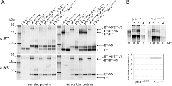
In most pestiviruses the Erns protein contains nine cysteines, eight of which form intramolecular disulfide bonds. The ninth cysteine residue is located close to the carboxy-terminal end at position 171 of the Erns protein (C438 or C441 in the polyprotein of CSFV or BVDV, respectively). This last cysteine is engaged in formation of Erns homodimers via disulfide bonds between two monomers of the protein (26). Since a very limited number of pestivirus isolates, most notably the BVDV-1 prototype strain NADL, do not contain C171, formation of Erns homodimers linked via this cysteine residue is not essential for pestivirus viability. To answer the question of whether Erns homodimers are in general dispensable for pestivirus propagation or whether noncovalently linked dimers are still present in the absence of C171, several Erns expression constructs were generated, coding for wt Erns of BVDV CP7 or a V5-tagged variant thereof, as well as constructs coding for mutants of these proteins with an arginine at position 171 instead of the cysteine. To determine the ability of these proteins to stably interact with each other, coprecipitation experiments were performed using a tagged variant and an untagged variant of Erns. The different proteins were transiently expressed alone or in combination in BHK-21 cells. Immunoprecipitated proteins were separated by SDS-PAGE under nonreducing conditions to preserve cystine-linked dimers (Fig. 1). All proteins were equally well expressed, and the presence of the V5 tag at the C terminus of the Erns protein did not prevent or severely impair dimer formation, since tagged as well as untagged Erns proteins containing C171 formed cysteine-linked dimers. The variants without the C171 migrated as monomers, as expected, in SDS-PAGE. The weak band visible for pB-ErnsC171R, which comigrates with the dimer band of pB-Erns (Fig. 1A, upper right panel), is unspecific, since this band is also visible when the corresponding tagged protein is expressed (Fig. 1A, upper right panel, pB-Erns-V5C171R).
FIG. 1.
(A) Coprecipitation experiments. Tagged (pB-Erns-V5 and pB-Erns-V5C171R) and untagged (pB-Erns and pB-Erns-C171R) variants of Erns from BVDV CP7 with or without C171 were (co)expressed in BHK-21 cells and metabolically labeled. Cells were lysed with a lysis buffer containing 1% Triton X-100 and proteins precipitated from the lysate (right panels) or the cell culture supernatant (left panels) with an Erns-specific serum (α-Erns, upper panels) or a V5 specific antibody (α-V5, lower panels) and deglycosylated before separation through SDS-PAGE under nonreducing conditions. The transfected plasmids or plasmid combinations are indicated at the top. The positions in the gel of the tagged and untagged monomers as well as the positions of the different dimers are indicated by arrows. The antisera used for precipitation are indicated on the left, together with the molecular masses of the size marker bands. (B) Determination of membrane binding/secretion of wt and mutant Erns. The upper part shows an example of SDS-PAGE under reducing conditions of the proteins precipitated from different fractions of cells transfected with expression plasmids given above the respective lanes. 1, secreted proteins; 2, debris; 3, membrane fraction; 4, soluble proteins. The lower panel shows for each construct the calculated level of membrane association in percent. Both constructs were tested at least three times, and the meridian, upper, and lower percentiles are shown. The wt membrane association is indicated by the horizontal line (mean of wt membrane association). The mutated protein was judged to have no significantly different membrane association compared to the wt Erns using the Welch test for unequal variances.
Coexpression of tagged and untagged Erns showed clear cystine-linked dimers of both variants, indicating that the tagged and untagged proteins can still interact with each other. It was not possible, however, to coprecipitate the untagged monomer with the anti-V5 antibody if one of the two proteins in the coexpression assays lacked C171 (Fig. 1A, lower right panel, first three lanes from the right). The detection of monomeric wt Erns after precipitation with the V5 antibody (Fig. 1A, lower right panel, lane 4 from the right) is most likely due to a low degree of reduction of disulfide-linked heterodimer after precipitation. These findings indicate that dimer formation depends entirely on the cystine structure or that the intermolecular binding force of dimers formed in the absence of the covalent linkage is not strong enough to withstand the lysis and precipitation conditions.
Analysis of the secreted proteins gave also no indication of coprecipitation of Erns monomers in the absence of C171. Secreted dimers were difficult to detect in some cases (Fig. 1A, left two panels). In general, dimers composed of tagged Erns seemed to be secreted slightly more efficiently than those composed of the wt protein. To analyze whether prevention of dimer formation exhibits by itself an influence on Erns membrane association/secretion, we fractionated cells expressing wt Erns or the C171R mutant and quantified the Erns proteins contained in the different fractions. Figure 1B shows a representative experiment and the result of a statistical analysis of at least three experiments, showing that there is no significant difference in membrane association/secretion between the two proteins.
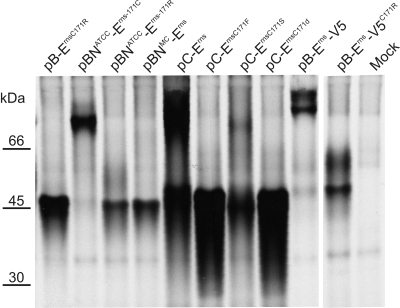
As cysteine 171 is widely conserved in different pestiviruses but notably lacking in the published BVDV NADL sequence (5), we decided to look at dimerization of Erns proteins of different viruses. The rationale behind this was that the Erns-coding sequences of naturally occurring viruses lacking C171 might have acquired mutations that stabilize the putative noncovalent homodimerization of the protein. Therefore, several additional expression constructs were generated, starting with the RNAs from two different NADL isolates and from the already-cloned cDNA sequences of CSFV Alfort/Tübingen (34). Sequence analysis of cDNA fragments revealed that one isolate of BVDV NADL (ATCC VR534) contained variants coding for cysteine or arginine at position 171, whereas the biologically cloned virus (MC [5]) expressed Erns that uniformly contained arginine at this position. The wt sequence of CSFV Alfort/Tübingen contains a cysteine, but mutants were established in which C171 was replaced by phenylalanine or serine or deleted. As described above, the proteins were transiently expressed, precipitated with an Erns-specific serum, and separated by SDS-PAGE under nonreducing conditions (Fig. 2). The gels showed clearly that none of the proteins lacking the C171 had formed disulfide-linked dimers.
FIG. 2.
Immunoprecipitation of Erns proteins from different pestiviruses. Proteins were transiently expressed in BHK-21 cells and metabolically labeled, and the cells were lysed with a lysis buffer containing 1% Triton X-100. Proteins were precipitated with an Erns-specific serum and the precipitates separated on SDS-PAGE under nonreducing conditions. The transfected plasmids are indicated above the lanes: pBNATCC-Erns-171C and pBNATCC-Erns-171R, Erns sequences from variants of BVDV NADL (ATCC VR534); pBNMC-Erns, Erns sequence derived from biologically cloned BVDV NADL (5); pC-Erns, Erns from CSFV Alfort/Tübingen, wt or with the indicated mutations. For the other constructs, see the legend to Fig. 1 and the text. Broad bands or double bands (pB-Erns-V5 and pB-Erns-V5C171R) are due to variations in the native glycosylation of the proteins.
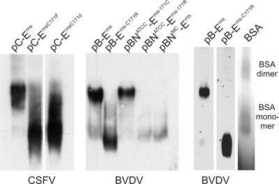
To further validate the results from the coprecipitation experiments, we tested for the ability to form dimers of Erns proteins in the presence or absence of C171 by a different approach. Cells expressing the different proteins were lysed under mild conditions in a lysis buffer containing 1% digitonin. The proteins in the resulting lysates were separated under native conditions in blue native PAGE with histidine (40), followed by transfer to nitrocellulose membranes under denaturing conditions. The Erns proteins were detected on the membrane with a specific rabbit anti-Erns serum (Fig. 3). The resulting Western blot showed that Erns dimers can be distinguished from the monomeric protein despite the glycosylation-dependent broad smear of the signals. Dimers were detected only in the lysates containing Erns with C171. The much larger amount of dimers versus monomers in these samples, compared to the results of the nonreducing SDS-PAGE (Fig. 1 and 2), is indicative of the mildness of the conditions used here for sample preparation. All mutants without C171 expressed only the monomeric form of the protein. The detection of BSA dimers (Fig. 3, rightmost lane) confirms that noncovalently linked protein complexes can be detected by the method used here. Thus, the results confirmed that the generation of stable Erns dimers is dependent on formation of the cystine bond between the monomers. Erns monomers will most likely interact via their protein interfaces even in the absence of C171, but the resulting association is obviously unstable, transient, or of such low affinity that it is disrupted even under very mild conditions as used for the coprecipitation experiments and the native PAGE.
FIG. 3.
Blue native PAGE of Erns proteins from different pestiviruses. Proteins were expressed in BHK-21 cells and lysed with a lysis buffer containing 1% digitonin. Proteins were separated through blue native PAGE with a histidine-containing buffer system (40) and blotted onto nitrocellulose membranes. The transfected plasmids are indicated above the lanes (see the legend to Fig. 2 for designations). Detection was carried out with an Erns-specific serum. On the right, the results of a control experiment are shown, with the left two lanes containing proteins expressed from pB-Erns and pB-Erns-C171R (equivalent to lanes 1 and 2 of the middle panel) and the right lane showing a Coomassie blue-stained BSA marker. The monomeric and dimeric bands of the marker protein are visible, corresponding to ca. 66 and 132 kDa, respectively.
Establishment and characterization of CSFV Erns dimerization-negative mutants.
In the above-described experiments, differences between the Erns from the field isolate BVDV NADL and the newly established mutated proteins from other pestiviruses were not observed. This indicates that Erns from NADL does not contain adaptive mutations allowing formation of stable noncovalently linked Erns homodimers. Accordingly, formation of stable dimers is not essential for BVDV. To further investigate this point, we established BVDV and CSFV mutants with alterations affecting the cysteine codon at position 171 of the Erns-coding sequence. These mutants were established in the genetic backgrounds of the BVDV isolate CP7 and the pathogenic CSFV strain Alfort/Tübingen. Three variants of the full-length CSFV cDNA clone were generated, two with point mutations leading to the exchange of phenylalanine or serine for cysteine (C171F or C171S, respectively) and a deletion mutant from which codon 171 was removed (C171d). Since the Erns protein of BVDV NADL contains an arginine at position 171, the BVDV CP7 sequence from construct pB-Erns-C171R was integrated into the full-length CP7 construct pA/BVDV (36) to give rise to pA/BVDV-Erns-C171R. Transfection of RNA transcribed in vitro from these constructs resulted in recovery of viable virus mutants. Reversion or second-site changes have not been found in the Erns-coding sequences of these mutants after seven tissue culture passages. Similar results have already been reported for a C171S mutant of the CSFV C strain (55).
For the CSFV mutants, growth curves were recorded in four independent experiments with wt Alfort/Tübingen and an RNase-negative mutant thereof (V-H297K) (57). As reported before, the RNase-negative mutant and the wt virus propagated equally well. The dimerization-negative mutant V-C171d showed slight growth retardation in the early phase of replication but reached final titers similar to those of the wt virus or the RNase-negative mutant (Fig. 4A). The growth rate of mutant V-C171F was basically identical to that of V-C171d and is therefore not shown in Fig. 4. We also established a full-length BVDV cDNA clone with a mutation affecting the Cys171 codon of the Erns gene, from which the BVDV dimerization-negative mutant CP7/C171R was recovered. Like the CSFV mutants, this BVDV mutant grew slightly slower than wt BVDV CP7 (not shown). It can therefore be concluded that mutations affecting C171 of the Erns protein reduce pestivirus replication to a limited extent. This agrees with published data for a C171S mutant of the CSFV C strain (55). Possible explanations for the titer reduction in dimerization-negative viruses could be reduced specific infectivity or decreased stability of the altered virions. Comparing the relationship of RNA content and infectious titers for wt virus and deletion mutant preparations revealed only small differences, showing that the mutant did not produce considerably increased amounts of noninfectious virus particles. Analysis of the inactivation kinetics of wt CSFV and the mutant V-C171d during incubation at 37°C revealed no significant differences, showing that the titer reduction is not due to lower stability of the virus mutant (Fig. 4B).
FIG. 4.
(A) Growth curves of the recombinant CSFV mutants and the wt virus. SK6 cells were infected with CSFV Alfort/Tübingen (wt), V-H297K (RNase negative), or V-C171d (dimerization negative) at an MOI of 0.1 and harvested by freezing and thawing at the indicated time points. Titers were determined by immunofluorescence staining with monoclonal antibody a18 at 72 h p.i. The curves represent the mean values from four individual experiments. Standard deviations are indicated. (B) Inactivation kinetics of wt CSFV and mutant V-C171d. Virus stocks (titers adjusted with tissue culture medium) were incubated at 37°C. Samples were taken for titration at the indicated time points. The graph shows the mean values from two independent experiments.
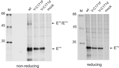
The inability of the C171 CSFV Erns mutants to dimerize after transient expression of the proteins has already been shown in Fig. 2 and 3. To demonstrate that Erns dimerization was indeed destroyed in the recovered virus mutants, porcine kidney (SK6) cells were infected with two of the CSFV mutants and the Erns protein analyzed after metabolic labeling and immunoprecipitation by SDS-PAGE for the presence of disulfide-linked dimers. To obtain sharper bands on the gel, the precipitates were deglycosylated with PNGase F prior to electrophoresis. As can be seen in Fig. 5, an Erns dimer band of about 50 kDa was detected for the wt virus, whereas the two mutants with changes at position 171 expressed only monomeric Erns. To prove that the 50-kDa band represented a disulfide-linked dimer, parts of the precipitates were also run under reducing conditions. As expected, the 50-kDa band observed before for wt CSFV was gone, and sharper bands of the monomeric protein were detected (Fig. 5, right panel).
FIG. 5.
Immunoprecipitation of Erns proteins from SK6 cells infected with different CSFVs. Infected cells were metabolically labeled and the cells lysed with a lysis buffer containing 1% Triton X-100. Proteins were precipitated with an Erns-specific serum and treated with PNGase F, and the deglycosylated precipitates were separated by SDS-PAGE under nonreducing (left) or reducing (right) conditions. The viruses used for infection are indicated above the lanes. The positions of protein size marker bands are given on the left.
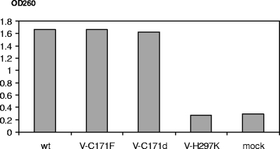
It was published before that both monomeric and dimeric Erns represent active RNases (49). Sequence comparison and structure modeling revealed that the region of the protein showing homology to T2 RNases is made up by the amino-terminal two-thirds of Erns and does not include the C-terminal part of the protein, which seems to represent a separate domain that might adopt different structures (26) and notably contains the membrane anchor (51). However, it could not be excluded that the mutations introduced in the C-terminal region here nevertheless influenced the RNase function of the protein. For an Erns C171S mutant expressed in the baculovirus system, somewhat reduced RNase activity was reported. The significance of this finding, however, is questionable, since another fraction of the enzyme preparation showed wt activity (55). To address this question for virus-infected cells, extracts of cells infected with wt or mutant CSFV were tested for enzymatic activity in an RNase assay. As is obvious from Fig. 6, wt CSFV as well as the mutants V-C171F and V-C171d showed comparable RNase activity, whereas the mutant with an exchange of one of the RNase active-site histidine residues (V-H297K) was equivalent to the mock control.
FIG. 6.
Determination of RNase activity present in crude extracts of SK6 cells infected with wt CSFV Alfort/Tübingen or the recombinant virus V-C171F (dimerization negative), V-C171d (dimerization negative), or V-H297K (RNase negative). Noninfected SK6 cells served as a negative control (mock). The enzymatic degradation of poly(U) was determined by measuring the optical density at 260 nm (OD260) as a marker of the release of small acid-soluble RNA fragments (27, 47).
V-C171d exhibits an attenuated phenotype in the natural host.
To see if the prevention of Erns dimerization showed any obvious effect in the natural host, we performed an infection experiment in pigs. The objective of this experiment was to compare clinical signs observed after infection with the CSFV Alfort/Tübingen mutant V-C171d with those seen in wt CSFV Alfort/Tübingen-infected animals. The latter virus was recovered from the infectious cDNA clone that was used for construction of the virus mutant. As a further control, an RNase-negative variant of Alfort/Tübingen (V-H297K) was used, which exhibited a significantly attenuated phenotype in earlier experiments (57). Twelve animals with weight of about 20 kg were split in three groups of four animals each. The viruses were applied via the intramuscular and intranasal routes on day 0 (group 1, V-H297K; group 2, V-C171d; group 3, wt virus). After infection, animals were monitored for general health status for 22 days, and rectal temperatures were recorded daily or, in the late stage of the experiment, every second day. Blood was taken from the jugular vein for buffy coat isolation, leukocyte counting, fluorescence-activated cell sorting (FACS) analysis, and serum preparation.
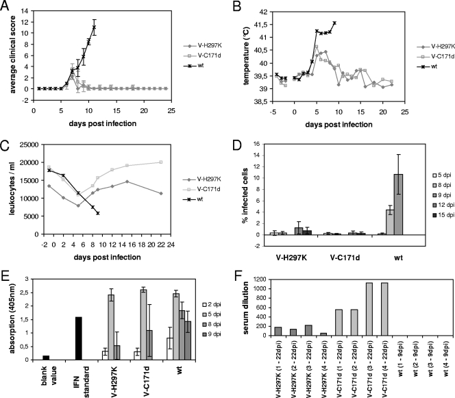
The animals of group 3, infected with the wt virus, had to be killed prematurely on day 9 p.i. because of severe signs of disease typical for CSF. The animals of groups 1 and 2, infected with the RNase-negative mutant and the dimerization-incompetent mutant, respectively, showed only mild signs of disease during a short period of time, as demonstrated by the clinical scores (Fig. 7A). The difference between wt and mutant viruses is also obvious from the body temperature curves (Fig. 7B). In groups 1 and 2, the animals developed mild fever (temperature of just above 40°C) for several days starting around day 4 p.i. In contrast, animals infected with wt virus reached higher temperatures, and no decrease was detectable until euthanasia on day 9 p.i.
FIG. 7.
Results of an animal experiment in which groups of four pigs were infected with the RNase-negative CSFV V-H297K (group 1), the dimerization-negative mutant V-C171d (group 2), or wt Alfort/Tübingen recovered from the infectious cDNA clone (group 3). Clinical scores, according to the system published by Mittelholzer et al. (38), are given as mean values for each group (A). Also shown are the average body temperature (B), the mean values of leukocyte counts (C), the percentage of virus-positive cells in complete blood samples (as described in reference 57) (D), the amount of type I IFN in plasma samples compared to an IFN-α standard (1 ng per well) (E), and the titers of virus-neutralizing serum antibodies of the pigs, given as the maximal reciprocal dilution of the sera that is able to neutralize 100 50% tissue culture infective doses of CSFV Alfort/Tübingen (F). Standard deviations are indicated in panels A, D, and E.
The degree of leukopenia and the virus load in the blood of infected pigs represent reliable parameters for the virulence of CSFV isolates. We therefore determined leukocyte numbers and looked for viremia through preparation of buffy coats and virus isolation on tissue culture cells. All animals showed leukopenia, but in the pigs infected with the mutants, the decrease was considerably less pronounced and the numbers of white blood cells increased again after day 5 p.i. (Fig. 7C). Similarly, viremia was detected for all animals, but once again virus-positive cells were only transiently detected in the animals infected with the mutants, whereas wt virus was present until termination (Table 1). To gain further insight into the degree of virus load, the amount of virus-positive cells in whole blood samples was determined via FACS analysis (Fig. 7D). This analysis allows a simple quantification of the virus load but is less sensitive than the virus isolation from purified buffy coats. Virus-positive cells could not definitely be detected with this method in the animals infected with the mutants, but all animals infected with wt CSFV were found to be clearly positive, containing up to 14% of virus-positive white blood cells. Thus, it is obvious that the virus load is considerably reduced in the animals infected with the mutants.
TABLE 1.
Detection of CSFV in the blood of infected animals
| Time of bleeding (day p.i.) | Detection of CSFVa in blood from the indicated animal
|
|||||||||||
|---|---|---|---|---|---|---|---|---|---|---|---|---|
| V-H297K
|
V-C171d
|
wt
|
||||||||||
| 841 | 842 | 843 | 844 | 847 | 848 | 849 | 850 | 574 | 576 | 593 | 598 | |
| −1 | −/− | −/− | −/− | −/− | −/− | −/− | −/− | −/− | −/− | −/− | −/− | −/− |
| 2 | −/− | −/− | −/− | −/− | −/− | −/− | −/− | −/− | −/− | −/− | −/− | −/− |
| 5 | +/− | −/− | +/− | +/− | +/− | +/− | −/− | +/+ | +/+ | +/+ | +/+ | +/+ |
| 8 | +/− | −/− | +/+ | +/+ | +/+ | −/− | +/+ | +/+ | +/+ | +/+ | +/+ | +/+ |
| 9 | ND | ND | ND | ND | ND | ND | ND | ND | +/+ | +/+ | +/+ | +/+ |
| 12 | −/− | −/− | −/− | −/− | −/− | −/− | −/− | −/− | ||||
| 15 | −/− | −/− | −/− | −/− | −/− | −/− | −/− | −/− | ||||
| 22 | −/− | −/− | −/− | −/− | −/− | −/− | −/− | −/− | ||||
Viruses used for infection: V-H297K, RNase-negative virus (group 1); V-C171d, dimerization-negative mutant (group 2); wt, CSFV Alfort/Tübingen (group 3). The animals in group 3 had to be euthanized on day 9 p.i.; blood samples were taken on this day only from these animals. +/+, virus positive in both wells of culture plate; +/−, only one well positive; −/−, both wells negative; ND, not done.
Both in vitro analyses and experiments with BVDV-infected fetuses showed that Erns is involved in pestiviral interference with the type I IFN response to virus infection (21, 31, 33). It was therefore interesting to investigate whether differences in the type I IFN levels could be detected in the blood of infected pigs from groups 1, 2, and 3. Plasma samples obtained during preparation of buffy coats were tested in an Mx promoter/chloramphenicol acetyltransferase reporter assay (11). The blood of all animals contained significant amounts of IFN on day 5 p.i., and in all cases a decrease of these values was observed on days 8 and 9 (day 9 was tested only for wt virus-infected animals) (Fig. 7E). Importantly, wt virus-infected animals induced a type I IFN response in the animals, and there was no significant difference between the three groups. This finding is reminiscent of results published for noncytopathogenic BVDV, which also induces an IFN response in acutely infected animals, despite the presence of Npro and Erns RNase (2).
To get an impression of the efficiency of virus replication in the animals, the development of CSFV-neutralizing antibodies was analyzed. The animals from group 3 infected with the wt virus did not live long enough to mount a significant antibody response, and accordingly, neutralization activity was not detected (Fig. 7F). However, the sera taken at day 22 p.i. from the animals infected with the mutants showed efficient neutralization activity. Interestingly, the neutralization titers determined for the animals infected with the dimerization-negative mutant were considerably higher than those for the pigs infected with the RNase-negative mutant, which is an indication of better replication of the former virus in the animal.
Attenuation is also observed for the point mutant V-C171S.
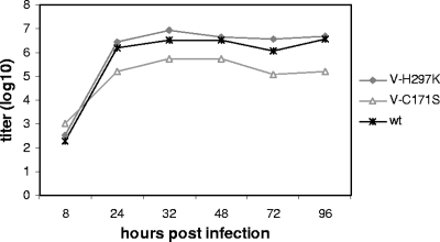
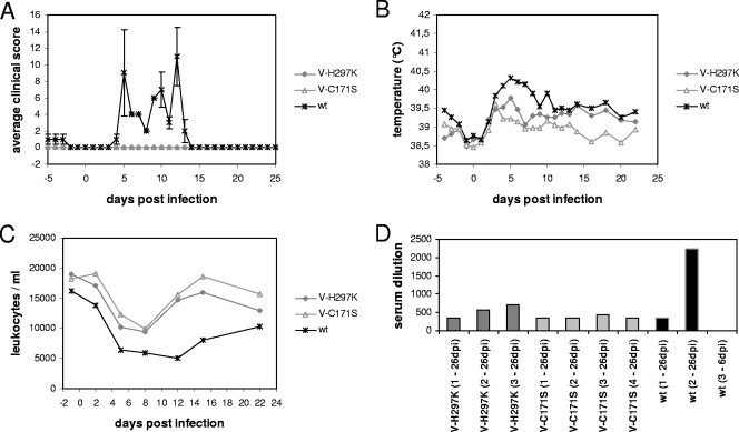
To exclude side effects that might result from the structural impact of the deletion of Cys 171, a further animal experiment was conducted with a mutant carrying a less invasive change. We decided to use the point mutant V-C171S, since the S-for-C exchange represents the most conservative mutation that can abolish disulfide-dependent dimer formation (Fig. 2). Despite the conservative exchange, the point mutation induced a reduction of growth rate similar to that detected for the deletion mutant (Fig. 8). Infection was done as described above. Back titration of the residual virus material brought back from the stable showed that the control groups infected with wt and RNase-negative viruses had received slightly less virus than expected (105.4 instead of 106 per animal), whereas the animals of group 2 were infected with nearly the intended dose (105.8 instead of 106 per animal) of V-C171S. Clinical scores, temperatures, and leukocyte numbers were recorded as described above (Fig. 9). The signs of disease in the wt controls were less prominent than in the first experiment but were clearly detected. One animal of this group was found dead at day 5 p.i. Pathological evaluation revealed that the animal most likely died of heart and circulation failure induced by a serious stenosis of the aorta valve that presumably resulted from an earlier infection. In contrast to the wt controls, neither the RNase-negative nor the dimerization-negative viruses induced any signs of disease in the animals (Fig. 9A). Only a transient temperature elevation soon after infection indicated the presence of the virus in these animals (Fig. 9B). Viremia was also transient in these animals, with one pig in each group in which virus was not detected at all (Table 2). Viremia lasted longer in the animals infected with the wt virus, and these pigs also showed a more pronounced reduction of leukocyte numbers than the animals infected with either of the mutants (Fig. 9C). Nevertheless, the pigs of all three groups were clearly productively infected, since they mounted a considerable neutralizing antibody response, with titers of at least 1:355 at day 21 p.i. (Fig. 9D). Thus, even though the animals infected with the wt virus developed less severe signs of disease in this experiment, it can be concluded that a point mutant with an exchange of Ser for Cys at position 171 is also considerably attenuated. This finding strongly supports the conclusion that prevention of Erns dimerization leads to attenuation of CSFV.
FIG. 8.
Growth curve of the recombinant CSFV mutant V-C171S (dimerization negative) compared with those of the wt virus Alfort/Tübingen (wt) and mutant V-H297K (RNase negative). SK6 cells were infected with the CSFV variants at an MOI of 0.1 and harvested by freezing and thawing at the indicated time points. Titers were determined by immunofluorescence staining with monoclonal antibody a18 at 72 h p.i. The curves represent the mean values from two individual experiments.
FIG. 9.
Results of an animal experiment in which pigs were infected with the RNase-negative CSFV V-H297K (group 1, 3 pigs), the dimerization-negative point mutant V-C171S (group 2, 4 pigs) or wt Alfort/Tübingen recovered from the infectious cDNA clone (group 3, 3 pigs). (A to C) Mean values of clinical scores (A), body temperature (B), and leukocyte numbers (C) for each group of animals. (D) Titers of virus-neutralizing serum antibodies of the pigs are given as the maximal reciprocal dilution of the sera that are able to neutralize 65 TCID50 of CSFV Alfort/Tübingen. Standard deviations are indicated in panel A.
TABLE 2.
Detection of CSFV in the blood of infected animals
| Time of bleeding (day p.i.) | Detection of CSFVa in blood from the indicated animal
|
|||||||||
|---|---|---|---|---|---|---|---|---|---|---|
| V-H297K\
|
V-C171S
|
wt
|
||||||||
| 1186 | 1187 | 1188 | 1190 | 1191 | 1192 | 1193 | 1196 | 1197 | 1198 | |
| −1 | −/− | −/− | −/− | −/− | −/− | −/− | −/− | −/− | −/− | −/− |
| 2 | −/− | −/− | −/− | −/− | −/− | −/− | −/− | −/− | −/− | −/− |
| 5 | −/− | −/− | −/− | −/− | −/− | −/− | −/− | +/+ | +/+ | +/+ |
| 8 | +/+ | +/+ | −/− | +/+ | −/− | +/+ | +/− | +/+ | +/+ | |
| 12 | +/− | +/− | −/− | −/− | −/− | −/− | −/− | +/+ | +/+ | |
| 15 | −/− | −/− | −/− | −/− | −/− | −/− | −/− | +/+ | −/− | |
| 21 | −/− | −/− | −/− | −/− | −/− | −/− | −/− | −/− | −/− | |
Viruses used for infection: V-H297K, RNase-negative virus (group 1); V-C171S, dimerization-negative mutant (group 2); wt, CSFV Alfort/Tübingen (group 3). One animal in group 3 had to be euthanized on day 5 p.i. +/+, virus positive in both wells of culture plate; +/−, only one well positive; −/−, both wells negative.
DISCUSSION
Pestiviruses have evolved unique mechanisms to interfere with the host's immune response in order to persist within their host populations. As best understood for BVDV, establishment of persistent infection represents a key point in this strategy. Transmission of virus to the fetus upon infection of pregnant host animals early in pregnancy eliminates the virus-specific adaptive immune response, since the virus is regarded as self antigen by the immune system (52). In addition, recent studies have shown that the two viral proteins Npro and Erns have the ability to repress the host cellular type I IFN response and thereby interfere with innate immune responses to pestivirus infection (13, 16, 21, 27, 33, 43, 44). Experiments with pregnant animals revealed that both of these viral functions are important for establishment of persistent BVDV infections (33). Transplacental transmission of CSFV is also commonly observed after infection of pregnant sows, but the requirements for viral persistence in piglets and basic features of the persistent infection are much less defined for CSFV (39, 52).
Npro interacts both with the IFN regulatory factor 3 (1, 16, 50) and the α form of inhibitor of nuclear factor κB (IκBα) (9). While the function of the latter is not fully understood, the interaction with IFN regulatory factor 3 results in proteasomal degradation of this factor and thereby repression of the early type I IFN response of the infected cell.
The role of Erns with regard to interference with the innate immune response is much less clear. It has been put forward that secreted Erns could be responsible for blocking an IFN response to extracellular dsRNA. Evidence for this hypothesis was found in in vitro experiments, and it was shown that the RNase activity of Erns is essential for this function (21, 31). However, the putative origin of this so-far hypothetical extracellular RNA is obscure, and it might be that Erns (also) serves other functions.
BS RNase represents a secreted cytotoxic RNase with a high degree of sequence similarity with pancreatic RNase A. In contrast to the monomeric RNase A, BS RNase forms homodimers, and it was shown that only the dimers were biologically active even though the monomers were still active RNases. Moreover, synthetic homodimerization of RNase A rendered this enzyme cytotoxic (3, 22, 23, 28). It was therefore interesting to analyze the role of dimerization in the function of the pestivirus Erns. Since a few naturally occurring viruses express Erns proteins that lack C171, it was clear that disulfide-mediated dimerization of the protein is not necessary for virus viability. As reported previously for CSFV (55) and reported here for the first time for BVDV, viable viruses can also be obtained when the C171 codon is mutated in viruses that usually display this residue in their Erns proteins. The mutant viruses showed only rather weak growth retardation, and passages in tissue culture did not reveal a tendency for reversion or second-site mutations. Taken together, our results indicate that no adaptive mutations are necessary to allow virus growth in tissue culture cells in the absence of C171 and that changes affecting this residue do not represent a significant disadvantage for the viruses in tissue culture.
Stable protein dimerization can also occur without disulfide bonds. As a typical example, the glycoproteins E1 and E2 of human HCV form stable noncovalently linked heterodimers (30). It was therefore important to test whether pestivirus Erns is still able to form homodimers when C171 is absent. In our experiments we did not find indications for dimer formation when disulfide bonding was prevented by mutation of residue 171. This was also true for BVDV NADL, a naturally occurring isolate encoding an Erns with arginine at position 171. We employed two different experimental setups to analyze this point, but even though we used very mild conditions for cell lysis and sample preparation, neither coprecipitation analysis nor native protein gel electrophoresis indicated the presence of dimers. It therefore seems unlikely that Erns homodimers that are stable under biologically relevant conditions can be formed in the absence of C171.
Taken together, our experiments showed that formation of stable Erns dimers is not necessary for virus viability. Moreover, the ability to establish these homodimers seems to represent no significant advantage for the viruses during tissue culture propagation. Nevertheless, the cysteine codon at position 171 of the Erns-coding sequence and, thus, the ability to express Erns homodimers has been conserved during the evolution of pestiviruses, since the vast majority of isolates contain this codon. As a matter of fact, there are only very few pestivirus sequences in the databases with altered sequences at this position. In addition to BVDV NADL, two further BVDV-1 sequences, ILLC and Ind S1449, as well as the CSFV vaccine strains Riems and LPC, were found to lack cysteine at position 171 (Table 3). Interestingly, for the CSFV vaccine strains and BVDV NADL there exist also GenBank entries containing C171, indicating that there could be different variants in the isolates, as we found for the isolate BVDV NADL ATCC VR534. The high degree of conservation of C171 in pestiviruses indicates that Erns homodimers have a positive effect for the virus during replication in its natural host. Clear evidence that this idea is correct was obtained in our animal experiment, which showed that the deletion or mutation of cysteine 171 and thereby the prevention of Erns dimer formation leads to considerable attenuation of CSFV. This attenuation can hardly be due to a general growth retardation of the virus mutant, since only a slight reduction of the growth rates was found for the dimerization-negative mutants in comparison with wt virus. Mutation of C171 to serine was reported to influence the interaction of the protein with heparin/heparan sulfate-like polysaccharide chains (55). The significance of this finding is difficult to interpret, since CSFV dependency on such polysaccharides was reported to result from in vitro propagation and to be without in vivo relevance (19, 20, 56). Thus, even though side effects of the deletion or exchange of C171 not related to loss of dimerization cannot be formally excluded, it is very likely that the virus attenuation observed here is due to the absence of stable Erns dimers.
TABLE 3.
Pestivirus sequences in databases that show replacement of Cys 171
| Accession no. | Virus strain | Comment(s) |
|---|---|---|
| NC_001461 | NADL | Mildly virulent; some sequences for this virus contain a cysteine codon at position 171 |
| U86599 and U86600 | BVDV-1 ILLC and ILLNC | Virus pair isolated from an American animal with mucosal disease |
| AY911670 | BVDV-1 Ind S1449 | Virus isolated from an Indian cow |
| AY259122 | CSFV Riems | Vaccine strain, some sequences for this virus contain a cysteine codon at position 171 |
| AF352565 | CSFV LPC | Vaccine strain, some sequences for this virus contain a cysteine codon at position 171 |
The development and intensity of the clinical scores and fever curves as well as the degree of leukopenia were very similar for the virus mutant lacking the Erns RNase activity and the variants with the deletion or mutation preventing stable Erns dimer formation. Importantly, the dimerization-negative mutants induced in the animals levels of neutralizing antibodies comparable to those induced by the RNase-negative mutant, so efficient replication of the virus in the pigs is very likely. As mentioned above, dimerization has been found to be crucial for the biological functions but not enzymatic activity of BS RNase. There is good evidence that the dimeric form of the enzyme is not efficiently blocked by intracellular RNase inhibitors, so that internalized BS RNase provokes cell death as a consequence of degradation of cellular RNA (22, 28). In the light of these results, it is tempting to speculate that the attenuation of CSFV as a consequence of the prevention of Erns dimer formation and the abrogation of the RNase activity rely on the same principles of the virus-host interaction, namely, preventing the RNase from becoming active at its so-far-unknown place of biological function. Thus, the prevention of dimer formation could lead to effects equivalent to abrogation of the RNase activity, even though the virus is able to express an active RNase as shown in our enzymatic tests.
The mechanisms behind the function of pestivirus Erns with regard to virus virulence and establishment of persistent infections are still obscure. It has been shown that the RNase activity of the protein is important for this function, but the genuine substrate of the enzyme has not yet been identified. The present work indicates that the RNase activity of the protein alone is not sufficient for its virulence factor function but that the enzyme obviously has to be present as a homodimer. This finding adds a new facet to this fascinating scenario of virus-host interplay.
Acknowledgments
We thank Maren Ziegler, Janett Wieseler, and Petra Wulle for excellent technical assistance. We are especially grateful to Heiner Voigt and Gaby Kuebart for help with the FACS analyses.
This study was supported by grants from Boehringer Ingelheim Vetmedica GmbH and the Deutsche Forschungsgemeinschaft (DFG-ME1367/4).
Footnotes
Published ahead of print on 4 March 2009.
REFERENCES
- 1.Bauhofer, O., A. Summerfield, Y. Sakoda, J. D. Tratschin, M. A. Hofmann, and N. Ruggli. 2007. Classical swine fever virus Npro interacts with interferon regulatory factor 3 and induces its proteasomal degradation. J. Virol. 813087-3096. [DOI] [PMC free article] [PubMed] [Google Scholar]
- 2.Brackenbury, L. S., B. V. Carr, and B. Charleston. 2003. Aspects of the innate and adaptive immune responses to acute infections with BVDV. Vet. Microbiol. 96337-344. [DOI] [PubMed] [Google Scholar]
- 3.Ciglic, M. I., P. J. Jackson, S. A. Raillard, M. Haugg, T. M. Jermann, J. G. Opitz, N. Trabesinger-Ruf, and S. A. Benner. 1998. Origin of dimeric structure in the ribonuclease superfamily. Biochemistry 374008-4022. [DOI] [PubMed] [Google Scholar]
- 4.Cocquerel, L., C. Wychowski, F. Minner, F. Penin, and J. Dubuisson. 2000. Charged residues in the transmembrane domains of hepatitis C virus glycoproteins play a major role in the processing, subcellular localization, and assembly of these envelope proteins. J. Virol. 743623-3633. [DOI] [PMC free article] [PubMed] [Google Scholar]
- 5.Colett, M. S., R. Larson, C. Gold, D. Strick, D. K. Anderson, and A. F. Purchio. 1988. Molecular cloning and nucleotide sequence of the pestivirus bovine viral diarrhea virus. Virology 165191-199. [DOI] [PubMed] [Google Scholar]
- 6.Corapi, W. V., R. O. Donis, and E. J. Dubovi. 1990. Characterization of a panel of monoclonal antibodies and their use in the study of the antigenic diversity of bovine viral diarrhea virus. Am. J. Vet. Res. 511388-1394. [PubMed] [Google Scholar]
- 7.Deleersnyder, V., A. Pillez, C. Wychowski, K. Blight, J. Xu, Y. S. Hahn, C. M. Rice, and J. Dubuisson. 1997. Formation of native hepatitis C virus glycoprotein complexes. J. Virol. 71697-704. [DOI] [PMC free article] [PubMed] [Google Scholar]
- 8.Devereux, J., P. Haeberli, and O. A. Smithies. 1984. A comprehensive set of sequence analysis programs for the VAX. Nucleic Acids Res. 12387-395. [DOI] [PMC free article] [PubMed] [Google Scholar]
- 9.Doceul, V., B. Charleston, H. Crooke, E. Reid, P. P. Powell, and J. Seago. 2008. The Npro product of classical swine fever virus interacts with IκBα, the NF-κB inhibitor. J. Gen. Virol. 891881-1889. [DOI] [PubMed] [Google Scholar]
- 10.Fetzer, C., B. A. Tews, and G. Meyers. 2005. The carboxy-terminal sequence of the pestivirus glycoprotein E(rns) represents an unusual type of membrane anchor. J. Virol. 7911901-11913. [DOI] [PMC free article] [PubMed] [Google Scholar]
- 11.Fray, M. D., G. E. Mann, and B. Charleston. 2001. Validation of an Mx/CAT reporter gene assay for the quantification of bovine type-I interferon. J. Immunol. Methods 249235-244. [DOI] [PubMed] [Google Scholar]
- 12.Fricke, J., C. Voss, M. Thumm, and G. Meyers. 2004. Processing of a pestivirus protein by a cellular protease specific for light chain 3 of microtubule-associated proteins. J. Virol. 785900-5912. [DOI] [PMC free article] [PubMed] [Google Scholar]
- 13.Gil, L. H., I. H. Ansari, V. Vassilev, D. Liang, V. C. Lai, W. Zhong, Z. Hong, E. J. Dubovi, and R. O. Donis. 2006. The amino-terminal domain of bovine viral diarrhea virus Npro protein is necessary for alpha/beta interferon antagonism. J. Virol. 80900-911. [DOI] [PMC free article] [PubMed] [Google Scholar]
- 14.Hausmann, Y., G. Roman-Sosa, H. J. Thiel, and T. Rümenapf. 2004. Classical swine fever virus glycoprotein E rns is an endoribonuclease with an unusual base specificity. J. Virol. 785507-5512. [DOI] [PMC free article] [PubMed] [Google Scholar]
- 15.Heinz, F. X., M. S. Collett, R. H. Purcell, E. A. Gould, C. R. Howard, M. Houghton, J. M. Moormann, C. M. Rice, and H.-J. Thiel. 2000. Family Flaviviridae, p. 859-878. In M. H. V. van Regenmortel, C. M. Fauquet, D. H. L. Bishop, E. B. Carstens, M. K. Estes, S. M. Lemon, J. Maniloff, M. A. Mayo, D. J. McGeoch, C. R. Pringle, and R. B. Wickner (ed.), Virus taxonomy: classification and nomenclature of viruses. Seventh report of the International Committee on Taxonomy of Viruses. Academic Press, San Diego, CA.
- 16.Hilton, L., K. Moganeradj, G. Zhang, Y. H. Chen, R. E. Randall, J. W. McCauley, and S. Goodbourn. 2006. The NPro product of bovine viral diarrhea virus inhibits DNA binding by interferon regulatory factor 3 and targets it for proteasomal degradation. J. Virol. 8011723-11732. [DOI] [PMC free article] [PubMed] [Google Scholar]
- 17.Horiuchi, H., K. Yanai, M. Takagi, K. Yano, E. Wakabayashi, A. Sanda, S. Mine, K. Ohgi, and M. Irie. 1988. Primary structure of a base non-specific ribonuclease from Rhizopus niveus. J. Biochem. (Tokyo) 103408-418. [DOI] [PubMed] [Google Scholar]
- 18.Hulst, M. M., G. Himes, E. Newbigin, and R. J. M. Moormann. 1994. Glycoprotein E2 of classical swine fever virus: expression in insect cells and identification as a ribonuclease. Virology 200558-565. [DOI] [PubMed] [Google Scholar]
- 19.Hulst, M. M., H. G. van Gennip, and R. J. Moormann. 2000. Passage of classical swine fever virus in cultured swine kidney cells selects virus variants that bind to heparan sulfate due to a single amino acid change in envelope protein E(rns). J. Virol. 749553-9561. [DOI] [PMC free article] [PubMed] [Google Scholar]
- 20.Hulst, M. M., H. G. van Gennip, A. C. Vlot, E. Schooten, A. J. de Smit, and R. J. Moormann. 2001. Interaction of classical swine fever virus with membrane-associated heparan sulfate: role for virus replication in vivo and virulence. J. Virol. 759585-9595. [DOI] [PMC free article] [PubMed] [Google Scholar]
- 21.Iqbal, M., E. Poole, S. Goodbourn, and J. W. McCauley. 2004. Role for bovine viral diarrhea virus Erns glycoprotein in the control of activation of beta interferon by double-stranded RNA. J. Virol. 78136-145. [DOI] [PMC free article] [PubMed] [Google Scholar]
- 22.Kim, J. S., J. Soucek, J. Matousek, and R. T. Raines. 1995. Mechanism of ribonuclease cytotoxicity. J. Biol. Chem. 27031097-31102. [DOI] [PubMed] [Google Scholar]
- 23.Kim, J. S., J. Soucek, J. Matousek, and R. T. Raines. 1995. Structural basis for the biological activities of bovine seminal ribonuclease. J. Biol. Chem. 27010525-10530. [DOI] [PubMed] [Google Scholar]
- 24.König, M., T. Lengsfeld, T. Pauly, R. Stark, and H.-J. Thiel. 1995. Classical swine fever virus: independent induction of protective immunity by two structural glycoproteins. J. Virol. 696479-6486. [DOI] [PMC free article] [PubMed] [Google Scholar]
- 25.Lackner, T., A. Muller, A. Pankraz, P. Becher, H. J. Thiel, A. E. Gorbalenya, and N. Tautz. 2004. Temporal modulation of an autoprotease is crucial for replication and pathogenicity of an RNA virus. J. Virol. 7810765-10775. [DOI] [PMC free article] [PubMed] [Google Scholar]
- 26.Langedijk, J. P., P. A. van Veelen, W. M. Schaaper, A. H. de Ru, R. H. Meloen, and M. M. Hulst. 2002. A structural model of pestivirus E(rns) based on disulfide bond connectivity and homology modeling reveals an extremely rare vicinal disulfide. J. Virol. 7610383-10392. [DOI] [PMC free article] [PubMed] [Google Scholar]
- 27.La Rocca, S. A., R. J. Herbert, H. Crooke, T. W. Drew, T. E. Wileman, and P. P. Powell. 2005. Loss of interferon regulatory factor 3 in cells infected with classical swine fever virus involves the N-terminal protease, Npro. J. Virol. 797239-7247. [DOI] [PMC free article] [PubMed] [Google Scholar]
- 28.Lee, J. E., and R. T. Raines. 2005. Cytotoxicity of bovine seminal ribonuclease: monomer versus dimer. Biochemistry 4415760-15767. [DOI] [PubMed] [Google Scholar]
- 29.Lindenbach, B. D., and C. M. Rice. 2001. Flaviviridae: the viruses and their replication, p. 991-1042. In D. M. Knipe and P. M. Howley (ed.), Fields virology, vol. 1. Lippincott-Raven Publishers, Philadelphia, PA. [Google Scholar]
- 30.Lindenbach, B. D., H.-J. Thiel, and C. M. Rice. 2007. Flaviviridae: the viruses and their replication, p. 1101-1152. In D. M. Knipe and P. M. Howley (ed.), Fields virology, vol. 1. Lippincott-Raven Publishers, Philadelphia, PA. [Google Scholar]
- 31.Magkouras, I., P. Matzener, T. Rumenapf, E. Peterhans, and M. Schweizer. 2008. RNase-dependent inhibition of extracellular, but not intracellular, dsRNA-induced interferon synthesis by Erns of pestiviruses. J. Gen. Virol. 892501-2506. [DOI] [PubMed] [Google Scholar]
- 32.Meyer, C., M. Von Freyburg, K. Elbers, and G. Meyers. 2002. Recovery of virulent and RNase-negative attenuated type 2 bovine viral diarrhea viruses from infectious cDNA clones. J. Virol. 768494-8503. [DOI] [PMC free article] [PubMed] [Google Scholar]
- 33.Meyers, G., A. Ege, C. Fetzer, F. M. von, K. Elbers, V. Carr, H. Prentice, B. Charleston, and E. M. Schürmann. 2007. Bovine viral diarrhea virus: prevention of persistent fetal infection by a combination of two mutations affecting the Erns RNase and the Npro protease. J. Virol. 813327-3338. [DOI] [PMC free article] [PubMed] [Google Scholar]
- 34.Meyers, G., T. Rümenapf, and H.-J. Thiel. 1989. Molecular cloning and nucleotide sequence of the genome of hog cholera virus. Virology 171555-567. [DOI] [PubMed] [Google Scholar]
- 35.Meyers, G., A. Saalmüller, and M. Büttner. 1999. Mutations abrogating the RNase activity in glycoprotein E(rns) of the pestivirus classical swine fever virus lead to virus attenuation. J. Virol. 7310224-10235. [DOI] [PMC free article] [PubMed] [Google Scholar]
- 36.Meyers, G., N. Tautz, P. Becher, H.-J. Thiel, and B. Kümmerer. 1996. Recovery of cytopathogenic and noncytopathogenic bovine viral diarrhea viruses from cDNA constructs. J. Virol. 708606-8613. [DOI] [PMC free article] [PubMed] [Google Scholar]
- 37.Meyers, G., H.-J. Thiel, and T. Rümenapf. 1996. Classical swine fever virus: recovery of infectious viruses from cDNA constructs and generation of recombinant cytopathogenic defective interfering particles. J. Virol. 701588-1595. [DOI] [PMC free article] [PubMed] [Google Scholar]
- 38.Mittelholzer, C., C. Moser, J. D. Tratschin, and M. A. Hofmann. 2000. Analysis of classical swine fever virus replication kinetics allows differentiation of highly virulent from avirulent strains. Vet. Microbiol. 74293-308. [DOI] [PubMed] [Google Scholar]
- 39.Moennig, V., and P. G. W. Plagemann. 1992. The pestiviruses. Adv. Virus Res. 4153-98. [DOI] [PubMed] [Google Scholar]
- 40.Niepmann, M., and J. Zheng. 2006. Discontinuous native protein gel electrophoresis. Electrophoresis 273949-3951. [DOI] [PubMed] [Google Scholar]
- 41.Op De Beeck, A., R. Montserret, S. Duvet, L. Cocquerel, R. Cacan, B. Barberot, M. M. Le, F. Penin, and J. Dubuisson. 2000. The transmembrane domains of hepatitis C virus envelope glycoproteins E1 and E2 play a major role in heterodimerization. J. Biol. Chem. 27531428-31437. [DOI] [PubMed] [Google Scholar]
- 42.Reimann, I., I. Semmler, and M. Beer. 2007. Packaged replicons of bovine viral diarrhea virus are capable of inducing a protective immune response. Virology 366377-386. [DOI] [PubMed] [Google Scholar]
- 43.Rüggli, N., B. H. Bird, L. Liu, O. Bauhofer, J. D. Tratschin, and M. A. Hofmann. 2005. N(pro) of classical swine fever virus is an antagonist of double-stranded RNA-mediated apoptosis and IFN-alpha/beta induction. Virology 340265-276. [DOI] [PubMed] [Google Scholar]
- 44.Rüggli, N., J. D. Tratschin, M. Schweizer, K. C. McCullough, M. A. Hofmann, and A. Summerfield. 2003. Classical swine fever virus interferes with cellular antiviral defense: evidence for a novel function of N(pro). J. Virol. 777645-7654. [DOI] [PMC free article] [PubMed] [Google Scholar]
- 45.Rümenapf, T., R. Stark, M. Heimann, and H. J. Thiel. 1998. N-terminal protease of pestiviruses: identification of putative catalytic residues by site-directed mutagenesis. J. Virol. 722544-2547. [DOI] [PMC free article] [PubMed] [Google Scholar]
- 46.Rümenapf, T., G. Unger, J. H. Strauss, and H.-J. Thiel. 1993. Processing of the envelope glycoproteins of pestiviruses. J. Virol. 673288-3295. [DOI] [PMC free article] [PubMed] [Google Scholar]
- 47.Sainz, I. F., L. G. Holinka, Z. Lu, G. R. Risatti, and M. V. Borca. 2008. Removal of a N-linked glycosylation site of classical swine fever virus strain Brescia Erns glycoprotein affects virulence in swine. Virology 370122-129. [DOI] [PubMed] [Google Scholar]
- 48.Schägger, H., and G. V. Jagow. 1987. Tricine-sodium dodecyl sulfate-polyacrylamide gel electrophoresis for the separation of proteins in the range from 1 to 100 kDa. Anal. Biochem. 166368-379. [DOI] [PubMed] [Google Scholar]
- 49.Schneider, R., G. Unger, R. Stark, E. Schneider-Scherzer, and H.-J. Thiel. 1993. Identification of a structural glycoprotein of an RNA virus as a ribonuclease. Science 2611169-1171. [DOI] [PubMed] [Google Scholar]
- 50.Seago, J., L. Hilton, E. Reid, V. Doceul, J. Jeyatheesan, K. Moganeradj, J. McCauley, B. Charleston, and S. Goodbourn. 2007. The Npro product of classical swine fever virus and bovine viral diarrhea virus uses a conserved mechanism to target interferon regulatory factor-3. J. Gen. Virol. 883002-3006. [DOI] [PubMed] [Google Scholar]
- 51.Tews, B. A., and G. Meyers. 2007. The pestivirus glycoprotein Erns is anchored in plane in the membrane via an amphipathic helix. J. Biol. Chem. 28232730-32741. [DOI] [PubMed] [Google Scholar]
- 52.Thiel, H.-J., P. G. W. Plagemann, and V. Moennig. 1996. Pestiviruses, p. 1059-1073. In B. N. Fields, D. M. Knipe, and P. M. Howley (ed.), Fields virology, vol. 1. Lippincott-Raven Publishers, Philadelphia, PA. [Google Scholar]
- 53.Thiel, H.-J., R. Stark, E. Weiland, T. Rümenapf, and G. Meyers. 1991. Hog cholera virus: molecular composition of virions from a pestivirus. J. Virol. 654705-4712. [DOI] [PMC free article] [PubMed] [Google Scholar]
- 54.Tratschin, J. D., C. Moser, N. Rüggli, and M. A. Hofmann. 1998. Classical swine fever virus leader proteinase Npro is not required for viral replication in cell culture. J. Virol. 727681-7684. [DOI] [PMC free article] [PubMed] [Google Scholar]
- 55.van Gennip, H. G., A. T. Hesselink, R. J. Moormann, and M. M. Hulst. 2005. Dimerization of glycoprotein E(rns) of classical swine fever virus is not essential for viral replication and infection. Arch. Virol. 1502271-2286. [DOI] [PubMed] [Google Scholar]
- 56.van Gennip, H. G., A. C. Vlot, M. M. Hulst, A. J. de Smit, and R. J. Moormann. 2004. Determinants of virulence of classical swine fever virus strain Brescia. J. Virol. 788812-8823. [DOI] [PMC free article] [PubMed] [Google Scholar]
- 57.Von Freyburg, M., A. Ege, A. Saalmüller, and G. Meyers. 2004. Comparison of the effects of RNase-negative and wild-type classical swine fever virus on peripheral blood cells of infected pigs. J. Gen. Virol. 851899-1908. [DOI] [PubMed] [Google Scholar]
- 58.Weiland, E., R. Stark, B. Haas, T. Rümenapf, G. Meyers, and H.-J. Thiel. 1990. Pestivirus glycoprotein which induces neutralizing antibodies forms part of a disulfide linked heterodimer. J. Virol. 643563-3569. [DOI] [PMC free article] [PubMed] [Google Scholar]
- 59.Weiland, F., E. Weiland, G. Unger, A. Saalmüller, and H. J. Thiel. 1999. Localization of pestiviral envelope proteins E(rns) and E2 at the cell surface and on isolated particles. J. Gen. Virol. 801157-1165. [DOI] [PubMed] [Google Scholar]
- 60.Widjojoatmodjo, M. N., H. G. van Gennip, A. Bouma, P. A. van Rijn, and R. J. Moormann. 2000. Classical swine fever virus E(rns) deletion mutants: trans-complementation and potential use as nontransmissible, modified, live-attenuated marker vaccines. J. Virol. 742973-2980. [DOI] [PMC free article] [PubMed] [Google Scholar]
- 61.Windisch, J. M., R. Schneider, R. Stark, E. Weiland, G. Meyers, and H.-J. Thiel. 1996. RNase of classical swine fever virus: biochemical characterization and inhibition by virus-neutralizing monoclonal antibodies. J. Virol. 70352-358. [DOI] [PMC free article] [PubMed] [Google Scholar]
- 62.Wyatt, L. S., B. Moss, and S. Rozenblatt. 1995. Replication-deficient vaccinia virus encoding bacteriophage T7 RNA polymerase for transient gene expression in mammalian cells. Virology 210202-205. [DOI] [PubMed] [Google Scholar]


