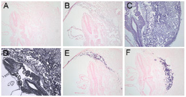
Fig. 4. In situ hybridization on E9.5 stage of mouse embryo development.
No probe (A), sense probe for LAT1 (B), anti-sense probe for LAT1 (C), anti-sense probe for PolydT (D), anti-sense probe for Pl1 (E), and anti-sense probe for Tpbpα (F). More homogenous expression of LAT1 is seen at E9.5 embryo development with slightly darker staining for TGC’s lining the implantation site (C). All negative controls (A, B) show no staining and positive controls (D, E, F) show either homogenous staining across all tissues for PolydT (D), or TGC specific staining for Pl1 (E) and Plf (not shown) and Spongiotrophoblast specific staining for Tpbpα (F).

