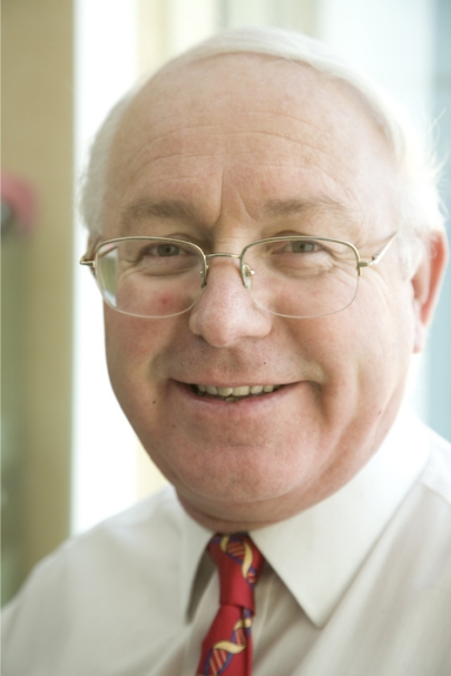
Sir Philip Cohen, a biochemist elected as a foreign associate to the National Academy of Sciences in 2008, remembers the moment in his distinguished career when his studies in cell signaling and protein phosphorylation took off. The moment arrived during a 1978 seminar given by Thomas Vanaman at the University of Dundee (Dundee, Scotland, UK). At that time, Cohen had been working on the first kinase to be studied, phosphorylase kinase, which helps to regulate the breakdown of glycogen. Cohen had purified the kinase to study how it was regulated by cAMP and calcium, but he could not untangle the mechanisms by which calcium activated it. That afternoon, Vanaman spoke about work he had done on calmodulin, a calcium-binding protein and a known intermediary in calcium-regulated processes. Vanaman's descriptions of calmodulin—its small size and heat stability—gave Cohen a better idea of the perplexing faint blue smear that he had observed at the bottom of his polyacrylamide gels. Cohen had been so focused on the massive kinase subunits controlled by cAMP that he had missed the small protein that ran off the end of the gel.
Philip Cohen
Rushing back to his laboratory after Vanaman's seminar, Cohen prepared a different type of gel and identified a small band ahead of the dye front—a band that appeared even after the sample had been boiled, indicating the component was heat-stable. “Then I knew what the answer was going to be,” Cohen said. “This must be the missing component.” Shortly after, Cohen's hypothesis was confirmed: calmodulin mediated the action of calcium ions on phosphorylase kinase.
Cohen has remained at the University of Dundee since 1971 and has worked on many areas of signal transduction, from protein phosphorylation to ubiquitination. His inaugural article, published in a recent issue of PNAS, identifies the phosphorylation sites on Pellino that activate this signaling protein that controls ubiquitination events in the innate immune system (1).
In the Beginning
Born just outside London in 1945, Philip Cohen spent much of his childhood exploring the woods around his house. His father worked as a printing ink technologist, designing color inks for use in several of Britain's major publications. The combination of an interest in natural history and an aptitude for chemistry led Cohen to pursue a career in biochemistry at University College London (London, UK). “Somehow I thought that biochemistry must be a cross between chemistry and bird-watching,” Cohen said, recalling that the subject initially did not hold much interest for him.
Only when he began his first independent laboratory projects in his third year at the university did he decide to continue with biochemistry. He discovered that, “with my own hands, I could actually find out something that no one had found out before.”
Cohen graduated from University College London with his Ph.D. in biochemistry in 1969, where he worked with Michael Rosemeyer in physical protein chemistry to identify the subunits of glucose-6-phosphate dehydrogenase. He then accepted a postdoctoral fellowship at the University of Washington (Seattle, WA) with future Nobel Laureate Edmond Fischer, examining protein phosphorylation and the regulation of glycogen metabolism.
At the time, protein phosphorylation was thought to be a specific mechanism confined to glycogen metabolism. In fact, the process is a near-universal regulatory system that evolved in early prokaryotes that regulates protein function in a simple, flexible, and—perhaps most importantly—reversible process. “When you attach a phosphate to a protein,” Cohen said, “it can do something simple like switch its activity on or off, but it can also stabilize a protein or allow it to be degraded or allow it to move from one part of the cell to another part of the cell.”
During his time in Seattle, Cohen primarily worked on glycogen phosphorylase. The subject began to draw more of his attention after he left Fischer's laboratory and took a position in the newly founded biochemistry department at the University of Dundee in 1971.
After his first eureka! moment when he discovered the connection between calmodulin and phosphorylase kinase, Cohen went on to discover other calmodulin-dependent enzymes, including the first calcium- and calmodulin-dependent phosphatase, calcineurin. Calcineurin was later shown by others to activate a transcription factor in T cells by removing phosphate groups, which then up-regulates the production of interleukin-2 and prompts T cell proliferation.
These immune-system effects took on increased medical importance when researchers discovered that the protein was the target of the immunosuppressant drug cyclosporine. Used mostly to prevent transplant rejection, cyclosporine had been on the market for 8 years before researchers discovered that it targeted calcineurin.
A Multiplicity Uncovered
Cohen's studies on phosphorylase kinase had unexpectedly led him to discover that it was phosphorylated at multiple sites. The other enzymes known to be regulated by phosphorylation only had one site. “It seemed to me an extra sophistication in the control system,” he said.
He stumbled across glycogen synthase in a literature review, and suspected that reports of a single phosphorylation site on the enzyme were wrong. As he investigated the protein, he found that not only did glycogen synthase have several phosphorylation sites, each phosphate group was introduced by a separate kinase. “So the more we studied this enzyme, actually, the more complicated it became,” Cohen said. “We eventually found it was phosphorylated on no less than 9 different sites by 7 different kinases.”
Cohen and his team identified a third kinase, glycogen synthase kinase 3 (GSK3), and showed that the phosphate groups added by GSK3 in muscle cells were removed within minutes after stimulating the cells with insulin. So, Cohen figured, either the insulin inactivated GSK3, or it turned on a phosphatase that removed the phosphate groups, which activated glycogen synthase.
He later established how insulin affected GSK3 through a series of chemical reactions regulated by phosphorylation. His research group showed that protein kinase B (PKB, also called Akt) switched off GSK3 activity and with Dario Alessi discovered that PKB was activated by 3-phosphoinositide-dependent protein kinase 1 (PDK1). These series of studies explained how insulin, acting through the “second messenger” phosphatidylinositol (3,4,5)-trisphosphate, activated glycogen synthase in muscle (2).
As he continued to work on glycogen metabolism, he also started to look at protein phosphorylation more globally, classifying the various protein phosphatases involved in different cellular processes. In 1983, he published a series of 7 articles that examined the different phosphatases, culminating in a review in Science that defined 4 different classes of protein phosphatases (3).
“Until we actually carried out that study,” Cohen said, “different people in the world were all working on what they thought were different phosphatases, each of which removed phosphate from a particular protein. However, what our studies showed is that actually everybody was working with the same set of 3 or 4 classes of enzyme.”
Another important finding that happened after this classification study, Cohen said, was a greater understanding of how 4 major phosphatase enzymes could recognize a myriad of enzymatic targets. He found a major part of the answer with the discovery that type 1 phosphatases have a glycogen recognition subunit that binds glycogen, thus localizing the phosphatase within a cell and bringing the enzyme into contact with potential substrates.
Research in the early 1990s by his laboratory and other groups showed that type 1 phosphatases have many other recognition subunits that localize them to different areas of the cell. “I think this has turned out to be very general, because we now know that many signaling proteins find their correct locations and substrates by analogous devices,” Cohen said.
A Clear Signal
While trying to understand the insulin signaling pathway, Cohen had wondered whether mitogen-activated protein kinases (MAPKs) played a role in this process. The classical MAPK signal transduction pathway links extracellular signals (mitogens) to downstream intracellular effects. Although the MAPK signaling cascade turned out not to relate specifically to insulin signaling, as Cohen had initially thought, it played a role in cellular growth and proliferation.
His work on MAPK signaling led to the identification and characterization of MAP kinase kinase 1 (MAP2K1), which integrates the extracellular signals by phosphorylating MAP kinase. Cohen's group then used a cell line whose stimulation by nerve growth factor (NGF) activated MAPK signaling and caused the cells to differentiate into a neuronal-like phenotype. The effect of NGF contrasted with previous studies showing that epidermal growth factor (EGF), which also activates the MAPK signaling pathway, does not cause neuronal differentiation.
“With my own hands, I could find out something no one had found before.”
“I found that there was an important difference between the way in which NGF and EGF were activating the pathway,” Cohen said. “The activation by EGF was actually very fast, but very transient, and it more or less finished after 30 min, whereas in the case of nerve growth factor, it kept up for many hours.” Neuronal differentiation, then, required the activation of the MAPK pathway for many hours, something Cohen went on to establish (4).
In their continued work on MAPK signaling pathways, Cohen and his colleagues identified a novel MAPK signaling module not activated by growth factors but rather by cell-damaging agents like UV radiation and heat shock.
Drug Discovery
In 1994, after Cohen was asked to serve on an advisory board for SmithKline Beecham, the pharmaceutical company presented data on a novel class of drug to treat rheumatoid arthritis—a drug that targeted the same p38 MAP kinase that Cohen had shown to be activated by cell-damaging agents. A later collaboration with Parke Davis Pharmaceuticals led to an understanding of how the first specific inhibitor of the classical MAPK pathway works to prevent the activation of MAP2K1 by the protein kinase Raf.
These collaborations opened Cohen's eyes not just to the therapeutic potentials of kinase inhibitors and their use as reagents in basic research, but also to the development of a relationship between industry and academia. Between 1996 and 1998, Cohen persuaded 6 pharmaceutical companies to help support his signal transduction unit, part of the protein phosphorylation unit he founded at the University of Dundee.
The corporate collaboration, which has recently been renewed for a third time, “led to the development of a number of new technologies, like kinase profiling, that have helped to accelerate the development of specific inhibitors of kinases, and to a lot of useful new tools and reagents for the cell signaling community,” Cohen said.
This research has resulted in the development of new therapeutic agents.
The “Next Big Thing”
A few years ago, Cohen switched his research to the study of the innate immune system, which led him to focus on a new area of cellular signaling: ubiquitination. Just as phosphorylation involves the reversible attachment of phosphate groups to enzymes, ubiquitination reversibly attaches to enzymes a small protein called ubiquitin. “The ubiquitination is actually more complicated than phosphorylation because you can form many types of polyubiquitin chains with huge numbers of those linked to each other,” he said.
Along with the ubiquitinases are de-ubiquitinases that remove the ubiquitin proteins from enzymes. Sequencing of the human genome revealed ≈500 protein kinases and 140 protein phosphatases, but >600 ubiquitin ligases and 100 de-ubiquitinating enzymes. “It's really clear that ubiquitination is going to be at least as important as phosphorylation as a control mechanism,” Cohen said.
Cohen received a grant from the Scottish government in 2008 for approximately $15 million (USD) to assemble a protein ubiquitination unit, similar to his current protein phosphorylation unit. He hopes to create another industry–academic partnership, because he believes that “the ubiquitin system is going to be the next big area for drug discovery.”
A Knightly Honor
Cohen still lives and works in Dundee, having seen the Scottish city transform into a bustling biotech center, in part, through his own laboratory and work. The University of Dundee currently employs >800 people in the life sciences, up from ≈20 when Cohen arrived in 1971.
Cohen's achievements have not gone unnoticed. He is widely cited in biology and biochemistry, was knighted in 1998, and received the Royal Medal for his work on protein phosphorylation in 2008.
Although he recently stepped down as director of research in the College of Life Sciences, he continues as Director of the Medical Research Council Protein Phosphorylation Unit and the Protein Ubiquitination Unit, with his work on the interplay between protein phosphorylation and ubiquitination and on building more partnerships between business and academia. “I'm still stumbling into lots of unexpected things in the innate immune system right now, which I wasn't expecting when I started working in this area,” he said.
Footnotes
This is a Profile of a recently elected member of the National Academy of Sciences to accompany the member's Inaugural Article on page 4584 in issue 12 of volume 106.
References
- 1.Smith H, et al. Identification of the phosphorylation sites on the E3 ubiquitin ligase Pellino that are critical for activation by IRAK1 and IRAK4. Proc Natl Acad Sci USA. 2009;106:4584–4590. doi: 10.1073/pnas.0900774106. [DOI] [PMC free article] [PubMed] [Google Scholar]
- 2.Alessi DR, et al. Characterisation of a 3-phos-phoinositide-dependent protein kinase which phosphorylates and actives protein kinase Bα. Curr Biol. 1997;7:261–269. doi: 10.1016/s0960-9822(06)00122-9. [DOI] [PubMed] [Google Scholar]
- 3.Ingebritsen TS, Cohen P. Protein phosphatases: Properties and role in cellular regulation. Science. 1983;221:331–338. doi: 10.1126/science.6306765. [DOI] [PubMed] [Google Scholar]
- 4.Traverse S, et al. Sustained activation of the mitogen-activated protein (MAP) kinase cascade may be required for differentiation of PC12 cells. Comparison of the effects of nerve growth factor and epidermal growth factor. Biochem J. 1992;288:351–355. doi: 10.1042/bj2880351. [DOI] [PMC free article] [PubMed] [Google Scholar]


