Abstract
Spiroplasma is widespread as a heritable bacterial symbiont in insects and some other invertebrates, in which it sometimes acts as a male-killer and causes female-biased sex ratios in hosts. Besides Wolbachia, it is the only heritable bacterium known from Drosophila, having been found in 16 of over 200 Drosophila species screened, based on samples of one or few individuals per species. To assess the extent to which Spiroplasma infection varies within and among species of Drosophila, intensive sampling consisting of 50–281 individuals per species was conducted for natural populations of 19 Drosophila species. Infection rates varied among species and among populations of the same species, and 12 of 19 species tested negative for all individuals. Spiroplasma infection never was fixed, and the highest infection rates were 60% in certain populations of D. hydei and 85% in certain populations of D. mojavensis. In infected species, infection rates were similar for males and females, indicating that these Spiroplasma infections do not confer a strong male-killing effect. These findings suggest that Spiroplasma has other effects on hosts that allow it to persist, and that environmental or host variation affects transmission or persistence leading to differences among populations in infection frequencies.
Introduction
Based on recent molecular surveys, heritable bacterial symbionts are widespread in arthropods, but, in most cases, their effects on hosts are unknown (e.g., [1], [2]. Drosophila species harbor only two types of heritable bacterial endosymbionts [3], [4]. The most widely studied, and the most common, is Wolbachia [5], [3]. The other heritable bacterial endosymbiont in Drosophila is Spiroplasma, now reported in a total of 16 species [6], [7], [8], [9], [3], [10] and, curiously, rarely found to coinfect with Wolbachia. In some Drosophila species, Spiroplasma causes male-killing [11], [12], [13], [8], while in others it does not [14], [3], [11]. Spiroplasma has been studied far less than Wolbachia, and factors underlying its distribution among and within Drosophila species are unknown.
Factors potentially affecting endosymbiont infection prevalence include the transmission fidelity of the bacteria and its effects on host fitness. Vertical transmission can exhibit high fidelity as evidenced by the decades-long persistence of Spiroplasma-positive strains of D. hydei and D. aldrichi in the Drosophila Species Stock Center [3]. Experimental studies show that temperature affects fidelity of maternal inheritance of Spiroplasma in Drosophila hosts, suggesting that infections may be influenced by climate or microhabitat [15], [16], [17]. Condition-dependent effects on host fitness or reproduction also can influence infection frequencies. Male-killing endosymbionts can be favored in conditions where female offspring benefit from reduced competition from their male siblings [18]. In other insects, heritable symbionts often provide defenses against temperature stress or natural enemies, leading to fitness advantages of infected lineages [19].
Field surveys from wild populations of D. hydei revealed infection rates of 23–66% of females, the highest levels yet reported for any Drosophila [14]. In contrast, infection of wild D. willistoni and D. nebulosa by male-killing Spiroplasma ranged from 1–6%, varying seasonally [6]. These earlier studies suggest interspecific differences in infection rates, but limitations in sampling design or extent prevent inference regarding infection patterns or dynamics. Rates of infection by male-killing compared to non-male-killing Spiroplasma within and among different Drosophila species need to be examined before the basis for infection and its persistence can be understood.
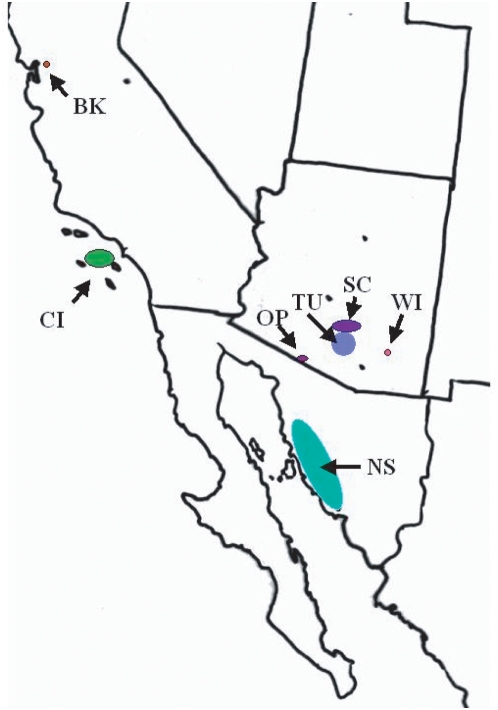
Drosophila species vary widely in their geographic distributions and ecologies [20]. The natural abundance of multiple Drosophila species at any given locality provides an opportunity to perform larger-scale screening in wild populations and to address questions about the ecological and evolutionary dynamics of Spiroplasma infections. We examined infection status in wild-caught females and males of 19 Drosophila species from localities (Figure 1) in the southwestern United States and northwestern Mexico in order to (1) ask how the incidence of infected flies varies in nature and (2) assess the sex ratio of infected flies in order to detect evidence of male killing infections. Our screen employed PCR primers universal for Spiroplasma, rather than those used to target male-killing strains, resulting in as complete detection as possible. Furthermore, a greater depth of sampling within each species allowed us to detect Spiroplasma infections at low frequencies.
Figure 1. Collection localities for Drosophila.
BK = Berkeley, CA, CI = Catalina Island, CA, OP = Organ Pipe Cactus Nat'l Mon, AZ, TU = Tucson, AZ, SC = Santa Catalina Mts, AZ, WI = Willcox, AZ, NS = Northwestern Sonora, MX.
Materials and Methods
Flies were collected at the localities shown in Figure 1 either directly from cactus (D. mojavensis), cave walls (D. macroptera, D. grisea), or from mushroom (D. tenebrosa) and banana baits (other species) (Table 1). Live flies were keyed to species and sex, maintained on species-appropriate culture medium for several days, and then frozen.
Table 1. Drosophila species screened, dates and locations of collection.
| Subgenus | Species | Collection Site | Date | Zone |
| Drosophila | D. aldrichi | Tucson, AZ | 2006–2007 | Desert |
| D. arizonae | Tucson, AZ | 2006–2007 | Desert | |
| NW Sonora, Mex. | 2006–2007 | Desert | ||
| Organ Pipe Natl. Mon. AZ | 2007 | Desert | ||
| D. carbonaria | Tucson, AZ | 2006–2008 | Desert | |
| D. grisea | Catalina Mts. AZ | 2007–2008 | Montane | |
| D. hamatofila | Catalina Isl., CA | 2002,2006–2007 | Coastal | |
| D. hydei | Tucson, AZ | 2006–2008 | Desert | |
| NW Sonora, Mex | 2006–2008 | Desert | ||
| Willcox, AZ | 2007 | Prairie | ||
| D. Immigrans | Berkeley, CA | 2007–2008 | Temperate | |
| Tucson, AZ | 2008 | Desert | ||
| D. macroptera | Catalina Mts., AZ | 2007 | Montane | |
| D. mettleri | Catalina Isl., CA | 2002, 2006–2007 | Coastal | |
| Tucson, AZ | 2006–2007 | Desert | ||
| NW Sonora, Mex | 2006–2007 | Desert | ||
| D. mojavensis | Catalina Isl., CA | 2007 | Coastal | |
| Organ Pipe Natl. Mon. AZ | 2007 | Desert | ||
| NW Sonora, Mex. | 2006–2007 | Desert | ||
| D. nigrospiracula | Organ Pipe Natl. Mon., AZ | 2007 | Desert | |
| Tucson, AZ | 2006–2007 | Desert | ||
| NW Sonora, Mex. | 2008 | Desert | ||
| D. pachea | Organ Pipe Natl. Mon., AZ | 2007 | Desert | |
| Tucson, AZ | 2007 | Desert | ||
| NW Sonora, Mex. | 2007 | Desert | ||
| D. rubrifrons | Catalina Mts., AZ | 2007 | Montane | |
| D. tenebrosa | Catalina Mts., AZ | 2007 | Montane | |
| D. wheeleri | Catalina Isl., CA | 2002, 2006 | Coastal | |
| Sophophora | D. simulans | Catalina Isl., CA | 2006–2007 | Coastal |
| Tucson, AZ | 2006–2008 | Desert | ||
| NW Sonora, Mex. | 2007–2008 | Desert | ||
| Catalina Mts., AZ | 2008 | Montane | ||
| D. melanogaster | NW Sonora, Mex. | 2007–2008 | Desert | |
| Tucson, AZ | 2006–2007 | Desert | ||
| D. pseudoobscura | Catalina Isl., CA | 2006 | Coastal | |
| Tucson, AZ | 2006–2008 | Desert | ||
| NW Sonora, Mex. | 2007 | Desert | ||
| Catalina Mts., AZ | 2008 | Montane | ||
| Dorsilopha | D. busckii | Berkeley, CA | 2007–2008 | Coastal |
| Tucson, AZ | 2008 | Desert |
DNA extraction from individual flies was carried out as previously described [3]. Briefly, whole flies were extracted with the single-fly squish prep protocol [21]. PCR screens for Spiroplasma were based on amplification of an approximately 410 base pair fragment of bacterial 16S rDNA using the spiroplasma-diagnostic primers 23f (5′-CTCAGGATGAACGCTGGCGGCAT-3′) and TKSSsp (TAGCCGTGGCTTTCTGGTAA [22]) and a touchdown thermal cycler program [3]. The initial screening PCR volume was 10 ul. These primers are expected to amplify almost all Spiroplasma strains and would amplify male-killing and non-male killing strains known from insects, based on comparison to sequence databases. The primers also have the potential to amplify some other groups of Bacteria.
To verify the identify of positive samples as Spiroplasma, each was re-amplified at larger volume (50 μl), and both strands were sequenced with an ABI 3700 at the University of Arizona's Genomics Analysis & Technology Core facility. As a check for DNA quality, all samples were screened for a fragment of mitochondrial cytochrome c oxidase I gene (COI) using primers HCO and LCO with an annealing temperature of 45°C [3]. Only samples that gave positive amplifications for COI were included in the survey. Sequences were edited and aligned using Mega 3.1 [23] and identified using Blastn [24] to query the nr database at GenBank.
Results
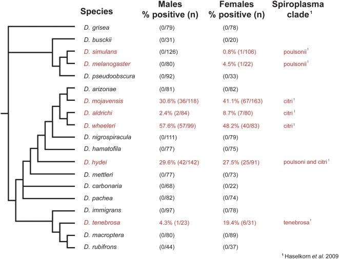
Of 19 Drosophila species screened, Spiroplasma was found in seven ( Figure 2). Infection incidence ranged from under 1% in D. simulans and D. melanogaster to an average of 37% in D. mojavensis. Some species are relatively rare in nature, such that fewer individuals were collected and screened. Sex differences in infection were not significant, although in the case of D. aldrichi the excess of infected females approached significance (X2 = 3.20, 0.10>p>0.05). In D. hydei, more than one Spiroplasma strain was distinguishable based upon 16S rDNA sequence, although no co-infections with distinct symbionts were observed within the same host [10], [3].
Figure 2. Frequency of Spiroplasma infection in wild-caught Drosophila.
The phylogenetic relationships of Drosophila are represented as a cladogram based on Markow & O'Grady [20] Spiroplasma-infected species are colored in red.
For two species, sampling permitted comparisons between localities (Table 2). For D. hydei, the proportion of infected flies was several times higher for samples from Willcox, Arizona than for samples from Sonora. For D. mojavensis, infection rate was higher at Santa Catalina Island than at Organ Pipe National Monument.
Table 2. Frequency of infection in populations of D hydei and of D mojavensis .
| Species | Population | Males | Females |
| D hydei | Northwestern Sonora, MX | 27.0% (34/126) | 24.7% (19/77) |
| Wilcox, AZ | 60.0% (6/10) | 60.0% (6/10) | |
| D mojavensis | Organ Pipe National Monument, AZ | 16.9% (13/77) | 14.0% (12/86) |
| Santa Catalina Island, CA | 84.6% (22/26) | 84.6% (55/65) |
Discussion
Our results represent the largest number of wild-caught insects screened to date for Spiroplasma. Over a third of the species screened showed Spiroplasma infection, though none of these species appeared to harbor a previously identified male-killing Spiroplasma strain. All of our positive samples were verified with sequencing. Although false negatives are possible (if our primers failed to amplify a novel strain), our screen would have detected known insect Spiroplasma strains, including male-killers and non-male-killers. A multi-locus sequence phylogenetic analysis of 69 of these Drosophila spiroplasmas revealed a large genetic diversity among Spiroplasma haplotypes. Based on this Bayesian phylogenetic analysis, the Drosophila spiroplasmas fall into four distinct, well-supported clades of the Spiroplasma phylogeny, with the most distantly related strain from the male-killing spiroplasmas having 14% sequence divergence at the 16S rDNA locus [10]. Furthermore, estimates of infection prevalence are likely to be conservative, as the sensitivity of our PCR screen may miss Drosophila with low Spiroplasma titer. Two infected species were in the subgenus Sophophora and five were in the subgenus Drosophila. Infection rates were considerably higher among infected species in the Drosophila subgenus compared to infected Sophophoran species. There was no pattern of infection related to geographic area.
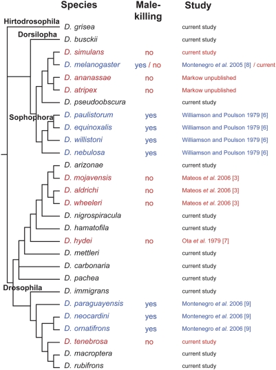
By screening both sexes for each species, we obtained indications as to whether Spiroplasma is acting as a male-killer, as known for some Drosophila [8]. In addition, each screening reaction had a positive control, the male-killing Spiroplasma infecting D. melanogaster [11]. Our primers were able to detect spiroplasmas up to 14% sequence divergent from the male-killing strain at the 16S rDNA locus. Other than for D. simulans and D. melanogaster, in which the infection frequency was under 1%, both sexes of infected species were found to be Spiroplasma-positive, indicating the absence of a strong male-killing phenotypes. Nor was the Spiroplasma found in the D. melanogaster female a male-killer, as the strain was established in culture and yielded infected flies of both sexes. Thus the male-killing effect does not appear to be a general explanation for the presence of Spiroplasma in these insects. Furthermore, as the number of Drosophila species found to be infected with Spiroplasma grows, the male-killing phenotype continues to be restricted to particular lineages, primarily the subgenus Sophophora and in the tripunctata radiation in the subgenus Drosophila (Figure 3).
Figure 3. Distribution of male-killing and non-male-killing Spiroplasma in natural populations of Drosophila species surveyed to date.
The phylogenetic relationships of Drosophila are represented as a cladogram based on Markow & O'Grady [20] Non-male-killing Spiroplasma-infected species are colored in red and male-killing Spiroplasma-infected species are in blue.
Host genotype clearly influences the distribution of Spiroplasma within as well as among Drosophila species. For example, D. willistoni shows intraspecific variation affecting Spiroplasma transmission [13], [25], [6]. Infection rates for natural populations of D. hydei in our study are similar to those reported by Kageyama et al. [14] reflecting a consistent pattern for this species from different global regions. Drosophila aldrichi, in which fewer than 10% of individuals were spiroplasma-positive, clearly shows a lower frequency of infected individuals of both sexes relative to D. hydei. In D. simulans, and D. melanogaster the infection level is even lower (Figure 2.) In contrast to the Wolbachia infections in D. innubila [26], infections with non-male-killing Spiroplasma appear to be more, as opposed to less, frequent than infections with male-killing types.
Though multiple factors likely affect spiroplasma prevalence, the fidelity of vertical transmission may play a role. Temperature affects maternal transmission of Spiroplasma in D. melanogaster and D. nebulosa [15], [17] and in D. hydei [16]. Similarly, field conditions including temperature influence maternal transmission efficiency of Wolbachia in Drosophila hosts [27], [28], [29], [30]. In our study, both D. mojavensis and D. hydei were collected from two locations and each showed a lower infection rate at the hotter site (Table 2). Transmission efficiency may be decreased at low temperatures, as shown experimentally for D. hydei [16], and also at the extreme high temperatures that occur at some desert localities sampled in our survey.
The variation in natural infection rates reported here, both among and within species, indicates a dynamic system in which infection, fitness effects and persistence of spiroplasmas in Drosophila are dependent upon the interplay of symbiont and host genotype and local environmental conditions. Given the ease of rearing and manipulating a range of evolutionarily, ecologically and genetically defined Drosophila species, our opportunities to disentangle and understand the roles of these factors are unparalleled.
Acknowledgments
We thank L. Matzkin, J. Bono, S. Castrezana, D. Gutilla, V. Corby-Harris, and the Catalina Island Conservancy for assistance in collecting flies, Steven Wasserman for comments on the manuscript and Becky Nankivell for assistance with Flyendo. Flies from México were obtained under permit FLOR 0030 from the Secretaría de Medio Ambiente y Recursos Naturales (SEMARNAT).
Footnotes
Competing Interests: The authors have declared that no competing interests exist.
Funding: This research was supported by NSF award DEB-0315815 to N.A.M. and T.A.M. The funders had no role in study design, data collection and analysis, decision to publish, or preparation of the manuscript.
References
- 1.Duron O, Bouchon D, Boutin S, Bellamy L, Zhou L, et al. The diversity of reproductive parasites among arthropods: Wolbachia do not walk alone. BMC Biol. 2008;6:27. doi: 10.1186/1741-7007-6-27. [DOI] [PMC free article] [PubMed] [Google Scholar]
- 2.Weinert LA, Tinsley MC, Temperley M, Jiggins FM. Are we underestimating the diversity and incidence of insect bacterial symbionts? A case study in ladybird beetles. Biol Lett. 2007;22:678–681. doi: 10.1098/rsbl.2007.0373. [DOI] [PMC free article] [PubMed] [Google Scholar]
- 3.Mateos M, Castrezana SJ, Nankivell BJ, Estes AM, Markow TA, et al. Heritable endosymbionts of Drosophila. . Genetics. 2006;174:363–376. doi: 10.1534/genetics.106.058818. [DOI] [PMC free article] [PubMed] [Google Scholar]
- 4.Flyendo: Drosophila endosymbiont database. Retrieved August 2008 from http://flyendoarlarizonaedu/ [Google Scholar]
- 5.Bourtzis K, Nirgianaki A, Markakis G, Savakis C. Wolbachia infection and cytoplasmic incompatibility in Drosophila species. Genetics. 1996;144:1063–1073. doi: 10.1093/genetics/144.3.1063. [DOI] [PMC free article] [PubMed] [Google Scholar]
- 6.Williamson DL, Poulson DF. Sex ratio organisms (Spiroplasmas) of Drosophila. . In: Whitcomb RF, Tully JG, editors. The Mycoplasmas. New York: Academic Press; 1979. pp. 175–208. [Google Scholar]
- 7.Ota T, Kawabe M, Oishi K, Poulson DF. Non-male-killing spiroplasmas in Drosophila hydei. . J Hered. 1979;70:211–213. [Google Scholar]
- 8.Montenegro H, Solferini VN, Klackzo LB, Hurst GDD. Male-killing Spiroplasma naturally infecting Drosophila melanogaster. . Insect Mol Biol. 2005;14:281–287. doi: 10.1111/j.1365-2583.2005.00558.x. [DOI] [PubMed] [Google Scholar]
- 9.Montenegro H, Hatadani LM, Medeiros HF, Klaczko LB. Male-killing in three species of the tripuncata radiation of Drosophila (Diptera: Drosophilidae). Journal of Zoological Systematics and Evolutionary Research. 2006;44:130–135. [Google Scholar]
- 10.Haselkorn TS, Markow TA, Moran NA. Multiple introductions of the Spiroplasma bacterial endosymbiont into Drosophila. . Molecular Ecology. 2009;18:1294–1305. [Google Scholar]
- 11.Pool JE, Wong A, Aquadro CF. Finding of male-killing Spiroplasma infecting Drosophila melanogaster in Africa implies transatlantic migration of this endosymbiont. Heredity. 2006;97:27–32. doi: 10.1038/sj.hdy.6800830. [DOI] [PMC free article] [PubMed] [Google Scholar]
- 12.Anbutsu H, Fukatsu T. Population dynamics of male-killing and non-male-killing spiroplasmas in Drosophila melanogaster. . Appl Environ Microbiol. 2003;69:1428–1434. doi: 10.1128/AEM.69.3.1428-1434.2003. [DOI] [PMC free article] [PubMed] [Google Scholar]
- 13.Ebbert MA. The interaction phenotype in the Drosophila willistoni spiroplasma symbiosis. Evolution. 1991;45:971–988. doi: 10.1111/j.1558-5646.1991.tb04364.x. [DOI] [PubMed] [Google Scholar]
- 14.Kageyama D, Anbutsu H, Watada M, Hosokawa T, Shimada M, et al. Prevalence of a non-male-killing Spiroplasma in natural populations of Drosophila hydei. . Appl Environ Microbiol. 2006;72:6667–6673. doi: 10.1128/AEM.00803-06. [DOI] [PMC free article] [PubMed] [Google Scholar]
- 15.Montenegro H, Klackzo LB. Low temperature cure of a male-killing agent in Drosophila melanogaster. . J Invert Pathology. 2004;86:50–51. doi: 10.1016/j.jip.2004.03.004. [DOI] [PubMed] [Google Scholar]
- 16.Osaka R, Nomura M, Watada M, Kagaeyama D. Negative effects of low temperatures on the vertical transmission and infection density of a Spiroplasma endosymbiont in Drosophila hydei. . Curr Microbiol. 2008;57:335–339. doi: 10.1007/s00284-008-9199-4. [DOI] [PubMed] [Google Scholar]
- 17.Anbutsu H, Goto S, Fukatsu T. High and low temperatures differently affect infection density and vertical transmission of male-killing Spiroplasma symbionts in Drosophila hosts. Appl Environ Microbiol. 2008;74:6053–6059. doi: 10.1128/AEM.01503-08. [DOI] [PMC free article] [PubMed] [Google Scholar]
- 18.Hurst GD, Jiggins F. Male-killing bacteria in insects: mechanisms, incidence and implications. Emerg Infect Dis. 2000;6:329–336. doi: 10.3201/eid0604.000402. [DOI] [PMC free article] [PubMed] [Google Scholar]
- 19.Moran NA, McCutcheon JP, Nakabachi A. Evolution and genomics of heritable bacterial symbionts. Annu Rev Genet. 2008;42:165–190. doi: 10.1146/annurev.genet.41.110306.130119. [DOI] [PubMed] [Google Scholar]
- 20.Markow TA, O'Grady PM. London: Academic Press; 2005. Drosophila: A Guide to Species Identification and Use. p. 250. [Google Scholar]
- 21.Gloor G, Engels W. Single-fly DNA preps for PCR. Drosophila Information Service. 1992;71:148–149. [Google Scholar]
- 22.Fukatsu T, Tsuchida T, Nikoh N, Koga R. Spiroplasma symbiont of the pea aphid, Acyrthosiphon pisum (Insecta: Homoptera). Applied and Environmental Microbiology. 2001;67:1284–1291. doi: 10.1128/AEM.67.3.1284-1291.2001. [DOI] [PMC free article] [PubMed] [Google Scholar]
- 23.Kumar S, Tamura K, Nei M. MEGA3, Integrated software for Molecular Evolutionary Genetics Analysis and sequence alignment. Briefings in Bioinformatics. 2004;5:150–163. doi: 10.1093/bib/5.2.150. [DOI] [PubMed] [Google Scholar]
- 24.Altschul SF, Gish W, Miller W, Myers EW, Lipman DJ. Basic local alignment search tool. J Mol Biol. 1990;215:403–410. doi: 10.1016/S0022-2836(05)80360-2. [DOI] [PubMed] [Google Scholar]
- 25.Malogolowkin C, Poulson DF, Wright EY. Experimental transfer of maternally inherited abnormal sex ratio in Drosophila willistoni. . Genetics. 1959;44:59–74. doi: 10.1093/genetics/44.1.59. [DOI] [PMC free article] [PubMed] [Google Scholar]
- 26.Dyer KA, Jaenike J. Evolutionarily stable infection by a male-killing endosymbiont in Drosophila innubila: molecular evidence from the host and parasite genomes. Genetics. 2004;168:1443–1445. doi: 10.1534/genetics.104.027854. [DOI] [PMC free article] [PubMed] [Google Scholar]
- 27.Olsen K, Reynolds KT, Hoffman AA. A field cage test of the effects of the endosymbiont Wolbachia on Drosophila melanogaster. . Heredity. 2001;86:731–737. doi: 10.1046/j.1365-2540.2001.00892.x. [DOI] [PubMed] [Google Scholar]
- 28.Hoffman AA, Hercus M, Hayat D. Population dynamics of the Wolbachia infection causing cytoplasmic incompatibility in Drosophila melanogaster. . Genetics. 1998;148:221–231. doi: 10.1093/genetics/148.1.221. [DOI] [PMC free article] [PubMed] [Google Scholar]
- 29.Hurst GDD, Johnson AP, von der Shulenberg JHG, Fuyama Y. Male-killing Wolbachia in Drosophila: a temperature-sensitive trait with a threshold bacterial density. Genetics. 2001;156:699–709. doi: 10.1093/genetics/156.2.699. [DOI] [PMC free article] [PubMed] [Google Scholar]
- 30.Clancy DJ, Hoffmann AA. Environmental effects on cytoplasmic incompatibility and bacterial load in Wolbachia-infected Drosophila simulans. Entomol Exp Appl. 1998;86:13–24. [Google Scholar]


