Abstract
CD8+ cytotoxic T lymphocytes (CTL) play an important role in controlling virus replication in HIV- and SIV-infected humans and monkeys, respectively. Three well-studied SIV CTL determinants are the two Mamu A*01-restricted epitopes Gag CM9 and Tat SL8, and the Mamu B*17-restricted epitope Nef IW9. Point mutations leading to amino acid replacements in these epitopes have been reported to mediate SIV escape from CTL control. We found that synthetic peptides containing mutations in SIV Gag CM9 and Tat SL8 were no longer recognized by the respective CTL. On the other hand, the described I-to-T replacement at the N-terminal amino acid residue of the SIV Nef IW9 epitope only moderately affected CTL recognition of the variant peptide, TW9. In an attempt to dissect the mechanism of escape of the Nef TW9 mutation, we investigated the effect of this mutation on CTL recognition of CD4+ T cells infected with an engineered SIVmac239 that contained the TW9 mutation in Nef. Although, the wild type and mutant virus both infected and efficiently replicated in rhesus macaque CD4+ T cells, the TW9 mutant virus failed to induce IFN-γ expression in an SIV Nef IW9-specific CTL clone. Thus, unlike escape from Gag CM9- or Tat SL8-specfic CTL control presumably by loss of epitope binding, these results point to a defect at the level of processing and/or presentation of the variant TW9 epitope with resultant loss of triggering of the cognate TCR on CTL generated against the wild type peptide. Our data highlight the value of functional assays using virus-infected target cells as opposed to peptide-pulsed APC when assessing relevant escape mutations in CTL epitopes.
Keywords: AIDS, CD8+ T-cell clones, immune escape, Nef, SIV
Introduction
Although virus-specific CTL can control HIV or SIV replication in infected humans and monkeys, respectively, (Allen et al., 2002; Barouch et al., 2001; Borrow et al., 1994; Egan et al., 2000; Froebel et al., 1994; Gruters, van Baalen, and Osterhaus, 2002; Kuroda et al., 1999; Rowland-Jones et al., 2001) the virus eventually overwhelms the immune system. Mutations in immunodominant CTL epitopes have been reported to mediate viral escape from CTL control (Allen et al., 2004; Allen et al., 2000a; Allen et al., 2000b; Barouch et al., 2002; Borrow et al., 1997; Goulder et al., 2001; Johnson and Desrosiers, 2002b; Leslie et al., 2004; O'Connor et al., 2002). These mutations could prevent either peptide binding to restricting MHC class I molecules or peptide-MHC class I-complex recognition by the cognate TCR. Alternatively, mutations can also affect proteolytic processing required to generate epitope peptides, leading to loss of presentation (Milicic et al., 2005). These escape mutations pose a major challenge in the development of putative HIV-1 vaccines aimed at inducing virus-specific CTL responses, hence, the role of different mutations within major HIV or SIV CTL epitopes in immune escape is the subject of intense investigation (Allen et al., 2004; Allen et al., 2000a; Allen et al., 2000b; Barouch et al., 2002; Borrow et al., 1997; Goulder et al., 2001; Johnson and Desrosiers, 2002b; Leslie et al., 2004; O'Connor et al., 2002). However, the oligoclonality of the CTL response to the virus in vivo and the possibility that CTL responses can co-evolve with viral escape variants in infected individuals has complicated this effort (Haas et al., 1996).
Many experimental SIV vaccination regimens are focused on eliciting CTL responses to one or a combination of the early expressed viral accessory proteins Tat and Nef, and the late expressed structural protein Gag (Amara et al., 2001; Barouch et al., 2002; Gallimore et al., 1995). Immunodominant epitopes within each of these proteins have been described, the most widely reported being the Mamu-A*01-restricted SIV Gag181-189CM9 (CTPYDINQM, Gag CM9) and Tat28-35SL8 (STPESANL, Tat SL8) epitopes, and the Mamu-B*17-restricted Nef165-173IW9 (IRYPKTFGW, Nef IW9) epitope (Allen et al., 2000b; Friedrich et al., 2004c; Loffredo et al., 2007; O'Connor et al., 2003). Mutations have been reported to occur within the Tat SL8 epitope very early (3-4 weeks) during acute infection (Allen et al., 2000b; Borrow et al., 1997; O'Connor et al., 2002) while mutations in the Gag CM9 epitope tend to occur much later (>12 months) during chronic infection (Barouch et al., 2002; O'Connor et al., 2002). This difference is apparently due to the negative impact of mutations within the Gag CM9 epitope on the replicative fitness of the virus with resultant extraepitopic compensatory mutations necessary for viral persistence in vivo (Friedrich et al., 2004a; Friedrich et al., 2004b).
Nef-specific CTL activity is thought to be an important component of the cell-mediated immune response necessary to control viral replication and spread (Gallimore et al., 1995). Besides its central role in promoting efficient viral replication (Johnson and Desrosiers, 2002a), Nef displays immunoregulatory properties such as downregulation of CD4 and MHC class I expression on virally-infected cells (Bell et al., 1998; Hrecka et al., 2005; Mangasarian et al., 1999; Piguet et al., 1999; Schwartz et al., 1996; Venzke et al., 2006), the latter protecting virally-infected cells from MHC class I-restricted T-cell recognition. A substitution of an isoleucine (IW9) with a threonine (TW9) residue at the N-terminus (p1) of the immunodominant SIV Nef IW9 epitope has been described (Friedrich et al., 2004c). Unlike the mutations in the Gag CM9 epitope, there is very limited information on the impact of the Nef TW9 mutation on viral replicative fitness (Friedrich et al., 2004c; O'Connor et al., 2002). Furthermore, the mechanism of escape of SIV Nef TW9 mutant virus from SIV Nef IW9-specific CTL recognition is not known.
In this study, we investigated the mechanisms behind CTL escape in three well studied immunodominat SIV epitopes. Using WT and variant synthetic peptides corresponding to the SIV Gag CM9, Tat SL8 and Nef IW9 epitopes and a mutant Nef TW9 virus, we show that while mutations within the Gag CM9 and the Tat SL8 epitopes abrogate binding to MHC class I and/or cognate TCR, the SIV Nef TW9 mutation abrogates CTL recognition by interfering with processing and/or presentation without negatively impacting viral replicative capacity.
Results
Mutant SIV Gag CM9 and Tat SL8 peptides are not recognized by SIV-specific CTL clones while mutant Nef IW9 peptide induces similar levels of IFN-γ expression as the WT peptide
The SIV Gag CM9 T2A or T2S, Tat SL8 S1P and Nef IW9 I1T (TW9) epitope mutations have been reported to occur in vivo in SIV infected Mamu A*01/B*17 positive rhesus macaques (Barouch et al., 2002; Friedrich et al., 2004a; Friedrich et al., 2004b; Friedrich et al., 2004c; O'Connor et al., 2002). To test the potential cross reactivity between the WT and variant peptides, we compared induction of IFN-γ expression in SIV Gag CM9-, Tat SL8-, or Nef IW9-specific CTL clones following co-culture with an autologous B-cell line pulsed with either SIV WT or the variant peptides.
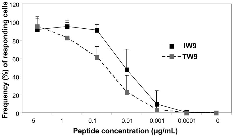
The SIV Gag CM9 T2A and Tat SL8 S1P mutations did not induce a measurable response at 5μg/mL (saturating peptide concentration) whereas the WT peptides produced strong responses (85.2% and 75.4% for Gag CM9 and Tat SL8, respectively) (Table 1). The Gag CM9 T2S variant peptide induced IFN-γ in only a small fraction (<5%) of the Gag CM9-specific CTL at the highest peptide concentration tested. This suggests that these mutations interfere with binding of the peptide to either the restricting MHC class I molecule or to the cognate T-cell receptor. In contrast, the variant Nef TW9 peptide induced IFN-γ expression in a Nef IW9-specific CTL clone at levels comparable to the WT IW9 peptide. Also when IFN-γ expression was evaluated against titrated amounts of the WT Nef IW9 or variant TW9 peptide in repeated experiments, the two peptides showed similar titration curves for IFN-γ induction in an SIV Nef IW9-specific CTL with a moderately lower frequency of cells responding to the variant peptide (Fig 1). Hence, both the WT Nef IW9 and variant TW9 peptides can efficiently induce cytokine expression in a CTL clone generated against the WT peptide, demonstrating that the variant TW9 peptide can bind the restricting MHC class I allele and the resulting complex can bind and trigger the cognate TCR of the WT IW9-specific CTL. We also observed that polyclonal CTL lines generated against the variant TW9-peptide showed a similar degree of cross-reactivity to WT IW9-peptide pulsed APC (data not shown), further showing a comparable ability of the TW9 peptide to bind both the restricting MHC class I molecule and the cognate TCR.
Table 1.
Intracellular cytokine responses to wild type and variant peptides
| SIV CTL epitope | Peptide sequencec | % IFN-γ producing cellsd |
|---|---|---|
| Gag CM9a | CTPYDINQM (wild type) | 85.2 |
| CAPYDINQM (T2A) | 0.1 | |
| CSPYDINQM (T2S) | 4.1 | |
| Tat SL8a | STPESANL (wild type) | 75.4 |
| PTPESANL (S1P) | 1.4 | |
| Nef IW9b | IRYPKTFGW (wild type) | 37.7 |
| TRYPKTFGW (I1T) | 39.2 |
Mamu A*01-restricted epitope.
Mamu B*17-restricted epitope.
Wild type peptide sequence and observed mutations in CTL epitopes in virus isolated from a chronically infected rhesus macaque.
Frequency of cells positive for IFN-γ in clonal population of CTL generated against WT peptide after stimulation with APC pulsed with 5 μg/mL of WT or variant peptide; analyses by ICS and flow cytometry.
Figure 1.
WT Nef IW9 and the variant Nef TW9 peptide elicit IFN-γ production in Nef IW9-specifc CTL. Herpesvirus papio-transformed B cells or CD4+ T cells pulsed with 10-fold serial dilutions of the IRYPKTFGW (IW9) or TRYPKTFGW (TW9) peptide (5 μg/mL down to 1 pg/mL) were used as APC to stimulate autologous SIV Nef IW9-specific CTL generated from an SIVmac239-infected rhesus macaque in a 5 h assay. The frequency of IFN-γ expressing CD8+ T cells was determined by flow cytometry. Shown are the means and SD at different peptide concentrations for three independent experiments
SIV Nef TW9 mutant virus retains its full capacity to infect and replicate in rhesus macaque CD4+ T cells but does not stimulate a Nef IW9-specific CTL clone
Given our finding that the WT Nef IW9-specific CTL can recognize APC pulsed with a Nef TW9 variant peptide, we next wanted to characterize a virus that expressed the Nef TW9 mutation in terms of infectivity, replicative capacity and recognition of mutant virus-infected cells by WT Nef IW9-specific CTL. To this end we engineered an SIVmac239 (Kestler et al., 1990) variant virus with the described I-to-T replacement at p1 of the Nef IW9 epitope (Fig. 2) and then infected rhesus macaque CD4+ T cells with either the WT or mutant virus. Viral infectivity, replication and spread were analyzed by PCR/RT-PCR and flow cytometry.
Figure 2.
Schematic representation of Nef gene sequence present in infectious molecular clone of SIVmac239. Nef reading frame begins at nucleotide 9348 and ends at 10139 (numbering of Regier and Desrosiers, 1990) (Regier and Desrosiers, 1990). A T to C point mutation was introduced at position 9841 in the downstream 3′ region (9840-9866) encoding the Mamu B*17-restricted SIV Nef IW9 epitope leading to an isoleucine to threonine switch at position 1 of the 9-mer Nef CTL epitope.
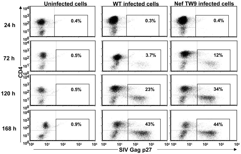
Rhesus macaque CD4+ T cells were effectively infected by the mutant SIV Nef TW9 virus (Fig 3). The kinetics of viral spread was similar for the Nef TW9 mutant and WT virus with the highest frequency of SIV Gag p27 positive CD4+ T cells seen around day 8 PI (Fig 3). In the absence of addition of more target cells, a massive loss of CD4+ T cells was observed in cultures of both the WT and mutant virus-infected cells beyond day 8 PI (data not shown). Similar levels of proviral DNA and viral RNA copies were observed in cultures of CD4+ T cells infected with the WT or mutant virus on day 8 PI (Table 2). Thus, the IW9 to TW9 switch in the immunodominant SIV Nef epitope did not have any observable effects on the virus infectivity and/or replication.
Figure 3.
Mutant SIVmac239 Nef TW9 infects and replicates in rhesus macaque CD4+ T cells at levels comparable with WT SIVmac239 Nef IW9 virus in vitro. CD4+ T cells from a rhesus macaque infected with WT SIVmac239 (Nef IW9) or the mutant Nef TW9 virus were cultured in 24-well tissue culture plates at a cell density of 1 × 10⁶ cells/mL. The frequency of SIV Gag p27 expressing CD4+ T cells was determined at 24, 72, 120 and 168 hours post infection by flow cytometry.
Table 2.
Quantification of virus
| Treatmenta | Proviral DNAb | Viral RNAc |
|---|---|---|
| Uninfected cells | d≤6.7×10² | d≤1.5×10³ |
| Nef IW9 infected cells | 1.5×10⁸ | 5.4×10⁹ |
| Nef TW9 infected cells | 2.2×10⁸ | 5.5×10⁹ |
Rhesus macaque CD4+ T cell clone, uninfected, infected with wild type SIVmac239 Nef IW9 or mutant Nef TW9.
DNA copies/100,000 cells determined by QPCR.
RNA copies Eq/mL cell culture supernatants determined by RT PCR. Analyses performed on day 8 post infection.
Assay sensitivity threshold.
Having demonstrated similar infectivity and replicative capacity of WT and mutant SIV Nef TW9 virus, we next investigated if the SIV-specific CTL clone generated against WT Nef IW9 peptide recognized CD4+ T cells infected with the mutant SIV Nef TW9 virus. Rhesus macaque CD4+ T cells infected with WT SIV Nef IW9 or mutant Nef TW9 virus were used as APC to stimulate an autologous SIV Nef IW9-specific CTL clone, and IFN-γ production by the virus-specific CTL assessed by flow cytometry.
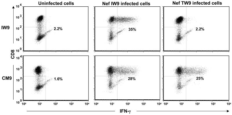
Although the frequency of SIV Gag p27 positive CD4+ T cells infected with the WT Nef IW9 or mutant Nef TW9 virus were similar in the day 8 PI cultures, only the WT Nef IW9-infected CD4+ T cells elicited intracellular IFN-γ expression in the SIV Nef IW9-specific CTL clone (Fig. 4). Hence, the CTL were capable of recognizing the mutant peptide sequence when presented as a preformed synthetic peptide, while the point mutation abrogated recognition of infected cells. An SIV Gag CM9-specific CTL clone responded to both the WT Nef IW9 and mutant Nef TW9 virus-infected CD4+ T cells.
Figure 4.
Rhesus macaque CD4+ T cells infected with the mutant SIVmac239 Nef TW9 virus fail to elicit IFN-γ production in a WT SIV Nef IW9-specific CD8+ T-cell clone. CD4+ T cells infected with either WT SIVmac239 (Nef IW9) or the mutant Nef TW9 virus were used as APC to stimulate an autologous SIV Nef IW9-specific CTL clone at a CD8+:CD4+ T cell ratio of 1:1. To allow for optimal numbers of infected target cells, day 8 post infection CD4+ T cells were used as stimulator cells. The frequency of IFN-γ-expressing CTL was determined by flow cytometry. An autologous SIV Gag CM9-specific CTL clone was included as a positive control.
Discussion
We have tested the effect on T-cell triggering by different reported SIV escape mutations using synthetic peptides corresponding to variant amino acid sequences and an engineered SIV mutant virus. Two different independent mutations in SIV Gag CM9 and one in Tat SL8 resulted in an almost complete loss of T cell triggering, demonstrating that these mutations impede peptide binding to either the MHC class I molecule or to the cognate TCR. In contrast, the I1T substitution in the SIV Nef IW9 CTL epitope only moderately affected CTL recognition as analyzed using peptide-pulsed APC. Further analysis of the effect of the SIV Nef TW9 mutation on viral replicative capacity using an engineered SIV Nef TW9 mutant virus showed that this single amino acid substitution had no discernible negative impact on viral replicative capacity. However, the I1T change was sufficient to abrogate recognition of SIV-infected cells by CTL generated against the WT SIV Nef IW9 sequence.
The efficient induction of intracellular IFN-γ expression in the CTL clone generated against the WT SIV Nef IW9 peptide by the variant SIV Nef TW9 peptide-pulsed APC suggests a preserved capacity of the variant peptide to bind the MHC class I molecule and effectively trigger the cognate T-cell receptor. This confirms and extends results of an in vivo study (Friedrich et al., 2004c) showing that Nef IW9-specific CTL lines generated from monkeys infected with WT virus recognized the altered sequences of the immunodominant peptide. In addition, the same group recently showed, using ELISpot, similar binding avidities of Nef IW9-specific CTL lines for WT and p1 variant peptides (I1A or I1T) but not peptide with concurrent variations at p1 and p6 (I1T + I6M) (Valentine et al., 2007). While the latter study involved IW9-specific CTL lines, the data together with our own current findings, suggest that the single amino acid change at p1 in the Nef IW9-epitope only moderately affects the binding avidity of the variant peptide to the MHC class I molecule or the cognate TCR. Thus, we reasoned that this single amino acid substitution mainly affects intracellular events occurring prior to MHC class I binding and subsequent interaction with the specific CTL.
To investigate the effect of the SIV Nef TW9 mutation on antigen processing and presentation in physiologically relevant SIV-infected autologous CD4+ T cells rather than using transformed B cell targets pulsed with synthetic peptides, we made a mutant virus expressing the Nef TW9 mutation and used infected CD4+ T cells as APC. Given reports of a fitness cost to the virus in vivo following mutations in immunodominant CTL epitopes in other SIV proteins such as Gag (Friedrich et al., 2004a; Friedrich et al., 2004b; Friedrich et al., 2004c), we assessed the replicative capacity of the engineered SIV Nef TW9 mutant virus in autologous CD4+ T cells prior to use as APC in vitro. We show here that the single amino acid replacement at the N-terminus of the immunodominant SIV Nef IW9 CTL epitope does not adversely affect the ability of the virus to infect and replicate in primary rhesus macaque CD4+ T cells. However, a very low number (<3%) of Nef IW9-specific CTL responded with IFN-γ production after exposure to cells infected with the mutant virus compared with WT virus. These data are in agreement with both an earlier study which showed that monkeys infected with an SIV mutant Nef virus did not generate a CTL response against the Nef IW9 WT epitope (Friedrich et al., 2004c), as well as with a recent paper by Valentine et al showing that cells infected with the Nef TW9 mutant virus were very poor APC for IW9-specific polyclonal T cell populations (Valentine et al., 2007). Together with the data using peptide-pulsed APC for CTL stimulation, these results point to a defect in the ability of SIV Nef TW9 mutant virus-infected cells to correctly generate and/or process the relevant epitope, rather than impaired binding to MHC class I or the TCR of the Nef IW9 specific T cells. It can not be ruled out, however, that the mutant TW9 peptide binds either the MHC or the TCR with slightly lower affinity, as indicated by the peptide titration. This could lead to a scenario where infected cells presenting low amounts of peptide would suboptimally present the TW9 epitope, leading to a lack of CTL activation. Alternatively, the variant TW9 epitope could be generated in virally-infected cells but at a lower efficiency with lower concentrations eventually being presented on the cell surface, leading to loss of TCR triggering. Regardless, the precise step/mechanism of the processing/presentation defect remains a subject worth investigating further. Bauer et al (Bauer et al., 1997) reported that structural constraints of HIV-1 Nef may curtail escape from HLA-B7-restricted CTL recognition. Our data showing lack of CTL recognition of mutant SIV Nef TW9-infected target cells, despite efficient recognition by the same CTL of APC pulsed with the variant TW9 peptide, suggest that such structural constrains do not apply for SIV Nef IW9 escape from Mamu B*17-restricted CTL.
Taken together, our data demonstrate that the I-to-T replacement at the N-terminal amino acid residue of the immunodominant SIV Nef IW9 epitope does not negatively impact viral replicative capacity but causes defective processing and/or presentation of the variant peptide with resultant loss of infected-cell recognition by the WT SIV Nef IW9-specific CTL. The ability to differentiate mutations that interfere directly with binding required for antigen-MHC class I complex formation and interaction with cognate TCR (e.g. SIV Gag CM9 T2A or T2S and Tat SL8 S1P) from those that prevent antigen processing and/or presentation (e.g. SIV Nef IW9 I1T) is an important tool for analyzing viral escape mutants. This highlights the importance of including functional assays using virus-infected target cells (Tsubota et al., 1989; Yang, 2003) as opposed to assays using peptide-pulsed APC in assessing CTL escape mutations.
Materials and Methods
Generation of SIV Gag181-189CM9-, Tat28-35SL8 and Nef165-173IW9-specific CD8+ and autologous CD4+ T-cell clones
SIV Gag181-189CM9- (CTPYDINQM, Gag CM9), Tat28-35SL8 (STPESANL, Tat SL8) and Nef165-173IW9 (IRYPKTFGW, Nef IW9)-specific CTL clones were isolated from a Mamu-A*01 and -B*17 double positive Indian rhesus macaque, Macaca mulatta, chronically infected with SIVmac239, as described previously (Andersen et al., 2007). Viral sequencing showed that in the chronic phase of infection the circulating plasma virus in this monkey contained the following mutations in the three immunodominant CTL epitopes; SIV Gag CM9 T2A and T2S, Tat SL8 S1P and Nef IW9 I1T (TW9) (Table 1 and unpublished data). PBMC from the monkey were stimulated for 1 week with irradiated autologous PBMC pulsed with CM9, SL8 or IW9 peptide (all from SynPep Corp., Dublin, CA). The cultures were restimulated weekly with CM9, SL8 or IW9 peptide-pulsed and irradiated autologous PBMC in the presence of recombinant human IL-2 (50 IU/mL: NIH AIDS Research and Reference Reagent Program, Germantown, MD). Following 2 rounds of stimulation, the CM9-, SL8- or IW9-specific CTL were cloned by limiting dilution and maintained essentially as described in Riddell et al., (Riddell and Greenberg, 1990) and Berger et al. (Berger et al., 2001), using bi-weekly anti-CD3 monoclonal antibody (mAb) (30 ng/mL; clone SP34-2; BD Biosciences, San Diego, CA, USA) stimulation with irradiated human PBMC and human Epstein Barr virus-transformed B-cells (TM B-LCL; kindly provided by Drs. S.R. Riddell and P.D. Greenberg, FHCRC, Seattle, WA) as feeder cells, but without anti-CD28 mAb stimulation. APC and feeder cells were irradiated in a Mark I Cs137 γ-irradiator (Shepherd & Associates, San Fernando, CA) at 6000 and 12500 rad for PBMC and TM B-LCL, respectively. Positive wells were tested for antigen specificity by flow cytometry using ICS for IFN-γ production following stimulation with cognate peptide-pulsed APC and by staining with CM9 (Beckman Coulter, Miami, FL), SL8 and IW9 (both from NIH AIDS Research and Reference Reagent Program) peptide/MHC-tetramers.
Autologous CD4+ T-cell clones were generated as described (Andersen et al., 2007). Briefly, highly enriched CD4+ T cells were isolated from PBMC or splenocytes by negative selection using Miltenyi LD columns and anti-CD8 microbeads followed by positive selection using MS columns and anti-CD4 microbeads (Miltenyi Biotec Inc., Auburn, CA). CD4+ T-cell clones were obtained after 2-weeks expansion in limiting dilution cultures containing irradiated human PBMC, IL-2 and anti-CD3 mAb and maintained as described above for CTL clones. In contrast to the CD8+ T-cell clones, the CD4+ T-cell clones did not have a defined antigenic specificity.
Virus stocks
The SIV isolate used in this study was derived from the molecular clone SIVmac239 (Genbank accession no. M33262.1) (Kestler et al., 1990). SIVmac239 Nef TW9 was generated by introducing a T to C point mutation at position 9841 in the downstream 3′ region (9840-9866) encoding the Mamu-B*17 restricted SIV Nef IW9 epitope leading to an isoleucine to threonine substitution at p1 of the 9-mer Nef CTL epitope. SIV virus stocks were produced by transfection of HEK293T cells with the wild type (Nef IW9) or mutant (Nef TW9) SIVmac239 using TransIt 293 reagent (Mirus Corp., Madison, WI) according to the manufacturer's recommendations. HEK293T cells were maintained in high glucose or Dulbecco modified Eagle medium (DMEM) supplemented with 10% heat-inactivated fetal calf serum, 2 mM L-glutamine, 50 U of penicillin per mL and 50 μg of streptomycin per mL (Invitrogen Corp., Carlsbad, CA, USA). Viral RNA content was determined as detailed below (see ‘Viral load analysis’).
Infection of a CD4+ T-cell clone with SIVmac239 Nef IW9 or Nef TW9
A clonal population of resting CD4+ T cells was activated with anti-CD3 mAb (T-25 flask-bound: 5 μg/mL) and IL-2 (50 IU/mL) for 48 h and then incubated with aliquots of freshly prepared virus stocks using the Viromag magnetofection reagents and procedures according to manufacturer's recommendation (OzBiosciences, Marseille, France). Approximately 0.25 mL virus stock containing 1.2 × 10⁹ viral RNA copies Eq/mL were added per 1×10⁶ CD4+ T cells. Incubations were carried out on a magnetic plate at 37°C in a humidified atmosphere of 5% CO2 for 2 h and the virus-exposed CD4+ T cells washed twice with PBS to remove residual non-incorporated viral material prior to use in assays.
Enumeration of virus-infected CD4+ T cells by intracellular staining for SIV Gag p27
Staining for surface markers (CD3 and CD4) and intracellular SIV Gag p27 was performed to assess the levels of SIV infection at different time points post infection (PI). All antibodies were obtained from BD Biosciences (San Diego, CA, USA) unless otherwise indicated. Cells were washed once in FACS surface buffer (PBS, 1% rat sera, 1% mouse sera, 0.05% sodium azide) and mAbs to the following surface markers conjugated to the indicated fluorochromes were added: CD4-PE or PerCP-Cy5.5 (clone L200) and CD3-APC (clone SP34-2). Cells were incubated for 30 minutes at 4°C, washed with sterile PBS and then fixed with 4% paraformaldehyde (PFA), 500 μL/sample tube for 30 minutes at 4°C. The cells were permeabilized (PBS, 0.1% saponin, 1% rat sera, 1% mouse sera and 0.05% sodium azide), 1 mL/sample tube for 5 minutes, washed (0.1% saponin in PBS) and incubated with FITC conjugated anti-SIV Gag p27 mAb (Clone 55-2F12, NIH AIDS Research and Reference Reagent Program). A final wash was performed and the cells resuspended in 0.1% PFA. Samples were acquired on a BD FACSCalibur flow cytometer (BD Bioscience) and subsequent data analyses performed using FCS Express Version 3 (De Novo Software, Thornhill, Ontario, Canada). Dead cells were excluded from the analyses based on forward versus side scatter gating, and at least 100,000 live cell events collected for each sample.
Measurement of IFN-γ expression by SIV-specific CTL clones in response to peptide-pulsed or SIV-infected target cells
SIV-specific CTL clones were co-cultured with autologous herpesvirus papio-transformed B-cell lines pulsed with dilutions of peptides in 5 mL polypropylene tubes. Alternatively, CD4+ T cells infected with WT Nef IW9 or mutant Nef TW9 virus were used as APC to stimulate the autologous SIV Nef IW9-specific CTL clone. The virus-infected CD4+ T cells had been in culture for 7 or 8 days prior to use as APC; in our system, the incubation period needed to achieve peak frequencies of SIV Gag p27 expressing target cells (Minang et al., In Press). A CTL:target cell ratio of 1:1 with a total of 1 × 10⁶ cells in a final volume of 2 ml per tube was used and non-pulsed autologous B cells or uninfected autologous CD4+ T cells were used as negative control APC. Monensin, 20 μL/test of a 1:20 dilution, (Golgi stop™; BD Bioscience) was added after an hour of incubation and the cultures incubated for additional 4 hours. Cells were washed, surface and intracellular stained, as described above, with PerCP-Cy5.5 conjugated anti-human CD8 mAb (clone SK1) and FITC conjugated antihuman IFN-γ mAb (clone 4S.B3), respectively. Samples were acquired and analyzed as described above.
Viral load analysis
Viral RNA was extracted from culture supernatant essentially as described previously (Cline et al., 2005) and viral replication quantified using FRET probe-based real time RT-PCR (TaqMan) as described (Cline et al., 2005; Lifson et al., 2001). All PCR reactions were run on ABI Prism 7700 Sequence Detection System and the fluorescent signal-based quantitation of viral RNA copy numbers in test samples were determined by ABI sequence detection software (Applied Biosystems, Foster City, CA).
Cell-associated DNA was extracted using the Qiagen DNA Mini Kit as recommended by the manufacturer (Qiagen, Valencia, CA, USA). The copy numbers of SIV DNA (gag-specific target) and that of the gene sequence for macaque CCR5, as reference for cell equivalents, were co-determined in a duplex format QPCR as described in Cline et al., (Cline et al., 2005) with omission of the reverse transcription step, and with the addition of primers (100 nM final concentration, each) and probe (100 nM final concentration) specific for the macaque CCR5 sequence and use of a cloned genomic fragment of the rhesus macaque CCR5 gene as standard (Genbank Acc. No. AF252567) (The Plasmid pR1-D, of the Macaca mulatta CCR5 gene, promoter region was a kind donation of Dr. Sunil K. Ahuja of the University of Texas Health Science Center San Antonio, San Antonio, Texas) (Mummidi et al., 2000). The duplex QPCR assay was run on an MX3000P™ instrument (Stratagene, La Jolla, CA) and results are reported as nominal SIV gag DNA copy numbers per 10,000 cell equivalents determined by copy numbers of the CCR5 sequence and based on 2 nominal copies of CCR5 per rhesus macaque cell (M. Piatak, unpublished results).
Acknowledgments
The authors thank Jeffrey D. Lifson for helpful discussions. The authors also thank Kelli Oswald and Lakeisha Galloway for help with viral load analyses as well as Yuan Li for help with sequencing virus from an infected rhesus macaque for mutations in the SIV Gag CM9, Tat SL8 and Nef IW9 epitopes. The following reagents were obtained through the AIDS Research and Reference Reagent Program, Division of AIDS, NIAID, NIH: IL-2 from Hoffman-La Roche Inc., NJ; IW9 and SL8 peptide/MHC tetramers from the NIH tetramer facility, Emory Vaccine Center, Emory University, Atlanta, GA. This project has been funded in whole or in part with federal funds from the National Cancer Institute, National Institutes of Health, under contract N01-CO-124000. The content of this publication does not necessarily reflect the views or policies of the Department of Health and Human Services, nor does mention of trade names, commercial products or organizations imply endorsement by the U.S. Government.
Footnotes
Publisher's Disclaimer: This is a PDF file of an unedited manuscript that has been accepted for publication. As a service to our customers we are providing this early version of the manuscript. The manuscript will undergo copyediting, typesetting, and review of the resulting proof before it is published in its final citable form. Please note that during the production process errors may be discovered which could affect the content, and all legal disclaimers that apply to the journal pertain.
References
- Allen TM, Altfeld M, Yu XG, O'Sullivan KM, Lichterfeld M, Le Gall S, John M, Mothe BR, Lee PK, Kalife ET, Cohen DE, Freedberg KA, Strick DA, Johnston MN, Sette A, Rosenberg ES, Mallal SA, Goulder PJ, Brander C, Walker BD. Selection, transmission, and reversion of an antigen-processing cytotoxic T-lymphocyte escape mutation in human immunodeficiency virus type 1 infection. J Virol. 2004;78(13):7069–78. doi: 10.1128/JVI.78.13.7069-7078.2004. [DOI] [PMC free article] [PubMed] [Google Scholar]
- Allen TM, Jing PC, Calore B, Horton H, O'Connor DH, Hanke T, Piekarczyk M, Ruddersdorf R, Mothe BR, Emerson C, Wilson N, Lifson JD, Belyakov IM, Berzofsky JA, Wang CX, Allison DB, Montefiori DC, Desrosiers RC, Wolinsky S, Kunstman KJ, Altman JD, Sette A, McMichael AJ, Watkins DI. Effects of cytotoxic T lymphocytes (CTL) directed against a single simian immunodeficiency virus (SIV) Gag CTL epitope on the course of SIVmac239 infection. J Virol. 2002;76(20):10507–10511. doi: 10.1128/JVI.76.20.10507-10511.2002. [DOI] [PMC free article] [PubMed] [Google Scholar]
- Allen TM, O'Connor DH, Jing P, Dzuris JL, Mothe BR, Vogel TU, Dunphy E, Liebl ME, Emerson C, Wilson N, Kunstman KJ, Wang X, Allison DB, Hughes AL, Desrosiers RC, Altman JD, Wolinsky SM, Sette A, Watkins DI. Tat-specific cytotoxic T lymphocytes select for SIV escape variants during resolution of primary viraemia. Nature. 2000a;407(6802):386–90. doi: 10.1038/35030124. [DOI] [PubMed] [Google Scholar]
- Allen TM, O'Connor DH, Jing PC, Dzuris JL, Mothe BR, Vogel TU, Dunphy E, Liebl ME, Emerson C, Wilson N, Kunstman KJ, Wang XC, Allison DB, Hughes AL, Desrosiers RC, Altman JD, Wolinsky SM, Sette A, Watkins DI. Tat-specific cytotoxic T lymphocytes select for SIV escape variants during resolution of primary viraemia. Nature. 2000b;407(6802):386–390. doi: 10.1038/35030124. [DOI] [PubMed] [Google Scholar]
- Amara RR, Villinger F, Altman JD, Lydy SL, O'Neil SP, Staprans SI, Montefiori DC, Xu Y, Herndon JG, Wyatt LS, Candido MA, Kozyr NL, Earl PL, Smith JM, Ma HL, Grimm BD, Hulsey ML, Miller J, McClure HM, McNicholl JM, Moss B, Robinson HL. Control of a mucosal challenge and prevention of AIDS by a multiprotein DNA/MVA vaccine. Science. 2001;292(5514):69–74. doi: 10.1126/science.1058915. [DOI] [PubMed] [Google Scholar]
- Andersen H, Barsov EV, Trivett MT, Trubey CM, Giavedoni LD, Lifson JD, Ott DE, Ohlen C. Transduction with human telomerase reverse transcriptase immortalizes a rhesus macaque CD8(+) T cell clone with maintenance of surface marker phenotype and function. AIDS Res Hum Retroviruses. 2007;23(3):456–65. doi: 10.1089/aid.2006.0194. [DOI] [PubMed] [Google Scholar]
- Barouch DH, Kunstman J, Kuroda MJ, Schmitz JE, Santra S, Peyerl FW, Krivulka GR, Beaudry K, Lifton MA, Gorgone DA, Montefiori DC, Lewis MG, Wolinsky SM, Letvin NL. Eventual AIDS vaccine failure in a rhesus monkey by viral escape from cytotoxic T lymphocytes. Nature. 2002;415(6869):335–9. doi: 10.1038/415335a. [DOI] [PubMed] [Google Scholar]
- Barouch DH, Santra S, Kuroda MJ, Schmitz JE, Plishka R, Buckler-White A, Gaitan AE, Zin R, Nam JH, Wyatt LS, Lifton MA, Nickerson CE, Moss B, Montefiori DC, Hirsch VM, Letvin NL. Reduction of simian-human immunodeficiency virus 89.6P viremia in rhesus monkeys by recombinant modified vaccinia virus Ankara vaccination. J Virol. 2001;75(11):5151–8. doi: 10.1128/JVI.75.11.5151-5158.2001. [DOI] [PMC free article] [PubMed] [Google Scholar]
- Bauer M, Lucchiari-Hartz M, Maier R, Haas G, Autran B, Eichmann K, Frank R, Maier B, Meyerhans A. Structural constraints of HIV-1 Nef may curtail escape from HLA-B7-restricted CTL recognition. Immunol Lett. 1997;55(2):119–22. doi: 10.1016/s0165-2478(96)02679-x. [DOI] [PubMed] [Google Scholar]
- Bell I, Ashman C, Maughan J, Hooker E, Cook F, Reinhart TA. Association of simian immunodeficiency virus Nef with the T-cell receptor (TCR) zeta chain leads to TCR down-modulation. J Gen Virol. 1998;79(Pt 11):2717–27. doi: 10.1099/0022-1317-79-11-2717. [DOI] [PubMed] [Google Scholar]
- Berger C, Huang ML, Gough M, Greenberg PD, Riddell SR, Kiem HP. Nonmyeloablative immunosuppressive regimen prolongs In vivo persistence of gene-modified autologous T cells in a nonhuman primate model. J Virol. 2001;75(2):799–808. doi: 10.1128/JVI.75.2.799-808.2001. [DOI] [PMC free article] [PubMed] [Google Scholar]
- Borrow P, Lewicki H, Hahn BH, Shaw GM, Oldstone MB. Virus-specific CD8+ cytotoxic T-lymphocyte activity associated with control of viremia in primary human immunodeficiency virus type 1 infection. J Virol. 1994;68(9):6103–10. doi: 10.1128/jvi.68.9.6103-6110.1994. [DOI] [PMC free article] [PubMed] [Google Scholar]
- Borrow P, Lewicki H, Wei X, Horwitz MS, Peffer N, Meyers H, Nelson JA, Gairin JE, Hahn BH, Oldstone MB, Shaw GM. Antiviral pressure exerted by HIV-1-specific cytotoxic T lymphocytes (CTLs) during primary infection demonstrated by rapid selection of CTL escape virus. Nat Med. 1997;3(2):205–11. doi: 10.1038/nm0297-205. [DOI] [PubMed] [Google Scholar]
- Cline AN, Bess JW, Piatak M, Jr, Lifson JD. Highly sensitive SIV plasma viral load assay: practical considerations, realistic performance expectations, and application to reverse engineering of vaccines for AIDS. J Med Primatol. 2005;34(56):303–12. doi: 10.1111/j.1600-0684.2005.00128.x. [DOI] [PubMed] [Google Scholar]
- Egan MA, Charini WA, Kuroda MJ, Schmitz JE, Racz P, Tenner-Racz K, Manson K, Wyand M, Lifton MA, Nickerson CE, Fu T, Shiver JW, Letvin NL. Simian immunodeficiency virus (SIV) gag DNA-vaccinated rhesus monkeys develop secondary cytotoxic T-lymphocyte responses and control viral replication after pathogenic SIV infection. J Virol. 2000;74(16):7485–95. doi: 10.1128/jvi.74.16.7485-7495.2000. [DOI] [PMC free article] [PubMed] [Google Scholar]
- Friedrich TC, Dodds EJ, Yant LJ, Vojnov L, Rudersdorf R, Cullen C, Evans DT, Desrosiers RC, Mothe BR, Sidney J, Sette A, Kunstman K, Wolinsky S, Piatak M, Lifson J, Hughes AL, Wilson N, O'Connor DH, Watkins DI. Reversion of CTL escape-variant immunodeficiency viruses in vivo. Nat Med. 2004a;10(3):275–81. doi: 10.1038/nm998. [DOI] [PubMed] [Google Scholar]
- Friedrich TC, Frye CA, Yant LJ, O'Connor DH, Kriewaldt NA, Benson M, Vojnov L, Dodds EJ, Cullen C, Rudersdorf R, Hughes AL, Wilson N, Watkins DI. Extraepitopic compensatory substitutions partially restore fitness to simian immunodeficiency virus variants that escape from an immunodominant cytotoxic-T-lymphocyte response. J Virol. 2004b;78(5):2581–5. doi: 10.1128/JVI.78.5.2581-2585.2004. [DOI] [PMC free article] [PubMed] [Google Scholar]
- Friedrich TC, McDermott AB, Reynolds MR, Piaskowski S, Fuenger S, De Souza IP, Rudersdorf R, Cullen C, Yant LJ, Vojnov L, Stephany J, Martin S, O'Connor DH, Wilson N, Watkins DI. Consequences of cytotoxic T-lymphocyte escape: common escape mutations in simian immunodeficiency virus are poorly recognized in naive hosts. J Virol. 2004c;78(18):10064–73. doi: 10.1128/JVI.78.18.10064-10073.2004. [DOI] [PMC free article] [PubMed] [Google Scholar]
- Froebel KS, Aldhous MC, Mok JY, Hayley J, Arnott M, Peutherer JF. Cytotoxic T lymphocyte activity in children infected with HIV. AIDS Res Hum Retroviruses. 1994;10 2:S83–8. [PubMed] [Google Scholar]
- Gallimore A, Cranage M, Cook N, Almond N, Bootman J, Rud E, Silvera P, Dennis M, Corcoran T, Stott J, et al. Early suppression of SIV replication by CD8+ nef-specific cytotoxic T cells in vaccinated macaques. Nat Med. 1995;1(11):1167–73. doi: 10.1038/nm1195-1167. [DOI] [PubMed] [Google Scholar]
- Goulder PJ, Brander C, Tang Y, Tremblay C, Colbert RA, Addo MM, Rosenberg ES, Nguyen T, Allen R, Trocha A, Altfeld M, He S, Bunce M, Funkhouser R, Pelton SI, Burchett SK, McIntosh K, Korber BT, Walker BD. Evolution and transmission of stable CTL escape mutations in HIV infection. Nature. 2001;412(6844):334–8. doi: 10.1038/35085576. [DOI] [PubMed] [Google Scholar]
- Gruters RA, van Baalen CA, Osterhaus AD. The advantage of early recognition of HIV-infected cells by cytotoxic T-lymphocytes. Vaccine. 2002;20(15):2011–5. doi: 10.1016/s0264-410x(02)00089-0. [DOI] [PubMed] [Google Scholar]
- Haas G, Plikat U, Debre P, Lucchiari M, Katlama C, Dudoit Y, Bonduelle O, Bauer M, Ihlenfeldt HG, Jung G, Maier B, Meyerhans A, Autran B. Dynamics of viral variants in HIV-1 Nef and specific cytotoxic T lymphocytes in vivo. J Immunol. 1996;157(9):4212–21. [PubMed] [Google Scholar]
- Hrecka K, Swigut T, Schindler M, Kirchhoff F, Skowronski J. Nef proteins from diverse groups of primate lentiviruses downmodulate CXCR4 to inhibit migration to the chemokine stromal derived factor 1. J Virol. 2005;79(16):10650–9. doi: 10.1128/JVI.79.16.10650-10659.2005. [DOI] [PMC free article] [PubMed] [Google Scholar]
- Johnson WE, Desrosiers RC. Viral persistance: HIV's strategies of immune system evasion. Annu Rev Med. 2002a;53:499–518. doi: 10.1146/annurev.med.53.082901.104053. [DOI] [PubMed] [Google Scholar]
- Johnson WE, Desrosiers RC. Viral persistence: HIV's strategies of immune system evasion. Annu Rev Med. 2002b;53:499–518. doi: 10.1146/annurev.med.53.082901.104053. [DOI] [PubMed] [Google Scholar]
- Kestler H, Kodama T, Ringler D, Marthas M, Pedersen N, Lackner A, Regier D, Sehgal P, Daniel M, King N, et al. Induction of AIDS in rhesus monkeys by molecularly cloned simian immunodeficiency virus. Science. 1990;248(4959):1109–12. doi: 10.1126/science.2160735. [DOI] [PubMed] [Google Scholar]
- Kuroda MJ, Schmitz JE, Charini WA, Nickerson CE, Lifton MA, Lord CI, Forman MA, Letvin NL. Emergence of CTL coincides with clearance of virus during primary simian immunodeficiency virus infection in rhesus monkeys. J Immunol. 1999;162(9):5127–33. [PubMed] [Google Scholar]
- Leslie AJ, Pfafferott KJ, Chetty P, Draenert R, Addo MM, Feeney M, Tang Y, Holmes EC, Allen T, Prado JG, Altfeld M, Brander C, Dixon C, Ramduth D, Jeena P, Thomas SA, St John A, Roach TA, Kupfer B, Luzzi G, Edwards A, Taylor G, Lyall H, Tudor-Williams G, Novelli V, Martinez-Picado J, Kiepiela P, Walker BD, Goulder PJ. HIV evolution: CTL escape mutation and reversion after transmission. Nat Med. 2004;10(3):282–9. doi: 10.1038/nm992. [DOI] [PubMed] [Google Scholar]
- Lifson JD, Rossio JL, Piatak M, Parks T, Li L, Kiser R, Coalter V, Fisher B, Flynn BM, Czajak S, Hirsch VM, Reimann KA, Schmitz JE, Ghrayeb J, Bischofberger N, Nowak MA, Desrosiers RC, Wodarz D. Role of CD8(+) lymphocytes in control of simian immunodeficiency virus infection and resistance to rechallenge after transient early antiretroviral treatment. J Virol. 2001;75(21):10187–10199. doi: 10.1128/JVI.75.21.10187-10199.2001. [DOI] [PMC free article] [PubMed] [Google Scholar]
- Loffredo JT, Burwitz BJ, Rakasz EG, Spencer SP, Stephany JJ, Vela JP, Martin SR, Reed J, Piaskowski SM, Furlott J, Weisgrau KL, Rodrigues DS, Soma T, Napoe G, Friedrich TC, Wilson NA, Kallas EG, Watkins DI. The antiviral efficacy of simian immunodeficiency virus-specific CD8+ T cells is unrelated to epitope specificity and is abrogated by viral escape. J Virol. 2007;81(6):2624–34. doi: 10.1128/JVI.01912-06. [DOI] [PMC free article] [PubMed] [Google Scholar]
- Mangasarian A, Piguet V, Wang JK, Chen YL, Trono D. Nef-induced CD4 and major histocompatibility complex class I (MHC-I) down-regulation are governed by distinct determinants: N-terminal alpha helix and proline repeat of Nef selectively regulate MHC-I trafficking. J Virol. 1999;73(3):1964–73. doi: 10.1128/jvi.73.3.1964-1973.1999. [DOI] [PMC free article] [PubMed] [Google Scholar]
- Milicic A, Price DA, Zimbwa P, Booth BL, Brown HL, Easterbrook PJ, Olsen K, Robinson N, Gileadi U, Sewell AK, Cerundolo V, Phillips RE. CD8+ T cell epitope-flanking mutations disrupt proteasomal processing of HIV-1 Nef. J Immunol. 2005;175(7):4618–26. doi: 10.4049/jimmunol.175.7.4618. [DOI] [PubMed] [Google Scholar]
- Mummidi S, Bamshad M, Ahuja SS, Gonzalez E, Feuillet PM, Begum K, Galvis MC, Kostecki V, Valente AJ, Murthy KK, Haro L, Dolan MJ, Allan JS, Ahuja SK. Evolution of human and non-human primate CC chemokine receptor 5 gene and mRNA. Potential roles for haplotype and mRNA diversity, differential haplotype-specific transcriptional activity, and altered transcription factor binding to polymorphic nucleotides in the pathogenesis of HIV-1 and simian immunodeficiency virus. J Biol Chem. 2000;275(25):18946–61. doi: 10.1074/jbc.M000169200. [DOI] [PubMed] [Google Scholar]
- O'Connor DH, Allen TM, Vogel TU, Jing PC, DeSouza IP, Dodds E, Dunphy EJ, Melsaether C, Mothe B, Yamamoto H, Horton H, Wilson N, Hughes AL, Watkins DI. Acute phase cytotoxic T lymphocyte escape is a hallmark of simian immunodeficiency virus infection. Nature Medicine. 2002;8(5):493–499. doi: 10.1038/nm0502-493. [DOI] [PubMed] [Google Scholar]
- O'Connor DH, Mothe BR, Weinfurter JT, Fuenger S, Rehrauer WM, Jing P, Rudersdorf RR, Liebl ME, Krebs K, Vasquez J, Dodds E, Loffredo J, Martin S, McDermott AB, Allen TM, Wang C, Doxiadis GG, Montefiori DC, Hughes A, Burton DR, Allison DB, Wolinsky SM, Bontrop R, Picker LJ, Watkins DI. Major histocompatibility complex class I alleles associated with slow simian immunodeficiency virus disease progression bind epitopes recognized by dominant acute-phase cytotoxic-T-lymphocyte responses. J Virol. 2003;77(16):9029–40. doi: 10.1128/JVI.77.16.9029-9040.2003. [DOI] [PMC free article] [PubMed] [Google Scholar]
- Piguet V, Gu F, Foti M, Demaurex N, Gruenberg J, Carpentier JL, Trono D. Nef-induced CD4 degradation: a diacidic-based motif in Nef functions as a lysosomal targeting signal through the binding of beta-COP in endosomes. Cell. 1999;97(1):63–73. doi: 10.1016/s0092-8674(00)80715-1. [DOI] [PubMed] [Google Scholar]
- Regier DA, Desrosiers RC. The complete nucleotide sequence of a pathogenic molecular clone of simian immunodeficiency virus. AIDS Res Hum Retroviruses. 1990;6(11):1221–31. doi: 10.1089/aid.1990.6.1221. [DOI] [PubMed] [Google Scholar]
- Riddell SR, Greenberg PD. The use of anti-CD3 and anti-CD28 monoclonal antibodies to clone and expand human antigen-specific T cells. J Immunol Methods. 1990;128(2):189–201. doi: 10.1016/0022-1759(90)90210-m. [DOI] [PubMed] [Google Scholar]
- Rowland-Jones SL, Pinheiro S, Kaul R, Hansasuta P, Gillespie G, Dong T, Plummer FA, Bwayo JB, Fidler S, Weber J, McMichael A, Appay V. How important is the ‘quality’ of the cytotoxic T lymphocyte (CTL) response in protection against HIV infection? Immunol Lett. 2001;79(12):15–20. doi: 10.1016/s0165-2478(01)00261-9. [DOI] [PubMed] [Google Scholar]
- Schwartz O, Marechal V, Le Gall S, Lemonnier F, Heard JM. Endocytosis of major histocompatibility complex class I molecules is induced by the HIV-1 Nef protein. Nat Med. 1996;2(3):338–42. doi: 10.1038/nm0396-338. [DOI] [PubMed] [Google Scholar]
- Tsubota H, Lord CI, Watkins DI, Morimoto C, Letvin NL. A cytotoxic T lymphocyte inhibits acquired immunodeficiency syndrome virus replication in peripheral blood lymphocytes. J Exp Med. 1989;169(4):1421–34. doi: 10.1084/jem.169.4.1421. [DOI] [PMC free article] [PubMed] [Google Scholar]
- Valentine LE, Piaskowski SM, Rakasz EG, Henry NL, Wilson NA, Watkins DI. Recognition of Escape Variants in Elispot Does Not Always Predict CD8+ T-Cell Recognition of SIV-Infected Cells Expressing the Same Variant Sequences. J Virol. 2007 doi: 10.1128/JVI.00275-07. [DOI] [PMC free article] [PubMed] [Google Scholar]
- Venzke S, Michel N, Allespach I, Fackler OT, Keppler OT. Expression of Nef downregulates CXCR4, the major coreceptor of human immunodeficiency virus, from the surfaces of target cells and thereby enhances resistance to superinfection. J Virol. 2006;80(22):11141–52. doi: 10.1128/JVI.01556-06. [DOI] [PMC free article] [PubMed] [Google Scholar]
- Yang OO. Will we be able to ‘spot’ an effective HIV-1 vaccine? Trends Immunol. 2003;24(2):67–72. doi: 10.1016/s1471-4906(02)00034-0. [DOI] [PubMed] [Google Scholar]


